. 2022 Dec 15;140(24):2626-2643.
doi: 10.1182/blood.2021014966.

S100A8/A9 drives the formation of procoagulant platelets through GPIbα

Affiliations

S100A8/A9 drives the formation of procoagulant platelets through GPIbα

Martina Colicchia et al. Blood. .

Abstract

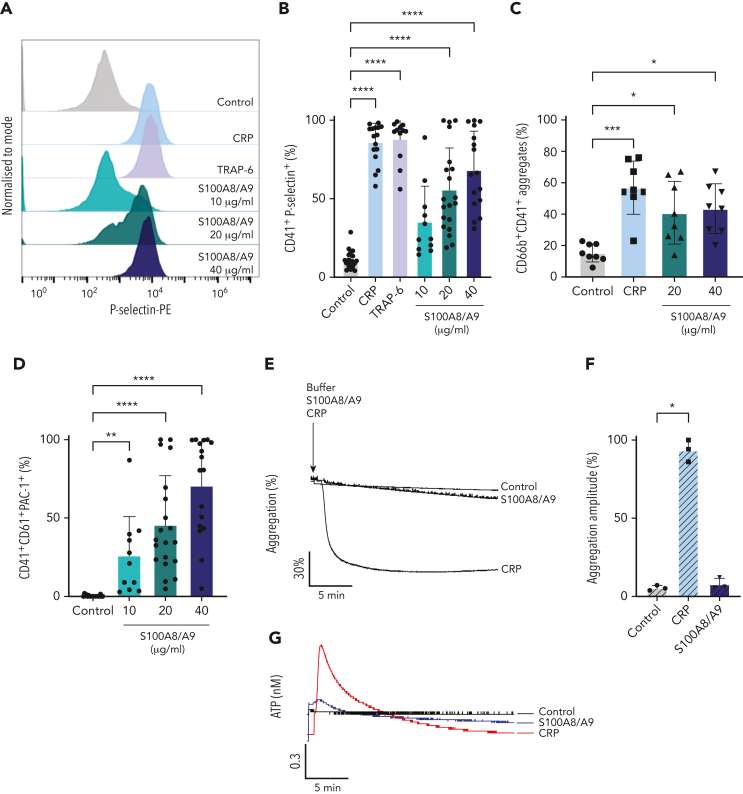
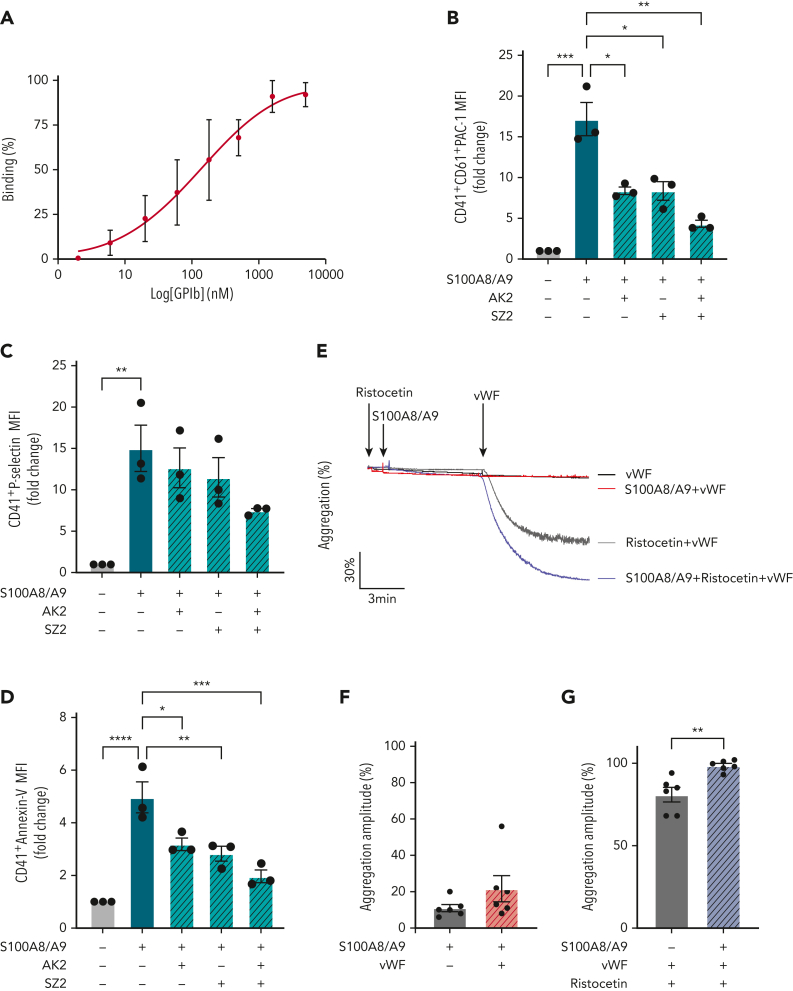
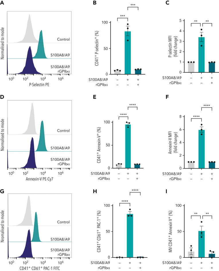
S100A8/A9, also known as "calprotectin" or "MRP8/14," is an alarmin primarily secreted by activated myeloid cells with antimicrobial, proinflammatory, and prothrombotic properties. Increased plasma levels of S100A8/A9 in thrombo-inflammatory diseases are associated with thrombotic complications. We assessed the presence of S100A8/A9 in the plasma and lung autopsies from patients with COVID-19 and investigated the molecular mechanism by which S100A8/A9 affects platelet function and thrombosis. S100A8/A9 plasma levels were increased in patients with COVID-19 and sustained high levels during hospitalization correlated with poor outcomes. Heterodimeric S100A8/A9 was mainly detected in neutrophils and deposited on the vessel wall in COVID-19 lung autopsies. Immobilization of S100A8/A9 with collagen accelerated the formation of a fibrin-rich network after perfusion of recalcified blood at venous shear. In vitro, platelets adhered and partially spread on S100A8/A9, leading to the formation of distinct populations of either P-selectin or phosphatidylserine (PS)-positive platelets. By using washed platelets, soluble S100A8/A9 induced PS exposure but failed to induce platelet aggregation, despite GPIIb/IIIa activation and alpha-granule secretion. We identified GPIbα as the receptor for S100A8/A9 on platelets inducing the formation of procoagulant platelets with a supporting role for CD36. The effect of S100A8/A9 on platelets was abolished by recombinant GPIbα ectodomain, platelets from a patient with Bernard-Soulier syndrome with GPIb-IX-V deficiency, and platelets from mice deficient in the extracellular domain of GPIbα. We identified the S100A8/A9-GPIbα axis as a novel targetable prothrombotic pathway inducing procoagulant platelets and fibrin formation, in particular in diseases associated with high levels of S100A8/A9, such as COVID-19.


Conflict of interest statement

Conflict-of-interest disclosure: This work is protected by UoB patent (Patent Application Number: PCT/GB2022/052007). The authors declare no competing financial interests.

Figures

None
Graphical abstract
Figure 1.
Figure 1.
S100A8/A9 accelerates the recruitment of annexin-V–positive platelets and fibrin generation at a venous shear rate. (A-C) Plasma levels of S100A8/A9 were measured by ELISA in healthy donors and patients with uncomplicated and complicated (ICU survivors/nonsurvivors) COVID-19 over 7 consecutive days after inclusion in the study. (A) S100A8/A9 levels in the plasma of healthy donors (control, n = 10), uncomplicated COVID-19 patients (n = 48), ICU survivors (n = 26), and ICU nonsurvivors (n = 13) on the first day of patient inclusion in the study. (B-C) S100A8/A9 levels in the plasma over 7 consecutive days after patient recruitment. (D-G) Representative immunofluorescence staining of lung autopsies obtained from COVID-19 patients or age-matched control. Images were captured using Epi fluorescence microscope and slide scanner Axioscan Z1. (D) Platelet CD42b and heterodimeric S100A8/A9 staining in lung parenchyma and microcirculation in COVID-19 and control lung autopsies. (E) Staining for S100A8/A9, MPO, and nuclei (DAPI) of a large vessel in the lung of a patient with COVID-19 with thrombotic complications. (F) Staining for platelet CD42b, fibrin, and nuclei (DAPI) in large thrombi of a patient with COVID-19. (G) S100A9, CD42b, and fibrin staining in COVID-19 lung. Arrow shows platelet-fibrin microaggregates. (H) Whole blood under recalcified conditions was perfused at a venous shear rate (100 s−1) over S100A8/A9 (40 μg/mL), collagen (100 μg/mL), or combination of S100A8/A9 and collagen-coated chambers. The presence of PS-positive platelets and fibrin were assessed using annexin-V-Alexa Fluor-647 and FITC-anti-fibrinogen/fibrin antibody, respectively. (H) Representative images taken at 15 minutes. (I) Quantification of annexin-V signal (3 minutes) (n = 5) and (J) fibrin signal time course (0-15 minutes) (n = 5) using predefined semiautomated scripts in Fiji as detailed in the supplemental Methods (available on the Blood website). Results are shown as mean ± standard error of the mean (SEM). The statistical significance was analyzed using two-way ANOVA with Tukey’s multiple comparison test between all groups. ∗P < .05, ∗∗P < .005, ∗∗∗P < .001, ∗∗∗∗P < .0001.
Figure 1.
Figure 1.
S100A8/A9 accelerates the recruitment of annexin-V–positive platelets and fibrin generation at a venous shear rate. (A-C) Plasma levels of S100A8/A9 were measured by ELISA in healthy donors and patients with uncomplicated and complicated (ICU survivors/nonsurvivors) COVID-19 over 7 consecutive days after inclusion in the study. (A) S100A8/A9 levels in the plasma of healthy donors (control, n = 10), uncomplicated COVID-19 patients (n = 48), ICU survivors (n = 26), and ICU nonsurvivors (n = 13) on the first day of patient inclusion in the study. (B-C) S100A8/A9 levels in the plasma over 7 consecutive days after patient recruitment. (D-G) Representative immunofluorescence staining of lung autopsies obtained from COVID-19 patients or age-matched control. Images were captured using Epi fluorescence microscope and slide scanner Axioscan Z1. (D) Platelet CD42b and heterodimeric S100A8/A9 staining in lung parenchyma and microcirculation in COVID-19 and control lung autopsies. (E) Staining for S100A8/A9, MPO, and nuclei (DAPI) of a large vessel in the lung of a patient with COVID-19 with thrombotic complications. (F) Staining for platelet CD42b, fibrin, and nuclei (DAPI) in large thrombi of a patient with COVID-19. (G) S100A9, CD42b, and fibrin staining in COVID-19 lung. Arrow shows platelet-fibrin microaggregates. (H) Whole blood under recalcified conditions was perfused at a venous shear rate (100 s−1) over S100A8/A9 (40 μg/mL), collagen (100 μg/mL), or combination of S100A8/A9 and collagen-coated chambers. The presence of PS-positive platelets and fibrin were assessed using annexin-V-Alexa Fluor-647 and FITC-anti-fibrinogen/fibrin antibody, respectively. (H) Representative images taken at 15 minutes. (I) Quantification of annexin-V signal (3 minutes) (n = 5) and (J) fibrin signal time course (0-15 minutes) (n = 5) using predefined semiautomated scripts in Fiji as detailed in the supplemental Methods (available on the Blood website). Results are shown as mean ± standard error of the mean (SEM). The statistical significance was analyzed using two-way ANOVA with Tukey’s multiple comparison test between all groups. ∗P < .05, ∗∗P < .005, ∗∗∗P < .001, ∗∗∗∗P < .0001.
Figure 2.
Figure 2.
Recombinant S100A8/A9 induces human platelet activation in vitro. Human washed platelets (106 platelets/condition) were incubated with different concentrations of recombinant heterodimeric S100A8/A9 (10, 20, and 40 μg/mL) for 30 minutes at 37°C. (A-B) Platelet activation was determined by flow cytometry using anti–P-selectin antibody. (A) Representative plots from a healthy donor; histograms normalized to mode (each peak normalized to its mode for each condition). (B) Percentage of CD41+P-selectin+ platelets. (C) The percentage of platelet-neutrophil aggregates (CD66b+/CD41+) in whole blood after addition of S100A8/A9 for 30 minutes at 37°C (n = 7). Blood was diluted 1:5. (D) Anti-CD41/CD61 PAC-1 antibody (against activated GPIIb/IIIa) binding to platelets was assessed by flow cytometry and presented as percentage of CD41+ platelets positive for PAC-1 (n = 17). (E-F) Washed platelets (2×10⁸/mL) were incubated with S100A8/A9 (40 μg/mL) or CRP (10 μg/mL), and platelet aggregation was assessed for 20 minutes by light transmission aggregometry (n = 3). (G-H) Washed platelets (2 × 10⁸/mL) were incubated with S100A8/A9 (40 μg/mL) or CRP (10 μg/mL), and ATP generation was assessed for 6 minutes with the CHRONO-LUME luciferin:luciferase assay kit from Chronolog (n = 3). (E,G) Representative traces. (I-J) Washed platelets (2 × 108/mL) were incubated with S100A8/A9 (20 μg/mL or 40 μg/mL) for 6 minutes under stirring condition followed by the addition of exogenous fibrinogen (200 μg/mL). Platelet aggregation was assessed for 6 minutes by light transmission aggregometry (n = 3). (K-L) Alexa-Fluor 488-labeled fibrinogen binding to platelets. (K) Representative plot for fibrinogen binding. (L) Percentage of platelets positive for fibrinogen-Alexa Fluor 488, CRP (10 μg/mL) and TRAP-6 (100 μM) were used as positive control. Data are shown as mean ± SD. The statistical significance was analyzed using ordinary one-way ANOVA. ∗P < .05, ∗∗P < .005, ∗∗∗P < .001, ∗∗∗∗P < .0001.
Figure 2.
Figure 2.
Recombinant S100A8/A9 induces human platelet activation in vitro. Human washed platelets (106 platelets/condition) were incubated with different concentrations of recombinant heterodimeric S100A8/A9 (10, 20, and 40 μg/mL) for 30 minutes at 37°C. (A-B) Platelet activation was determined by flow cytometry using anti–P-selectin antibody. (A) Representative plots from a healthy donor; histograms normalized to mode (each peak normalized to its mode for each condition). (B) Percentage of CD41+P-selectin+ platelets. (C) The percentage of platelet-neutrophil aggregates (CD66b+/CD41+) in whole blood after addition of S100A8/A9 for 30 minutes at 37°C (n = 7). Blood was diluted 1:5. (D) Anti-CD41/CD61 PAC-1 antibody (against activated GPIIb/IIIa) binding to platelets was assessed by flow cytometry and presented as percentage of CD41+ platelets positive for PAC-1 (n = 17). (E-F) Washed platelets (2×10⁸/mL) were incubated with S100A8/A9 (40 μg/mL) or CRP (10 μg/mL), and platelet aggregation was assessed for 20 minutes by light transmission aggregometry (n = 3). (G-H) Washed platelets (2 × 10⁸/mL) were incubated with S100A8/A9 (40 μg/mL) or CRP (10 μg/mL), and ATP generation was assessed for 6 minutes with the CHRONO-LUME luciferin:luciferase assay kit from Chronolog (n = 3). (E,G) Representative traces. (I-J) Washed platelets (2 × 108/mL) were incubated with S100A8/A9 (20 μg/mL or 40 μg/mL) for 6 minutes under stirring condition followed by the addition of exogenous fibrinogen (200 μg/mL). Platelet aggregation was assessed for 6 minutes by light transmission aggregometry (n = 3). (K-L) Alexa-Fluor 488-labeled fibrinogen binding to platelets. (K) Representative plot for fibrinogen binding. (L) Percentage of platelets positive for fibrinogen-Alexa Fluor 488, CRP (10 μg/mL) and TRAP-6 (100 μM) were used as positive control. Data are shown as mean ± SD. The statistical significance was analyzed using ordinary one-way ANOVA. ∗P < .05, ∗∗P < .005, ∗∗∗P < .001, ∗∗∗∗P < .0001.
Figure 3.
Figure 3.
S100A8/A9 induces the formation of procoagulant platelets. Human washed platelets (106 platelets/condition) were incubated with different concentrations of S100A8/A9 (10, 20 and 40 μg/mL) for 30 minutes at 37°C. (A-C) PS exposure was determined by flow cytometry using PE-Cy7–labeled annexin-V (n = 17). CRP (10 μg/mL) and TRAP-6 (100 μM) were used as positive control. (A) Representative histogram for annexin-V staining. Representative histograms normalized to mode (each peak normalized to its mode for each condition). (B) Percentage of CD41+annexin-V+ platelets. (C) Fold change in the mean fluorescent intensity (MFI) of agonist-activated platelets over control. (D) The percentage of CD41+MVs positive for annexin-V (n = 6). (E-F) Human washed platelets spreading on collagen or S100A8/A9. Representative differential interference contrast (DIC) images of spread platelets. (F) Representative annexin-V (magenta) and P-selectin (green) staining for adherent platelets. (G) Quantification of annexin-V–positive, P-selectin–positive, or annexin-V and P-selectin–double positive platelets adherent on collagen and S100A8/A9 (positive platelets/total platelets). The statistical significance was analyzed using two-way ANOVA with Tukey’s multiple comparison test between all groups. (H-I) Assessment of intracellular Ca2+ release from platelets spread over surfaces coated with S100A8/A9 (20 and 40 μg/mL), fibrinogen, or bovine serum albumin assessed using BAPTA-1-AM Ca2+ sensitive dye. (H) Representative Ca2+ traces assessed using Oregon Green-488 BAPTA-1-AM. (I) Peak fluorescence at time point zero (F0/Fmax). Data are shown as mean ± SD. The statistical significance was analyzed using a nonparametric test (Kruskal-Wallis test). ∗P < .05, ∗∗P < .005, ∗∗∗P < .001, ∗∗∗∗P < .0001.
Figure 3.
Figure 3.
S100A8/A9 induces the formation of procoagulant platelets. Human washed platelets (106 platelets/condition) were incubated with different concentrations of S100A8/A9 (10, 20 and 40 μg/mL) for 30 minutes at 37°C. (A-C) PS exposure was determined by flow cytometry using PE-Cy7–labeled annexin-V (n = 17). CRP (10 μg/mL) and TRAP-6 (100 μM) were used as positive control. (A) Representative histogram for annexin-V staining. Representative histograms normalized to mode (each peak normalized to its mode for each condition). (B) Percentage of CD41+annexin-V+ platelets. (C) Fold change in the mean fluorescent intensity (MFI) of agonist-activated platelets over control. (D) The percentage of CD41+MVs positive for annexin-V (n = 6). (E-F) Human washed platelets spreading on collagen or S100A8/A9. Representative differential interference contrast (DIC) images of spread platelets. (F) Representative annexin-V (magenta) and P-selectin (green) staining for adherent platelets. (G) Quantification of annexin-V–positive, P-selectin–positive, or annexin-V and P-selectin–double positive platelets adherent on collagen and S100A8/A9 (positive platelets/total platelets). The statistical significance was analyzed using two-way ANOVA with Tukey’s multiple comparison test between all groups. (H-I) Assessment of intracellular Ca2+ release from platelets spread over surfaces coated with S100A8/A9 (20 and 40 μg/mL), fibrinogen, or bovine serum albumin assessed using BAPTA-1-AM Ca2+ sensitive dye. (H) Representative Ca2+ traces assessed using Oregon Green-488 BAPTA-1-AM. (I) Peak fluorescence at time point zero (F0/Fmax). Data are shown as mean ± SD. The statistical significance was analyzed using a nonparametric test (Kruskal-Wallis test). ∗P < .05, ∗∗P < .005, ∗∗∗P < .001, ∗∗∗∗P < .0001.
Figure 4.
Figure 4.
CD36 blockade partially decreases GPIIb/IIIa activation and PS exposure, whereas immunoreceptor tyrosine-based activation motif receptor inhibition reduces P-selectin expression. Human washed platelets (106 platelets/condition) were incubated with S100A8/A9 (20 μg/mL) with or without different inhibitors for 30 minutes at 37°C. Inhibitors were preincubated with platelets for 10 minutes before addition of S100A8/A9: Paquinimod (blocks S100A9 binding to TLR4) (10 μM), RAGE inhibitor Azeliragon (1 μM), CD36 inhibitor SSO (25 μM), Syk inhibitor PRT-060318 (10 μM), Src inhibitor PP2 (20 μM), and BTK inhibitor Ibrutinib (500 nM) were used. Platelet activation was determined by flow cytometry using (A,D,G) anti-CD41/CD61 PAC-1 antibody, (B,E,H) anti–P-selectin antibody, and (C,F,I) annexin-V binding. Data are shown as the percentage of platelets positive (CD41+) for these markers. Data are shown as mean ± SD. Statistical significance was analyzed using ordinary one-way ANOVA. ∗P < .05, ∗∗P < .005, ∗∗∗P < .001, ∗∗∗∗P < .0001.
Figure 5.
Figure 5.
Murine S100A8/A9 activates mouse platelets through GPIbα and CD36. Washed murine platelets (106 platelets/condition) were incubated with different doses of recombinant mouse S100A8/A9 for 30 minutes at 37°C. Platelet activation was determined by flow cytometry using (A) anti–P-selectin antibody and (B) annexin-V binding. (C-K) Washed platelets isolated from WT, GPVI (GPVI−/−), CLEC-2 (CLEC-2−/−), GPVI/CLEC-2 knock-out (GPVI−/−/CLEC-2−/−), or GPIbα−/− mice (IL4R/GPIbα−Tg mice) were incubated with S100A8/A9 (10 μg/mL) for 30 minutes at 37°C. The percentage of platelets positive for P-selectin, GPIIb/IIIa activation (JON/A), and annexin-V binding were measured by flow cytometry. Data are shown as percentage platelets (CD41+) positive for different markers. (K) Platelets isolated from WT or IL4R/GPIbα−Tg mice were pretreated with SSO (25 μM) before addition of S100A8/A9 (10 μg/mL) for 30 minutes at 37°C, and annexin-V binding was assessed by flow cytometry. The statistical significance was analyzed using nonparametric test (Kruskal-Wallis test) (A-B), ordinary two-way ANOVA with Sidak’s multiple comparison test (C,D,F,H,J), and two-way ANOVA with Tukey’s multiple comparison test (K). Data are shown as mean ± SD. ∗P < .05, ∗∗P < .005, ∗∗∗P < .001.
Figure 5.
Figure 5.
Murine S100A8/A9 activates mouse platelets through GPIbα and CD36. Washed murine platelets (106 platelets/condition) were incubated with different doses of recombinant mouse S100A8/A9 for 30 minutes at 37°C. Platelet activation was determined by flow cytometry using (A) anti–P-selectin antibody and (B) annexin-V binding. (C-K) Washed platelets isolated from WT, GPVI (GPVI−/−), CLEC-2 (CLEC-2−/−), GPVI/CLEC-2 knock-out (GPVI−/−/CLEC-2−/−), or GPIbα−/− mice (IL4R/GPIbα−Tg mice) were incubated with S100A8/A9 (10 μg/mL) for 30 minutes at 37°C. The percentage of platelets positive for P-selectin, GPIIb/IIIa activation (JON/A), and annexin-V binding were measured by flow cytometry. Data are shown as percentage platelets (CD41+) positive for different markers. (K) Platelets isolated from WT or IL4R/GPIbα−Tg mice were pretreated with SSO (25 μM) before addition of S100A8/A9 (10 μg/mL) for 30 minutes at 37°C, and annexin-V binding was assessed by flow cytometry. The statistical significance was analyzed using nonparametric test (Kruskal-Wallis test) (A-B), ordinary two-way ANOVA with Sidak’s multiple comparison test (C,D,F,H,J), and two-way ANOVA with Tukey’s multiple comparison test (K). Data are shown as mean ± SD. ∗P < .05, ∗∗P < .005, ∗∗∗P < .001.
Figure 6.
Figure 6.
GPIbα binds to S100A8/A9and increases VWF-dependent platelet agglutination. (A) Binding of rGPIbα to immobilized S100A8/A9. Data presented as mean ± SEM (n = 3). (B-D) Washed human platelets (106/condition) were preincubated with or without AK2 Ab (40 μg/mL), SZ2 (40 μg/mL), or both for 10 minutes at 37°C and then stimulated with S100A8/A9 (20 μg/mL) for 30 minutes at 37°C. Platelet activation was determined by flow cytometry using (B) anti-CD41/CD61 PAC-1 antibody, (C) anti–P-selectin antibody, and (D) annexin-V binding. Data are shown as MFI of treated platelets over control (unstimulated) for different markers. (E-G) Ristocetin-induced platelet aggregation was assessed by addition of VWF (1 μg/mL) and ristocetin (1.5 mg/mL) to washed platelets. Platelets were primed with S100A8/A9 and ristocetin for 6 minutes before addition of VWF. Platelet aggregation was monitored for 6 minutes after VWF addition (n = 6). (E) Representative trace. Data are shown as mean ± SEM. The statistical significance was analyzed using ordinary one-way ANOVA (B-D) and nonparametric Mann–Whitney t test (Kruskal-Wallis test) (F-G). ∗P < .05, ∗∗P < .01, ∗∗∗P<.001, ∗∗∗∗P < .0001.
Figure 7.
Figure 7.
Recombinant human GPIbα blocks platelet response to S100A8/A9, whereas Bernard-Soulier syndrome platelets failed to respond to S100A8/A9. Washed human platelets (106 platelets/condition) were incubated with S100A8/A9 (40 μg/mL) in the absence or presence of recombinant human GPIbα (rGPIbα; 1.7 μM) for 30 minutes at 37°C (n = 3). Platelet activation was assessed by flow cytometry using (A-C) anti–P-selectin, (D-F) PS exposure using annexin-V, and (G-I) GPIIb/IIIa activation using PAC-1 antibody. Human washed platelets from a healthy donor or a Bernard-Soulier syndrome patient were incubated with S100A8/A9 (20 and 40 μg/mL) for 30 minutes at 37°C (J-L). CRP (10 μg/mL) was used as positive control. (J) GPIIb/IIIa activation, (K) P-selectin, and (L) PS exposure were measured. The statistical significance was analyzed using one-way ANOVA. Data presented as mean ± SEM. ∗P < .05, ∗∗P < .005, ∗∗∗P < .001, ∗∗∗∗P < .0001.
Figure 7.
Figure 7.
Recombinant human GPIbα blocks platelet response to S100A8/A9, whereas Bernard-Soulier syndrome platelets failed to respond to S100A8/A9. Washed human platelets (106 platelets/condition) were incubated with S100A8/A9 (40 μg/mL) in the absence or presence of recombinant human GPIbα (rGPIbα; 1.7 μM) for 30 minutes at 37°C (n = 3). Platelet activation was assessed by flow cytometry using (A-C) anti–P-selectin, (D-F) PS exposure using annexin-V, and (G-I) GPIIb/IIIa activation using PAC-1 antibody. Human washed platelets from a healthy donor or a Bernard-Soulier syndrome patient were incubated with S100A8/A9 (20 and 40 μg/mL) for 30 minutes at 37°C (J-L). CRP (10 μg/mL) was used as positive control. (J) GPIIb/IIIa activation, (K) P-selectin, and (L) PS exposure were measured. The statistical significance was analyzed using one-way ANOVA. Data presented as mean ± SEM. ∗P < .05, ∗∗P < .005, ∗∗∗P < .001, ∗∗∗∗P < .0001.

References

    1. Russwurm S, Vickers J, Meier-Hellmann A, et al. Platelet and leukocyte activation correlate with the severity of septic organ dysfunction. Shock. 2002;17(4):263–268. - PubMed
    1. Nicolai L, Massberg S. Platelets as key players in inflammation and infection. Curr Opin Hematol. 2020;27(1):34–40. - PubMed
    1. Lievens D, Zernecke A, Seijkens T, et al. Platelet CD40L mediates thrombotic and inflammatory processes in atherosclerosis. Blood. 2010;116(20):4317–4327. - PMC - PubMed
    1. Zhou J, Xu E, Shao K, et al. Circulating platelet-neutrophil aggregates as risk factor for deep venous thrombosis. Clin Chem Lab Med. 2019;57(5):707–715. - PubMed
    1. Marquardt L, Anders C, Buggle F, et al. Leukocyte-platelet aggregates in acute and subacute ischemic stroke. Cerebrovasc Dis. 2009;28(3):276–282. - PubMed

Publication types