. 2022 Aug 26;12(1):14605.
doi: 10.1038/s41598-022-17967-6.

A biomechanical-based approach to scale blast-induced molecular changes in the brain

Affiliations

A biomechanical-based approach to scale blast-induced molecular changes in the brain

Jose E Rubio et al. Sci Rep. .

Abstract

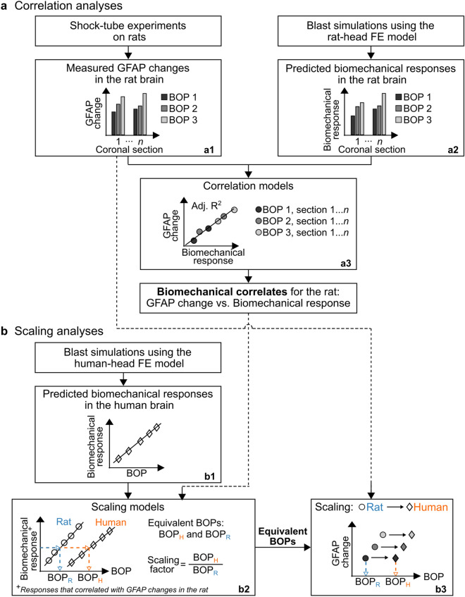
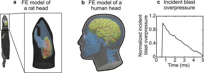
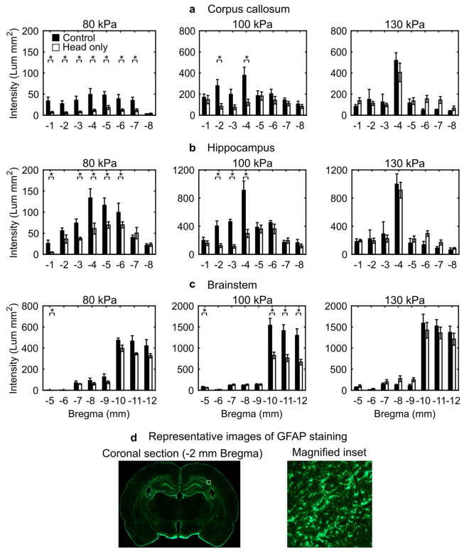
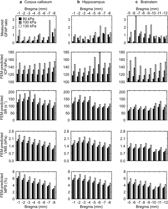
Animal studies provide valuable insights on how the interaction of blast waves with the head may injure the brain. However, there is no acceptable methodology to scale the findings from animals to humans. Here, we propose an experimental/computational approach to project observed blast-induced molecular changes in the rat brain to the human brain. Using a shock tube, we exposed rats to a range of blast overpressures (BOPs) and used a high-fidelity computational model of a rat head to correlate predicted biomechanical responses with measured changes in glial fibrillary acidic protein (GFAP) in rat brain tissues. Our analyses revealed correlates between model-predicted strain rate and measured GFAP changes in three brain regions. Using these correlates and a high-fidelity computational model of a human head, we determined the equivalent BOPs in rats and in humans that induced similar strain rates across the two species. We used the equivalent BOPs to project the measured GFAP changes in the rat brain to the human. Our results suggest that, relative to the rat, the human requires an exposure to a blast wave of a higher magnitude to elicit similar brain-tissue responses. Our proposed methodology could assist in the development of safety guidelines for blast exposure.


Conflict of interest statement

The authors declare no competing interests.

Figures

Figure 1
Figure 1
Flowchart of the biomechanical-based methodology to scale blast-induced molecular changes in the rat brain to the human brain. (a) Correlation analyses. We performed single shock-tube exposures on rats at different incident blast overpressures (BOPs) and characterized the brain-tissue changes in glial fibrillary acidic protein (GFAP) resulting from the blast-wave exposures (a1). Then, using a high-fidelity finite-element (FE) model of a rat head, we conducted blast simulations at the same BOPs as those of the experiments and computed the biomechanical responses (i.e., intracranial pressure, von Mises stress, maximum principal strain, and strain rate) in the rat brain (a2). Using the experimental and simulation data, we developed correlation models to identify correlates between the biomechanical responses and GFAP changes (a3). We performed these analyses for three brain regions: corpus callosum, hippocampus, and brainstem. (b) Scaling analyses. Using a high-fidelity FE model of a human head, we performed blast simulations at different BOPs and computed the biomechanical responses in the brain tissues (b1). Next, using the biomechanical correlates for the rat and the predicted responses for the human, we developed scaling models and determined equivalent BOPs in rats and humans that induced similar biomechanical responses in a given brain region of the two species (b2). In addition, using the equivalent BOPs, we computed the corresponding rat-to-human BOP scaling factors as the ratio of the equivalent BOPs (b2) and projected the experimentally measured GFAP changes in the rat brain to the human (b3). Adj. R2 adjusted R-squared, BOPH equivalent blast overpressure in the human, BOPR equivalent blast overpressure in the rat.
Figure 2
Figure 2
Representation of the previously validated high-fidelity, three-dimensional finite-element (FE) model of (a) a rat head, and (b) a human head used in the blast simulations. (c) Normalized pressure–time profile of the incident blast overpressure (BOP) used as the input to the blast simulations. To perform the simulations, we scaled the amplitude of this normalized profile by multiplying it by the target BOP magnitude.
Figure 3
Figure 3
GFAP-positive staining in the (a) corpus callosum, (b) hippocampus, and (c) brainstem resulting from a single head-only blast-wave exposure in rats for incident blast overpressures (BOPs) of 80, 100, and 130 kPa in a shock tube. We conducted immunohistochemical analyses (i.e., GFAP staining) on coronal brain sections serially cut from − 1 to − 12 mm relative to Bregma and harvested them at 24 h post-exposure. To identify changes in GFAP-positive staining between control and head-only-exposed rats, we compared the data for a given brain region across each coronal section using the Mann–Whitney test. Asterisks denote statistically significant differences (p < 0.05) between control (n = 5 for 80 and 100 kPa, and n = 4 for 130 kPa BOP) and blast-exposed groups (n = 5 for 80 kPa, and n = 10 for 100 and 130 kPa BOP). The bar height and vertical line length represent the mean and one standard error of the mean, respectively. The corpus callosum and hippocampus spanned from − 1 to − 8 mm relative to Bregma, while the brainstem spanned from − 5 to − 12 mm relative to Bregma. For the 80-kPa plots, we reduced the y-axis scale for each brain region to better illustrate the data. (d) Representative images of GFAP staining in a coronal brain section located at − 2 mm relative to Bregma. The magnified inset shows positively stained cells in the coronal section. GFAP glial fibrillary acidic protein.
Figure 4
Figure 4
Experimentally measured changes in GFAP and computationally predicted biomechanical responses in the (a) corpus callosum, (b) hippocampus, and (c) brainstem of a rat resulting from a single head-only blast-wave exposure for three blast overpressures. Using the immunohistochemical data for each brain region within a coronal section, we computed a GFAP ratio by dividing the corresponding mean value of the head-only-exposed rats by those of the controls. Using the blast-simulation data for each brain region within a coronal section, we determined the peak 90th percentile of each biomechanical response over the entire simulation time of 5 ms. The corpus callosum and hippocampus spanned from − 1 to − 8 mm relative to Bregma, while the brainstem spanned from − 5 to − 12 mm relative to Bregma. FEM finite-element model, GFAP glial fibrillary acidic protein, ICP intracranial pressure, MPS maximum principal strain, SR strain rate, VMS von Mises stress.
Figure 5
Figure 5
Scaling models of equivalent blast overpressures (BOPs) that elicited similar responses in the human brain and the rat brain. For a given biomechanical response value of strain rate (SR) or intracranial pressure (ICP) on the y-axis, the intercept with the two linear regression models provides the equivalent rat and human BOPs on the x-axis. To develop these scaling models, we only used biomechanical responses identified as correlates to GFAP changes in the rat. In these models, we considered the biomechanical correlates (i.e., SR and ICP) as the dependent variable and the incident BOP as the independent variable. The circles represent the rat data, while the diamonds show the human data. The solid lines represent the regression lines. In the SR graphs, we plotted the rat and human data on distinct y-axes. BOPH equivalent blast overpressure in the human, BOPR equivalent blast overpressure in the rat, FEM finite-element model, ICPH intracranial pressure in the human, ICPR intracranial pressure in the rat, SRH strain rate in the human, SRR strain rate in the rat.

References

    1. Elder GA, Cristian A. Blast-related mild traumatic brain injury: Mechanisms of injury and impact on clinical care. Mt. Sinai J. Med. 2009;76:111–118. doi: 10.1002/msj.20098. - DOI - PubMed
    1. Hicks RR, Fertig SJ, Desrocher RE, Koroshetz WJ, Pancrazio JJ. Neurological effects of blast injury. J. Trauma. 2010;68:1257–1263. doi: 10.1097/TA.0b013e3181d8956d. - DOI - PMC - PubMed
    1. Elder GA, Mitsis EM, Ahlers ST, Cristian A. Blast-induced mild traumatic brain injury. Psychiatr. Clin. N. Am. 2010;33:757–781. doi: 10.1016/j.psc.2010.08.001. - DOI - PubMed
    1. Ivanov I, et al. Blast exposure, white matter integrity, and cognitive function in Iraq and Afghanistan combat Veterans. Front. Neurol. 2017;8:127. doi: 10.3389/fneur.2017.00127. - DOI - PMC - PubMed
    1. Miller DR, Hayes JP, Lafleche G, Salat DH, Verfaellie M. White matter abnormalities are associated with overall cognitive status in blast-related mTBI. Brain Imaging Behav. 2017;11:1129–1138. doi: 10.1007/s11682-016-9593-7. - DOI - PMC - PubMed

Publication types