. 2022 Aug 26;12(1):14578.
doi: 10.1038/s41598-022-18771-y.

Inflammatory cytokines directly disrupt the bovine intestinal epithelial barrier

Affiliations

Inflammatory cytokines directly disrupt the bovine intestinal epithelial barrier

Charles K Crawford et al. Sci Rep. .

Abstract

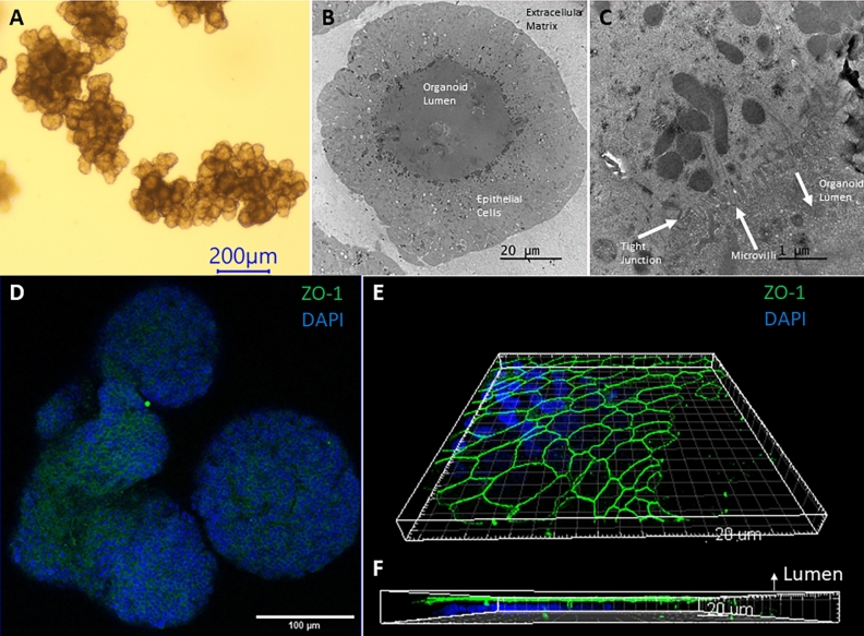
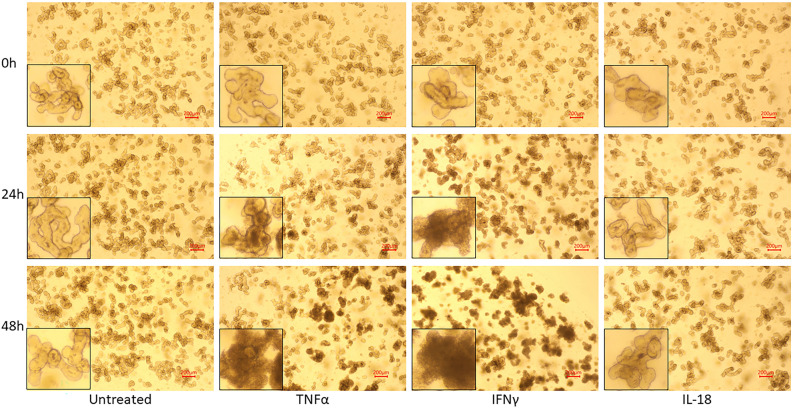
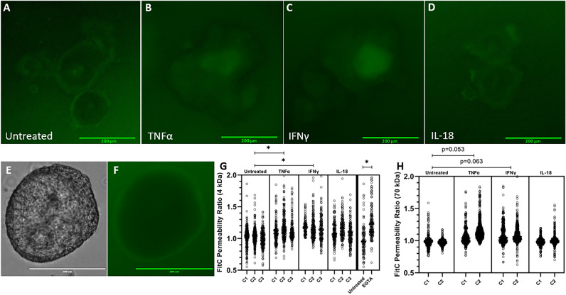
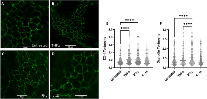
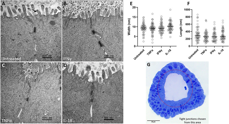
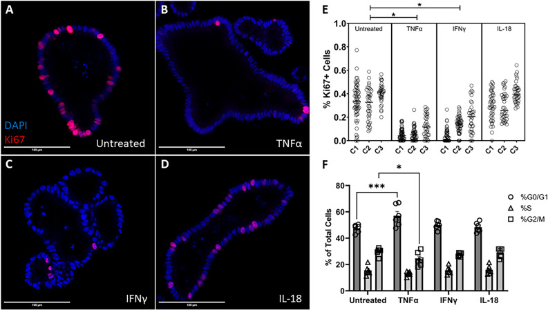
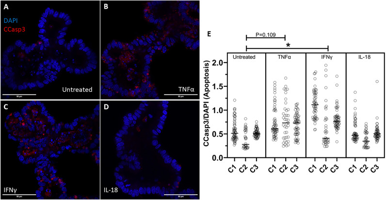
The small intestinal mucosa constitutes a physical barrier separating the gut lumen from sterile internal tissues. Junctional complexes between cells regulate transport across the barrier, preventing water loss and the entry of noxious molecules or pathogens. Inflammatory diseases in cattle disrupt this barrier; nonetheless, mechanisms of barrier disruption in cattle are poorly understood. We investigated the direct effects of three inflammatory cytokines, TNFα, IFNγ, and IL-18, on the bovine intestinal barrier utilizing intestinal organoids. Flux of fluorescein isothiocyanate (FITC)-labeled dextran was used to investigate barrier permeability. Immunocytochemistry and transmission electron microscopy were used to investigate junctional morphology, specifically tortuosity and length/width, respectively. Immunocytochemistry and flow cytometry was used to investigate cellular turnover via proliferation and apoptosis. Our study shows that 24-h cytokine treatment with TNFα or IFNγ significantly increased dextran permeability and tight junctional tortuosity, and reduced cellular proliferation. TNFα reduced the percentage of G2/M phase cells, and IFNγ treatment increased cell apoptotic rate. IL-18 did not directly induce significant changes to barrier permeability or cellular turnover. Our study concludes that the inflammatory cytokines, TNFα and IFNγ, directly induce intestinal epithelial barrier dysfunction and alter the tight junctional morphology and rate of cellular turnover in bovine intestinal epithelial cells.


Conflict of interest statement

The authors declare no conflicts of interest. Funding for the project was provided by the USDA National Institute of Food and Agriculture as well as Marcia Rivas Memorial Funds.

Figures

Figure 1
Figure 1
Bovine intestinal organoid characterization (A) Brightfield image of a bovine enteroid using a ×4 objective lens. Scale bar denotes 200 µm. (B,C) Transmission electron microscopy images of a bovine enteroid with annotated microvilli, tight junction, and lumen. (D) Whole mount Immunocytochemical imaging of ZO-1 (Alexa Fluor 488) and DAPI using a ×20 objective lens (E,F) 3D modeling (Imaris Image Software) of ZO-1 (Alexa Fluor 488) and DAPI staining using stacked confocal images taken with a ×100 objective lens, with annotated lumen, displaying the apical border of an enteroid.
Figure 2
Figure 2
Morphological changes induced by cytokine treatment. Brightfield microscopy images of bovine intestinal organoids using a ×4 objective lens, treated with inflammatory cytokines for 24 and 48 h. Bottom left corners display a zoomed-in enteroid from each image. Scale bars denote 200 µm.
Figure 3
Figure 3
Cytokine treatment increases bovine intestinal organoid barrier permeability, as measured by FITC Dextran permeability. (A–D) Representative images of bovine intestinal organoids exposed to 4 kDa FITC Dextran following 24 h cytokine treatment acquired using a ×10 objective lens and GFP (470/525 nm ex/em) LED light cube. Scale bars denote 200 µm. (E,F) Representative images of an untreated enteroid following 70 kDa FITC Dextran exposure collected using a ×20 objective lens and brightfield or GFP LED light cube. Scale bars denote 300 µm. (G,H) Luminal FITC intensity normalized to external FITC intensity following 24 h cytokine treatment or two-hour treatment with 2 mM EGTA as a positive control. C1, C2, and C3 indicate individual enteroid lines. Nested ANOVA (α = 0.05) determined a significant effect of treatment was present for both 4 kDa and 70 kDa FITC Dextran (p = 0.0097 and p = 0.039, respectively). TNFα and IFNγ increase 4 kDa FITC Dextran permeability (p = 0.0257, p = 0.0132, respectively) as determined by Holm-Šídák post hoc analysis. EGTA, a positive control, increases FITC Dextran permeability (p < 0.0001) determined by one-tailed Mann–Whitney non-parametric test (α = 0.05). Holm-Šídák post hoc analysis for 70 kDa FITC Dextran displayed trending, but not significant, effects of TNFα and IFNγ treatment (p = 0.052 and p = 0.063, respectively).
Figure 4
Figure 4
Cytokine treatment alters tight junction tortuosity. (A–D) Representative ZO-1 (Alexa Fluor 488) staining of bovine intestinal organoids using a ×100 objective lens following 24-h cytokine treatment. Scale bars denote 10 µm. (E) Quantification of ZO-1 tortuosity in cytokine-treated intestinal organoids. TNFα-treated bovine enteroids and IFNγ-treated bovine enteroids displayed a significant increase in junctional tortuosity (p < 0.0001 for each treatment) determined by Kruskal–Wallis non-parametric ANOVA (α = 0.05) utilizing Dunn’s post hoc analysis. (F) Quantification of Occludin tortuosity in cytokine-treated intestinal organoids. IFNγ-treated bovine enteroids displayed a significant increase in Occludin tortuosity (p < 0.0001) relative to all other treatments. **** denotes p < 0.0001.
Figure 5
Figure 5
Cytokine treatment does not alter junctional morphology as measured by transmission electron microscopy. (A–D) Representative TEM images of cytokine-treated intestinal organoid tight junctions. Scale bars denote 500 nm. (E) Tight junction width. (F) Tight junction length. (G) Light microscopy image of a bovine intestinal organoid highlighting the apical area from which tight junctions were analyzed. Scale bar denotes 20 µm. Cytokine treatment did not induce a significant change in junctional length or width as determined by Kruskal–Wallis non-parametric ANOVA (α = 0.05).
Figure 6
Figure 6
Cytokine treatment alters cellular proliferation. (A–D) Representative confocal images of cytokine-treated bovine intestinal organoids stained for Ki67 (Alexa Fluor 594) and DAPI taken on a ×40 objective. Scale bars denote 100 µm. (E) Percentage of proliferating cells as measured by confocal microscopy. C1, C2, and C3 indicate individual enteroid lines. (F) Cell cycle analysis of cytokine-treated bovine intestinal organoids. TNFα and IFNγ induce a reduction in cellular proliferation (p = 0.0031, p = 0.0072, respectively) evidenced by nested ANOVA (α = 0.05) Holm-Šídák post hoc analysis. Two-way ANOVA displays a significant interaction effect (p = 0.0002) and Dunnett’s multiple comparisons test show that TNFα-treatment induces a rise in the percentage of cells in the G0/G1 phase (p = 0.0003) and a reduction in the percentage of cells in the G2M phase (p = 0.0379). * denotes p < 0.05, *** denotes p < 0.001.
Figure 7
Figure 7
Cytokine treatment alters apoptosis. (A–D) Representative confocal images of cytokine-treated bovine intestinal organoids stained for Cleaved Caspase-3 (CCasp3) (Alexa Fluor 594) and DAPI using a ×63 objective lens. Scale bars denote 50 µm. (E) Area of intraluminal CCasp3 normalized to DAPI measured by confocal microscopy. C1, C2, and C3 indicate individual enteroid lines. IFNγ induces a rise in apoptotic cells (p = 0.0325) determined by nested ANOVA Holm-Šídák post hoc analysis.

References

    1. Collins JT, Nguyen A, Badireddy M. Anatomy, Abdomen and Pelvis, Small Intestine. StatPearls; 2021. - PubMed
    1. Barrett KE. New ways of thinking about (and teaching about) intestinal epithelial function. Adv. Physiol. Educ. 2008;32(1):25–34. doi: 10.1152/advan.00092.2007. - DOI - PubMed
    1. Hamilton I, et al. Small intestinal permeability in dermatological disease. Q. J. Med. 1985;56(221):559–567. - PubMed
    1. Harris CE, et al. Intestinal permeability in the critically ill. Intensive Care Med. 1992;18(1):38–41. doi: 10.1007/BF01706424. - DOI - PubMed
    1. Ukabam SO, Mann RJ, Cooper BT. Small intestinal permeability to sugars in patients with atopic eczema. Br. J. Dermatol. 1984;110(6):649–652. doi: 10.1111/j.1365-2133.1984.tb04699.x. - DOI - PubMed

Publication types

LinkOut - more resources