. 2022 Aug 26;12(1):14576.
doi: 10.1038/s41598-022-18885-3.

PERM1 regulates genes involved in fatty acid metabolism in the heart by interacting with PPARα and PGC-1α

Affiliations

PERM1 regulates genes involved in fatty acid metabolism in the heart by interacting with PPARα and PGC-1α

Chun-Yang Huang et al. Sci Rep. .

Abstract

PERM1 (PGC-1/ERR-induced regulator in muscle 1) is a muscle-specific protein induced by PGC-1 and ERRs. Previous studies have shown that PERM1 promotes mitochondrial biogenesis and metabolism in cardiomyocytes in vitro. However, the role of endogenous PERM1 in the heart remains to be investigated with loss-of-function studies in vivo. We report the generation and characterization of systemic Perm1 knockout (KO) mice. The baseline cardiac phenotype of the homozygous Perm1 KO mice appeared normal. However, RNA-sequencing and unbiased pathway analyses showed that homozygous downregulation of PERM1 leads to downregulation of genes involved in fatty acid and carbohydrate metabolism in the heart. Transcription factor binding site analyses suggested that PPARα and PGC-1α are involved in changes in the gene expression profile. Chromatin immunoprecipitation assays showed that PERM1 interacts with the proximal regions of PPAR response elements (PPREs) in endogenous promoters of genes involved in fatty acid oxidation. Co-immunoprecipitation and reporter gene assays showed that PERM1 promoted transcription via the PPRE, partly in a PPARα and PGC-1α dependent manner. These results suggest that endogenous PERM1 is involved in the transcription of genes involved in fatty acid oxidation through physical interaction with PPARα and PGC-1α in the heart in vivo.


Conflict of interest statement

The authors declare no competing interests.

Figures

Figure 1
Figure 1
Perm1 knockouts displayed survival rate, Mendelian rule, and body weight growth similar to those of wild type. (a) Schematic representation of genomic DNA deletion in Line 29 and Line 80 Perm1 KO mice. The deletion sequence was designed in exon 2. (b) DNA electrophoresis for genotyping. Left panel shows 361 bp in WT mice and 270 bp in KO for Line 29 mice. Right panel shows 361 bp in WT and 260 bp for Line 80 mice. The primer sequences for genotyping are shown. (c) Western blot analysis of PERM1, with densitometry for WT, heterozygous KO (He), and homozygous KO (Ho) shown individually. Left panel: Line 29; Right panel: The statistical significance was determined with Student’s t test, *p < 0.05 was considered significant. Line 29: WT (3 male + 3 female); He (3 male + 3 female); Ho (3 male + 3 female). Line 80: WT (3 male + 3 female); He (3 male + 3 female); Ho (3 male + 3 female). (d) Exon level visualization revealing Perm1 mRNA partial exon 2 deletion in homogenous Perm1 KO mice. WT (2 male + 2 female); KO (2 male + 2 female). (e) Upper and lower panels show 2-month survival rates for Line 29 and Line 80 individually. Line 29: WT (4 male + 4 female); He (16 male + 19 female); Ho (6 male + 8 female). Line 80: WT (7 male + 9 female); He (21 male + 19 female); Ho (11 male + 8 female).
Figure 2
Figure 2
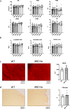
Perm1 knockout displayed cardiac function similar to that of wild type. (a) WT, Line 29 He, Line 80 He, Line 29 Ho, and Line 80 Ho mice at 3 months old were subjected to echocardiographic analyses. Bar graphs show heart rate (HR), IVSd (diastolic interventricular septum thickness), PWd (diastolic posterior wall thickness), LVIDd (end-diastolic LV internal dimension), LVIDs (end-systolic LV internal dimension), and EF (ejection fraction) for WT (10 male + 11 female), Line 29 He (10 male + 10 female), Line 80 He (7 male + 6 female), Line 29 Ho (7 male + 7 female), and Line 80 Ho (10 male + 10 female). (b) Lung, heart and LV weight to tibial bone length ratios for WT (11 male + 11 female), Line 29 He (7 male + 8 female), Line 80 He (10 male + 9 female), Line 29 Ho (9 male + 6 female), and Line 80 Ho (9 male + 10 female) after sacrifice at 3 months old. (c) WGA histology showing comparison of cardiomyocyte size between WT (3 male + 3 female) and Line 80 Ho (3 male + 3 female) at 3 months old. (d) PSR histology showing comparison of the fibrotic area between WT (3 male + 2 female) and Line 80 Ho (3 male + 2 female) at 3 months old. The statistical significance was determined with 1 way ANOVA (a,b), Student’s t test (c,d), *p < 0.05 was considered significant (ad).
Figure 3
Figure 3
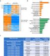
PERM1 regulates genes involved in metabolism in the mouse heart. (a) Illumina-based RNA-seq showing genes differentially expressed in Perm1 KO mice and WT mice, including 99 upregulated genes and 195 downregulated genes (n = 4/group, all male, 2 months old, The statistical significance was determined with Student’s t test, *p < 0.05 was considered significant. cantly regulated genes showed that the pathway and protein function affected in Perm1 KO mice is metabolism. (c) Transcription factor analysis by Ingenuity Pathway Analysis (IPA, Qiagen Inc). The significant p-value indicates likely inhibition or activation states of an upstream regulator in Perm1 KO mice compared with WT mice.
Figure 4
Figure 4
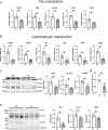
Perm1 knockout downregulated fat and carbohydrate metabolism-related proteins. (a) Expression levels of fat metabolism genes in Perm1 KO mice. Expression levels of the indicated fat metabolism genes were examined by RT-PCR. Samples were normalized by 15S gene expression, including Cpt1β, Cpt2, Mcad, Lcad, and Vlcad. The mean value from WT mice was expressed as 1 (N = 6), WT (3 male + 3 female); KO (3 male + 3 female), 2 months old. (b) Expression levels of carbohydrate metabolism genes in Perm1 KO mice. Expression levels of the indicated fat metabolism genes were examined by RT-PCR. Samples were normalized by 15S gene expression, including Slc2a1, Slc2a4, Hk1, Hk2, Pdk4, and Aldoa. The mean value from WT mice was expressed as 1 (N = 6), WT (3 male + 3 female); KO (3 male + 3 female), 2 months old. (c) Western blots showing CPT1β, VLCAD, and MCAD protein levels. The intensity was normalized by GAPDH (N = 6), WT (3 male + 3 female); KO (3 male + 3 female), 2 months old. (d) The heart lysate of WT and Perm1 KO mice were analyzed with a fatty acid oxidation (FAO) assay kit. The level of FAO was normalized by that in WT mice. WT (4 male + 4 female); KO (4 male + 4 female), 2 months old. (e) Western blots showing HK2, GLUT1, and GLUT4 protein levels. The intensity was normalized by GAPDH (N = 6), WT (3 male + 3 female); KO (3 male + 3 female), 2 months old. The statistical significance was determined with Student’s t test, *p < 0.05 was considered significant.
Figure 5
Figure 5
PERM1 promotes PPARα/PGC-1α-mediated transcription. (a) (Upper panels) Flag-PERM1 adenovirus was transduced in neonatal rat ventricular cardiomyocytes. Immunoprecipitation was conducted with anti-Flag antibody. Note that endogensou PERM1 in inputs is not well seen due to a short exposure time to avoid overexposures of exogenous PERM1. NS* indicates a non-specific band around 110 kD. (Lower panels) Co-immunoprecipiatation assays were performed using mouse heart lysagte with anti-PPARα antibody and control IgG. Immunoblot analyses were conducted with anti-PERM1 or PPARα antibody. (b) PPRE-Luciferase (PPRE-Luc) reporter gene assays were performed with indicated expression vectors and siRNAs. Scrabmled siControl was used as control. N = 6. (c) (Left panel) A schematic representation of endogenous PPRE in PPAR target gene promoters including Acox1, Cpt2, Acadm, and Cpt1β. Arrows indicate primers used for ChIP assays. (Right panel) ChIP assays were were conducted using mouse heart tissue with anti-PERM1 antibody and control IgG. N = 6. (d) A luciferase reporter gene horboring the endogenous promoter sequence of either Acox1 andAdcam was co-transfected with increasing concentrations of mammalin expression vector harbaring PERM1 in cultured neonatal rat ventricular myocytes. N = 6. (e) A luciferase reporter gene horboring the intact or mutated PPRE sequences on the endogenous Acox1 or Acadm was co-transfected with increasing concentrations of mammalin expression vector harbaring PERM1 in cultured neonatal rat ventricular myocytes. N = 6. (f) (Left panel) Representative Western blot image demonstrating that PERM1 interacts with PGC-1α in rat ventricular cardiomyocytes. Cells were transduced with Flag-PERM1 adenovirus for 1 day. Immunoprecipitation (IP) with Flag antibody and immunoblot with PGC-1α antibody. (Right panel) A representative Western blot image demonstrating that endogenous PERM1 interacts with PGC-1α in the heart. Co-immunoprecipiatation assay was performed using mouse heart lysate with anti-PERM1 antibody and control IgG. Immunoblot analyses were conducted with anti-PERM1 or PGC-1α antibody. (g) Luciferase reporter assays demonstrating that PERM1-induced reporter gene activation via PPRE was inhibited by knockdown of PGC-1α with siPGC-1α. N = 6. (h) The effect of siRNA used by this study including siPerm1, siPPARα and siPGC-1αin neonatal rat ventricular cardiomyocytes. Experiments are repeated 3 times. (i) Scheme demonstrating how PERM1 regulates PPARα/PGC-1α-mediated transcription of fatty acid oxidation genes. The statistical significance was determined with 1 way ANOVA (b (left), d,e), Student’s t test (c) and 2 way ANOVA (b (middle and right) and g). *p < 0.05 was considered significant (be,g).

References

    1. Cho Y, Hazen BC, Russell AP, Kralli A. Peroxisome proliferator-activated receptor gamma coactivator 1 (PGC-1)- and estrogen-related receptor (ERR)-induced regulator in muscle 1 (Perm1) is a tissue-specific regulator of oxidative capacity in skeletal muscle cells. J. Biol. Chem. 2013;288:25207–25218. doi: 10.1074/jbc.M113.489674. - DOI - PMC - PubMed
    1. Oka SI, et al. Perm1 regulates cardiac energetics as a downstream target of the histone methyltransferase Smyd1. PLoS One. 2020;15:e0234913. doi: 10.1371/journal.pone.0234913. - DOI - PMC - PubMed
    1. Cho Y, et al. Perm1 enhances mitochondrial biogenesis, oxidative capacity, and fatigue resistance in adult skeletal muscle. FASEB J. 2016;30:674–687. doi: 10.1096/fj.15-276360. - DOI - PMC - PubMed
    1. Huss JM, Kopp RP, Kelly DP. Peroxisome proliferator-activated receptor coactivator-1alpha (PGC-1alpha) coactivates the cardiac-enriched nuclear receptors estrogen-related receptor-alpha and -gamma. Identification of novel leucine-rich interaction motif within PGC-1alpha. J. Biol. Chem. 2002;277:40265–40274. doi: 10.1074/jbc.M206324200. - DOI - PubMed
    1. Huss JM, Torra IP, Staels B, Giguere V, Kelly DP. Estrogen-related receptor alpha directs peroxisome proliferator-activated receptor alpha signaling in the transcriptional control of energy metabolism in cardiac and skeletal muscle. Mol. Cell Biol. 2004;24:9079–9091. doi: 10.1128/MCB.24.20.9079-9091.2004. - DOI - PMC - PubMed

Publication types

Substances