. 2022 Aug;54(8):1262-1276.
doi: 10.1038/s12276-022-00834-9. Epub 2022 Aug 26.

Local immune cell contributions to fracture healing in aged individuals - A novel role for interleukin 22

Affiliations

Local immune cell contributions to fracture healing in aged individuals - A novel role for interleukin 22

Christian H Bucher et al. Exp Mol Med. 2022 Aug.

Abstract

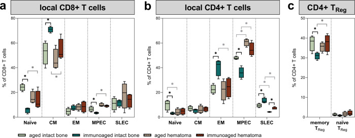
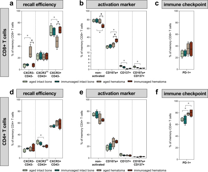
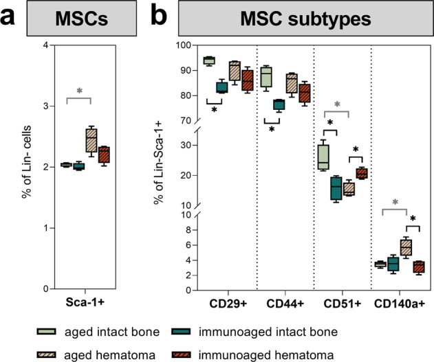
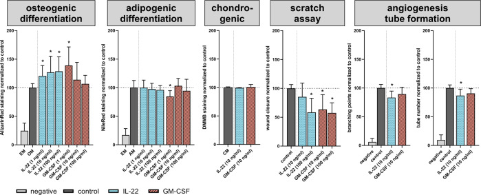
With increasing age, the risk of bone fractures increases while regenerative capacity decreases. This variation in healing potential appears to be linked to adaptive immunity, but the underlying mechanism is still unknown. This study sheds light on immunoaging/inflammaging, which impacts regenerative processes in aging individuals. In an aged preclinical model system, different levels of immunoaging were analyzed to identify key factors that connect immunoaged/inflammaged conditions with bone formation after long bone fracture. Immunological facets, progenitor cells, the microbiome, and confounders were monitored locally at the injury site and systemically in relation to healing outcomes in 12-month-old mice with distinct individual levels of immunoaging. Bone tissue formation during healing was delayed in the immunoaged group and could be associated with significant changes in cytokine levels. A prolonged and amplified pro-inflammatory reaction was caused by upregulated immune cell activation markers, increased chemokine receptor availability and a lack of inhibitory signaling. In immunoaged mice, interleukin-22 was identified as a core cell signaling protein that played a central role in delayed healing. Therapeutic neutralization of IL-22 reversed this specific immunoaging-related disturbed healing. Immunoaging was found to be an influencing factor of decreased regenerative capacity in aged individuals. Furthermore, a novel therapeutic strategy of neutralizing IL-22 may successfully rejuvenate healing in individuals with advanced immune experiences.


Conflict of interest statement

The authors declare no competing interests.

Figures

Fig. 1
Fig. 1. Analysis of the fracture gap after osteotomy in 12-month-old mice revealed a delayed fracture healing process in immunologically experienced (immunoaged) mice.
a Representative images of the newly formed bone (red) and cortical bone (gray) in the fracture gap 21 days post-surgery, which were analyzed via microcomputed X-ray tomography. b Callus volume (TV) was significantly decreased in the immunoaged group, whereas bone volume (BV), bone mineral density (BMD), trabecular number (Tb.N), and trabecular thickness (Tb.Th) were not significantly different between the two groups. The ratio of bone volume to total volume (BV/TV) was not affected by the immunological experience. However, the diminished bone formation in the immunoaged group led to a less stable fracture callus, as shown by a lower minimal polar moment of inertia (MMIp). N = 4 per group; box-and-whisker plot with a line at the median; Welch’s t-test.
Fig. 2
Fig. 2. Analysis of the gut microbiome showed substantial alterations post-surgery due to differences in the level of immunological experience.
The gut microbiota was analyzed in the cecum and sequenced by 16 S rRNA. a The microbiome diversity was significantly decreased post-surgery due to antibiotic treatment at the time of surgery. The reconstitution of the microbiome was significantly slower in the immunoaged group, whereas the aged mice reached presurgery levels 21 days post-surgery. b Phylogenic analysis revealed a strong dominance of Proteobacteria 3 days post-surgery. c Bacterial families significantly differed between immunoaged and aged mice pre-surgery as well as 21 days post-surgery. N = 3-4 per group; box-and-whisker plot with a line at the median and bar plots; Welch’s t-test.
Fig. 3
Fig. 3. Immune phenotype analysis of local T cells at the site of injury in comparison to the bone marrow from untreated mice 3 days post-surgery revealed significant differences in T-cell subtypes.
The fracture gap, including the hematoma and the adjacent bone marrow, was analyzed by flow cytometry 3 days post-surgery and compared to untreated bone marrow from the same groups. Subpopulations of CD8+ (a) and CD4+ (b) T cells were analyzed. Initially, memory formation was more advanced in immunoaged mice, as indicated by increases in memory populations and decreases in naïve T-cell numbers. However, these differences were equalized 3 days post-surgery at the fracture site. Significant differences at 3 days post-surgery could only be observed in short-lived effector cells (SLECs) in the CD4+ T-cell compartment around the fracture site. The osteotomy significantly changed the memory populations in CD4+ T cells: The percentage of effector memory (EM) T cells was diminished, whereas the percentage of memory precursor cells (MPEC) were significantly increased compared to that in the untreated bone marrow. c CD4+ regulatory T-cell numbers were increased in the immunoaged group after injury, and these cells were also less abundant in the untreated bone marrow of untreated mice. N = 4 per group; box-and-whisker plot with a line at the median; Welch’s t-test; black * = between immunological experience groups, gray * = between untreated and osteotomized groups.
Fig. 4
Fig. 4. Analysis of the local T cells at the site of injury 3 days post-surgery in comparison to the bone marrow from untreated mice revealed significant differences in T-cell activation states.
d, f Introducing a fracture decreased CXCR3 presentation on the cell surface in CD4+ T cells and increased the expression of PD-1 accordingly. a Interestingly, CXCR3 expression on CD8+ T cells was highly regulated by the initial experience level, as in aged mice, CXCR3 expression on CD8+ T cells 3 days post-surgery was downregulated, which was not seen in immunoaged mice. c However, there was no significant increase in PD-1 surface expression on memory and effector CD8+ T cells, and less PD-1 expression on memory/effector T cells resulted in escape from immune regulation. b, e Introducing a fracture activated CD8+ memory T cells, as shown by increased levels of CD107a on the cell surface. CD8+ T cells showed a significantly higher activation state in immunoaged mice 3 days post-surgery than in the aged-only group. N = 4 per group; box-and-whisker plot with a line at the median; Welch’s t-test; black * = between immunological experience groups, gray * = between untreated and osteotomized groups.
Fig. 5
Fig. 5. Analysis of the local macrophage and dendritic cell populations at the site of injury 3 days post-surgery revealed significant differences in macrophage polarization, activation, and dendritic cell subsets.
Macrophages and dendritic cells were analyzed via flow cytometry in the untreated bone marrow and at the fracture site in the immunoaged and aged groups. a Three days post-surgery, significantly more macrophages were present around the fracture gap than in the untreated bone marrow. b The activation state of macrophages at the fracture site differed, as significantly more M1-polarized macrophages could be found in the immunoaged group than in the aged group 3 days post-surgery. Additionally, an increased level of M2a-activated macrophages was observed in the immunoaged group. These data showed an increased activation state in the mice with higher adaptive immune experience and depicted a prolonged inflammatory phase. c Immunoaged mice showed an increased level of plasmacytoid dendritic cells (pDCs) at the fracture site compared to that in aged mice 3 days post-surgery. In contrast, increased levels of specified CD4+ and CD8+ lymphoid tissue-resident classical dendritic cells (lDCs) could be found in aged mice compared to immunoaged mice. d Increased adaptive immune experience significantly altered the innate immune system by increasing CD205+ expression on CD8+ lDCs. Higher expression levels of CD205+ on DCs facilitate communication with CD8+ T cells. N = 4 per group; box-and-whisker plot with a line at the median; Welch’s t-test; black * = between immunological experience groups, gray * = between untreated and osteotomized groups.
Fig. 6
Fig. 6. Analysis of local mesenchymal stromal cell subtypes at the site of injury 3 days post-surgery revealed significant differences in subtype distribution and priming.
Mesenchymal stromal cells (MSCs) were analyzed via flow cytometry in the untreated bone marrow and at the fracture site in the immunoaged and aged groups. a Flow cytometric analysis of bone marrow from the untreated bone versus the fracture hematoma three days post-surgery. Osteotomy increased the availability of MSCs (CD45-CD34-CD31-(Lin)Sca-1+) at the fracture site. The mobilization and recruitment of MSCs was significantly increased in the aged group. The immunoaged group, however, showed less proliferation and migration. b The heterogeneity of MSC subtypes also differed between groups with different immunological experience levels. Specialized MSCs were present at different frequencies, as a higher percentage of MSCs in the aged mice were PαS cells (Sca-1+ CD140a+), whereas a higher percentage were murine skeletal stem cells (mSSC, Sca-1+ CD51+) in the immunoaged mice. N = 4 per group; box-and-whisker plot with a line at the median; Welch’s t-test; black * = between immunological experience groups, gray * = between untreated and osteotomized groups.
Fig. 7
Fig. 7. Cytokine patterns in the basal state and in response to an injury showed increased reactivity in immunoaged mice.
Systemic cytokine levels in the blood were screened pre-surgery and 3 days post-surgery for immunoaged and less-experienced (aged) 12-month-old mice. The cytokine levels in the basal state revealed an inflammaged status in immunoaged mice, as indicated by increased systemic levels of TNFα, IL-6, and IL-10. Upon activation due to osteotomy, the cytokine levels varied based on the immune experience level: Increased levels of IL-22, GM-CSF, and IL-1b could be observed in immunoaged mice. Most importantly, IL-22 not only showed high basal expression levels in immunoaged compared to aged mice but also appeared to react distinctly in the respective immunoaged phenotype, with a significant downregulation upon injury in aged mice but an increase upon injury in immunoaged mice. N = 4 per group; bar plot with SD; Welch’s t-test; black * = between immunological experience groups, gray * = between untreated and osteotomized groups.
Fig. 8
Fig. 8. IL-22 and GM-CSF interfere with the regenerative potential of progenitor cells.
The two cytokines were tested individually at different concentrations to determine their effects on the differentiation and migratory/proliferative potential of mesenchymal stromal cells as well as the angiogenic potential of endothelial cells to form new tubes in vitro. IL-22 increased the mineralization of the extracellular matrix in the osteogenic differentiation assays, but higher concentrations of IL-22 decreased migratory and proliferative potential and inhibited the de novo formation of new vessels by reducing the tube number and branching points. GM-CSF predominantly interfered with the migratory and proliferative behavior of MSCs but did not inhibit the angiogenic potential of endothelial cells. EM expansion medium, OM osteogenic medium, AM adipogenic medium, CM chondrogenic medium; n = 5 MSC donors per assay, n = 5 pooled endothelial cell (HUVEC) donors, tested in triplicate; unpaired t-test.
Fig. 9
Fig. 9. Substitution of IL-22 in less-experienced (aged) mice impaired the fracture healing process, whereas neutralization of IL-22 accelerated or ameliorated the healing process in experienced (immunoaged) mice.
An osteotomy was introduced in the left femur, the endogenous IL-22 levels were altered via neutralization with antibodies or supplementation with recombinant IL-22 protein, and the healing outcome was assessed 21 days post-surgery with µCT. a, b In less-experienced (aged) 12-month-old mice, which had a lower level of IL-22 3 days post-surgery, IL-22 was supplemented during the first 7 days post-surgery, starting 24 h post-surgery. Fracture healing was almost completely abolished 21 days post-surgery under IL-22 supplementation. The callus volume (TV) and the bone volume (BV) were significantly decreased, and the microarchitecture (trabecular number Tb.N and trabecular thickness Tb.Th) and bone mineral density (BMD) significantly deteriorated. Immunoaged 12-month-old mice showed a higher level of IL-22 3 days post-surgery, and IL-22 was neutralized during the first 7 days post-surgery, starting 24 h post-surgery. Neutralization of IL-22 boosted the bone healing process, as an increased callus volume and bone volume could be observed 21 days post-surgery. The stability of the fracture gap was significantly increased (MMIp), and callus volume and stability reached the levels observed in less-experienced (aged) mice. N = 4–6 per group; box-and-whisker plot with a line at the median; Welch’s t-test.

References

    1. Minetto MA, et al. Common musculoskeletal disorders in the elderly: the star triad. J. Clin. Med. 2020;9:1216. doi: 10.3390/jcm9041216. - DOI - PMC - PubMed
    1. Prince MJ, et al. The burden of disease in older people and implications for health policy and practice. Lancet. 2015;385:549–562. doi: 10.1016/S0140-6736(14)61347-7. - DOI - PubMed
    1. Riggs BL, Melton LJ. The worldwide problem of osteoporosis: Insights afforded by epidemiology. Bone. 1995;17:S505–S511. doi: 10.1016/8756-3282(95)00258-4. - DOI - PubMed
    1. Blume SW, Curtis JR. Medical costs of osteoporosis in the elderly Medicare population. Osteoporos. Int. 2011;22:1835–1844. doi: 10.1007/s00198-010-1419-7. - DOI - PMC - PubMed
    1. Cummings SR, Melton LJ. Osteoporosis I: epidemiology and outcomes of osteoporotic fractures. Lancet. 2002;359:1761–1767. doi: 10.1016/S0140-6736(02)08657-9. - DOI - PubMed

Publication types