. 2022 Sep 30;12(10):jkac222.
doi: 10.1093/g3journal/jkac222.

The mIAA7 degron improves auxin-mediated degradation in Caenorhabditiselegans

Affiliations

The mIAA7 degron improves auxin-mediated degradation in Caenorhabditiselegans

Jorian J Sepers et al. G3 (Bethesda). .

Abstract

Auxin-inducible degradation is a powerful tool for the targeted degradation of proteins with spatiotemporal control. One limitation of the auxin-inducible degradation system is that not all proteins are degraded efficiently. Here, we demonstrate that an alternative degron sequence, termed mIAA7, improves the efficiency of degradation in Caenorhabditiselegans, as previously reported in human cells. We tested the depletion of a series of proteins with various subcellular localizations in different tissue types and found that the use of the mIAA7 degron resulted in faster depletion kinetics for 5 out of 6 proteins tested. The exception was the nuclear protein HIS-72, which was depleted with similar efficiency as with the conventional AID* degron sequence. The mIAA7 degron also increased the leaky degradation for 2 of the tested proteins. To overcome this problem, we combined the mIAA7 degron with the C. elegans AID2 system, which resulted in complete protein depletion without detectable leaky degradation. Finally, we show that the degradation of ERM-1, a highly stable protein that is challenging to deplete, could be improved further by using multiple mIAA7 degrons. Taken together, the mIAA7 degron further increases the power and applicability of the auxin-inducible degradation system. To facilitate the generation of mIAA7-tagged proteins using CRISPR/Cas9 genome engineering, we generated a toolkit of plasmids for the generation of dsDNA repair templates by PCR.

Keywords: C aenorhabditis elegans; AID system; TIR1; mIAA7; protein degradation.


Figures

Fig. 1.
Fig. 1.
The mIAA7 degron improves auxin-mediated degradation of DLG-1. a) Schematic overview of IAA proteins and the different AID degrons that have been derived from them. I = domain I; II = domain II; KR = conserved lysine and arginine residue; PB1 = Phox and Bem1p domain. b) Comparison between AID*- and mIAA7-mediated degradation kinetics of intestinal DLG-1 in L3 larvae using 1 mM auxin. Values in the graph are arbitrary units and each data point represents the average intensity at intestinal cell junctions for the given condition and timepoint. Error bars: mean ± SD; n: 7–20 animals. Images shown are representative maximum intensity projections that were acquired and displayed with the same settings for comparison, except for the indicated panel with enhanced brightness to show residual DLG-1. Arrowheads point to the apical junctional sites where 3 intestinal cells meet.
Fig. 2.
Fig. 2.
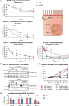
The mIAA7 degron robustly increases the efficiency of AID-induced protein degradation for several proteins across multiple tissues and cellular compartments. a) Comparison between AID*- and mIAA7-mediated degradation for indicated proteins and tissues. Drawing represents a generic polarized cell with the subcellular localization of the proteins investigated indicated. PAR-6 was measured at the apical domain of seam cells in L2 larvae on 5 μM auxin, BBLN-1 was measured at the apical domain in the intestine of L3 larvae treated with 50 μM auxin, SAX-7 was measured at the plasma membrane in the ALM neuron cell body of L3 larvae treated with 5 μM auxin, and RPS-26 was measured in the cytoplasm of body wall muscles of L2 larvae treated with 1 mM auxin. Values are normalized to the mean intensity levels at 0 h of auxin exposure, and each data point represents the average intensity for the given condition and timepoint. Due to the nature of the quantification for PAR-6 and RPS-26, the fluorescence levels did not reach zero as they did for the other targets. Nonzero baseline level values of wildtype animals are indicated, resembling a completely degraded protein pool. For SAX-7 degradation using the mIAA7 degron, exposure to auxin of 3 h or longer resulted in complete depletion and an inability to locate the ALM cell body. For these timepoints, values were plotted at 0 and no statistics were performed (light shaded dots). Error bars: mean ± SD; Statistical test: Mann–Whitney U test; n = 9–29 animals. b) Western blots detecting HIS-72: AID*::3xFLAG and HIS-72::mIAA7::3xFLAG in synchronized control or 4 mM auxin-treated L1 larvae. An anti-alpha-tubulin loading control and protein size standard markers in kilodaltons are provided. Time in hours in control media or 4 mM auxin containing media is indicated. c) Comparison of Larval growth between AID*- and mIAA7-mediated PAR-6 degradation in the epidermis using 5 μM and 1 mM auxin. d) Comparison of leaky degradation between AID* and mIAA7 degrons for the indicated proteins. Measurements for each protein were done as in panel A. Values are normalized to the mean intensity levels of animals expressing the same degron-tagged protein but not expressing TIR1. Each data point in the graph represents a single animal. Error bars: mean ± SD; statistical test: Mann–Whitney U test; n values are indicated in or above the bars.
Fig. 3.
Fig. 3.
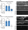
The mIAA7 degron is compatible with the C.e.AID2 system. mIAA7-mediated degradation of PAR-6 in the seam cells (a) and of SAX-7 in the ALM neuron (b) of L2 larvae using the C.e.AID2 system. Values are normalized to the mean intensity levels of animals expressing the same degron-tagged proteins but not expressing TIR1 and not treated with 5-Ph-IAA (i.e. the first bar). Each data point in the graph represents a single animal. Error bars: mean ± SD; statistical test: 1-way ANOVA with Šidák multiple testing correction; n values are indicated in or above the bars. Images shown are representative maximum intensity projections that were acquired and displayed with the same settings for comparison.
Fig. 4.
Fig. 4.
Tagging ERM-1 with multiple mIAA7 degrons improves auxin-mediated degradation. a) Comparison between AID*- and mIAA7-mediated degradation of intestinal ERM-1 throughout larval development using the C.e.AID2 system. Data points represent the mean intensity normalized to the mean intensity levels of animals with the same genotype and age that were not exposed to 5-Ph-IAA. Error bars: mean ± SD; Statistical test: Mann–Whitney U test; n = 12–16 animals. Images shown are representative maximum intensity projections that were acquired and displayed with the same settings for comparison. b) Schematic overview of the different ERM-1 alleles used for testing the effect of multiple degrons and degron location on auxin-mediated degradation efficiency. c) Comparison of intestinal degradation of ERM-1 tagged with 1 or multiple mIAA7 degrons at varying locations in L3 larvae using the C.e.AID2 system. Values are normalized to the mean intensity levels of animals with the same genotype that were not exposed to 5-Ph-IAA, and each data point represents a single animal. Images shown are representative maximum intensity projections that were acquired and displayed with the same settings for comparison. Error bars: mean ± SD; statistical test: 1-way ANOVA with Šidák multiple testing correction; n values are indicated in the bars.

References

    1. Armenti ST, Lohmer LL, Sherwood DR, Nance J.. Repurposing an endogenous degradation system for rapid and targeted depletion of C. elegans proteins. Development. 2014;141(23):4640–4647. 10.1242/dev.115048. - DOI - PMC - PubMed
    1. Ashley GE, Duong T, Levenson MT, Martinez MAQ, Johnson LC, Hibshman JD, Saeger HN, Palmisano NJ, Doonan R, Martinez-Mendez R, et al.An expanded auxin-inducible degron toolkit for Caenorhabditis elegans. Genetics. 2021;217(3):iyab006. 10.1093/genetics/iyab006. - DOI - PMC - PubMed
    1. Bhoi A, Palladino F, Fabrizio P.. Auxin confers protection against ER stress in Caenorhabditis elegans. Biol Open. 2021;10:bio057992. 10.1242/bio.057992. - DOI - PMC - PubMed
    1. Bossinger O, Klebes A, Segbert C, Theres C, Knust E.. Zonula adherens formation in Caenorhabditis elegans requires dlg-1, the homologue of the Drosophila gene discs large. Dev Biol. 2001;230(1):29–42. 10.1006/dbio.2000.0113. - DOI - PubMed
    1. Brenner S. The genetics of Caenorhabditis elegans. Genetics. 1974;77(1):71–94. - PMC - PubMed

Publication types