Observational Study
. 2022 Dec;10(12):1147-1159.
doi: 10.1016/S2213-2600(22)00259-4. Epub 2022 Aug 24.

Lung epithelial and myeloid innate immunity in influenza-associated or COVID-19-associated pulmonary aspergillosis: an observational study

Affiliations
Observational Study

Lung epithelial and myeloid innate immunity in influenza-associated or COVID-19-associated pulmonary aspergillosis: an observational study

Simon Feys et al. Lancet Respir Med. 2022 Dec.

Abstract

Background: Influenza-associated pulmonary aspergillosis (IAPA) and COVID-19-associated pulmonary aspergillosis (CAPA) affect about 15% of critically ill patients with influenza or COVID-19, respectively. These viral-fungal coinfections are difficult to diagnose and are associated with increased mortality, but data on their pathophysiology are scarce. We aimed to explore the role of lung epithelial and myeloid innate immunity in patients with IAPA or CAPA.

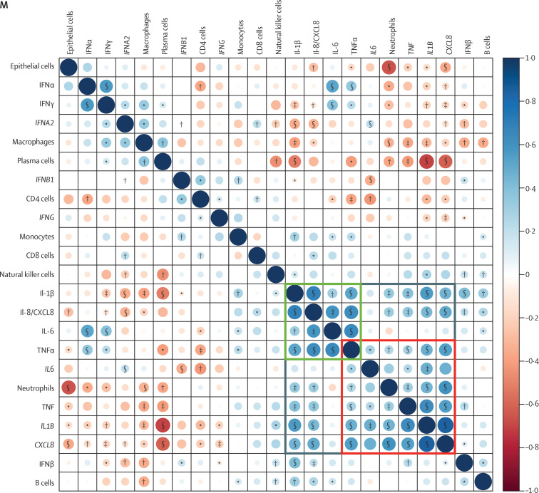
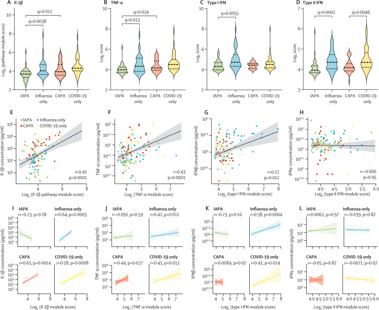
Methods: In this observational study, we retrospectively recruited patients who had been admitted to the intensive care unit (ICU) of University Hospitals Leuven, Belgium, requiring non-invasive or invasive ventilation because of severe influenza or COVID-19, with or without aspergillosis, between Jan 1, 2011, and March 31, 2021, whose bronchoalveolar lavage samples were available at the hospital biobank. Additionally, biobanked in vivo tracheobronchial biopsy samples from patients with IAPA or CAPA and invasive Aspergillus tracheobronchitis admitted to ICUs requiring invasive ventilation between the same dates were collected from University Hospitals Leuven, Hospital Network Antwerp (Belgium), and Amiens-Picardie University Hospital (France). We did nCounter gene expression analysis of 755 genes linked to myeloid innate immunity and protein analysis of 47 cytokines, chemokines, and growth factors on the bronchoalveolar lavage samples. Gene expression data were used to infer cell fractions by use of CIBERSORTx, to perform hypergeometric enrichment pathway analysis and gene set enrichment analysis, and to calculate pathway module scores for the IL-1β, TNF-α, type I IFN, and type II IFN (IFNγ) pathways. We did RNAScope targeting influenza virus or SARS-CoV-2 RNA and GeoMx spatial transcriptomics on the tracheobronchial biopsy samples.

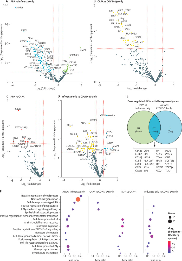
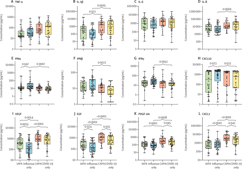
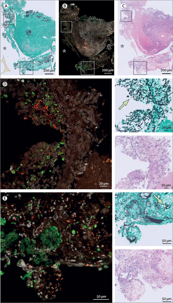
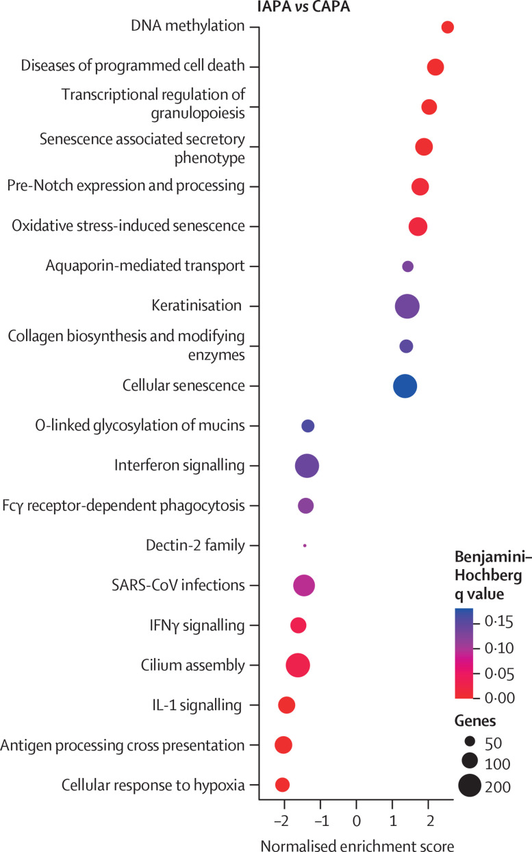
Findings: Biobanked bronchoalveolar lavage samples were retrieved from 166 eligible patients, of whom 40 had IAPA, 52 had influenza without aspergillosis, 33 had CAPA, and 41 had COVID-19 without aspergillosis. We did nCounter gene expression analysis on bronchoalveolar lavage samples from 134 patients, protein analysis on samples from 162 patients, and both types of analysis on samples from 130 patients. We performed RNAScope and spatial transcriptomics on the tracheobronchial biopsy samples from two patients with IAPA plus invasive Aspergillus tracheobronchitis and two patients with CAPA plus invasive Aspergillus tracheobronchitis. We observed a downregulation of genes associated with antifungal effector functions in patients with IAPA and, to a lesser extent, in patients with CAPA. We found a downregulated expression of several genes encoding proteins with functions in the opsonisation, recognition, and killing of conidia in patients with IAPA versus influenza only and in patients with CAPA versus COVID-19 only. Several genes related to LC3-associated phagocytosis, autophagy, or both were differentially expressed. Patients with CAPA had significantly lower neutrophil cell fractions than did patients with COVID-19 only. Patients with IAPA or CAPA had downregulated IFNγ signalling compared with patients with influenza only or COVID-19 only, respectively. The concentrations of several fibrosis-related growth factors were significantly elevated in the bronchoalveolar lavage fluid from patients with IAPA versus influenza only and from patients with CAPA versus COVID-19 only. In one patient with CAPA, we visualised an active or very recent SARS-CoV-2 infection disrupting the epithelial barrier, facilitating tissue-invasive aspergillosis.

Interpretation: Our results reveal a three-level breach in antifungal immunity in IAPA and CAPA, affecting the integrity of the epithelial barrier, the capacity to phagocytise and kill Aspergillus spores, and the ability to destroy Aspergillus hyphae, which is mainly mediated by neutrophils. The potential of adjuvant IFNγ in the treatment of IAPA and CAPA should be investigated.

Funding: Research Foundation Flanders, Coronafonds, the Max Planck Society, the Fundação para a Ciência e a Tecnologia, the European Regional Development Fund, "la Caixa" Foundation, and Horizon 2020.


Conflict of interest statement

Declaration of interests SF received travel grants from Pfizer. KL received consultancy fees from MRM Health, MSD, and Gilead; speaker fees from FUJIFILM WAKO, Pfizer, and Gilead; and a service fee from Thermo Fisher Scientific. LV received travel grants from Pfizer and Gilead. JW received investigator-initiated grants, speaker's fees, and travel fees from Pfizer, Gilead, and MSD and declares participation in advisory boards for Pfizer and Gilead and the receipt of study drugs from MSD. All other authors declare no competing interests.

Figures

Figure 1
Figure 1
Cell type fractions in bronchoalveolar lavage fluid samples from patients with IAPA, influenza only, CAPA, or COVID-19 only Box plots represent median and IQR, with whiskers set from minimum to maximum. Individual points represent each sample. q values were generated by Kruskall–Wallis tests, corrected by use of the Benjamini–Krieger–Yekutieli method. Only significant q values are shown. CAPA=COVID-19-associated pulmonary aspergillosis. IAPA=influenza-associated pulmonary aspergillosis.
Figure 2
Figure 2
Differentially expressed genes in bronchoalveolar lavage fluid samples from patients with IAPA, influenza only, CAPA, or COVID-19 only Volcano plots showing differentially expressed genes for the comparisons of IAPA versus influenza only (A), CAPA versus COVID-19 only (B), IAPA versus CAPA (C), and influenza only versus COVID-19 only (D). A dotted blue line marks a log2(fold change) value of zero. A dotted red line crossing the y-axis marks a Benjamini–Hochberg q value of 0·05. The two dotted red lines crossing the x-axis marks a log2(fold change) of 0·4 and –0·4, respectively. We selected genes to present if they were linked to interesting pathways related to antiviral and antifungal immunity. (E) Venn diagram showing the downregulated differentially expressed genes shared in the comparisons of IAPA versus influenza only and CAPA versus COVID-19 only. (F) Dot plot showing ClueGO hypergeometric enrichment pathway analysis based on the downregulated differentially expressed genes of the four disease comparisons, with Gene Ontology biological process as the pathway database. *For the comparison of IAPA versus CAPA, differentially expressed genes with q values (Benjamini–Hochberg) of less than 0·20 were included to generate the analyses. Differentially expressed genes with q values (Benjamini–Hochberg) of less than 0·05 were used for all other comparisons. CAPA=COVID-19-associated pulmonary aspergillosis. IAPA=influenza-associated pulmonary aspergillosis. NIK=NF-κB-inducing kinase.
Figure 3
Figure 3
Concentrations and correlations of cytokines, chemokines, and growth factors in bronchoalveolar lavage samples (A–L) Concentrations of major cytokines, chemokines, and growth factors. Box plots represent median and IQR, with whiskers set from minimum to maximum. Individual points represent each sample. q values were generated by Kruskall–Wallis tests, corrected by use of the Benjamini–Krieger–Yekutieli method. Only significant q values are shown. The scale between points is a log scale. (M) Correlogram showing the correlations between the CIBERSORTx-derived cell fractions, major cytokine concentrations, and gene expressions of the corresponding genes in bronchoalveolar lavage samples with all three types of results available. Components are hierarchically clustered. The grey box represents a major proinflammatory cluster. The green and red boxes represent two pro-inflammatory subclusters, with the green box representing proinflammatory cytokines and the red box representing the deconvoluted neutrophil fraction and the gene expression of proinflammatory cytokines. CAPA=COVID-19-associated pulmonary aspergillosis. EGF=epidermal growth factor. IAPA=influenza-associated pulmonary aspergillosis. PDGF-AA=platelet-derived growth factor AA. VEGF=vascular endothelial growth factor. *p<0·05. †p<0·01. ‡p<0·001. §p<0·0001.
Figure 3
Figure 3
Concentrations and correlations of cytokines, chemokines, and growth factors in bronchoalveolar lavage samples (A–L) Concentrations of major cytokines, chemokines, and growth factors. Box plots represent median and IQR, with whiskers set from minimum to maximum. Individual points represent each sample. q values were generated by Kruskall–Wallis tests, corrected by use of the Benjamini–Krieger–Yekutieli method. Only significant q values are shown. The scale between points is a log scale. (M) Correlogram showing the correlations between the CIBERSORTx-derived cell fractions, major cytokine concentrations, and gene expressions of the corresponding genes in bronchoalveolar lavage samples with all three types of results available. Components are hierarchically clustered. The grey box represents a major proinflammatory cluster. The green and red boxes represent two pro-inflammatory subclusters, with the green box representing proinflammatory cytokines and the red box representing the deconvoluted neutrophil fraction and the gene expression of proinflammatory cytokines. CAPA=COVID-19-associated pulmonary aspergillosis. EGF=epidermal growth factor. IAPA=influenza-associated pulmonary aspergillosis. PDGF-AA=platelet-derived growth factor AA. VEGF=vascular endothelial growth factor. *p<0·05. †p<0·01. ‡p<0·001. §p<0·0001.
Figure 4
Figure 4
Pathway module scores and their correlation with concentration for IL-1β, TNF-α, type I IFN, and type II IFN Pathway module scores for IL-1β (A), TNF-α (B), type I IFN (C), and type II IFN (D). q values were generated by Kruskall–Wallis tests, corrected by use of the Benjamini–Krieger–Yekutieli method. Only significant q values are shown. Spearman rank correlation between the major cytokine concentration and the corresponding module score for all patients for IL-1β (E), TNF-α (F), type I IFN (G), and type II IFN (H). Spearman rank correlation between the major cytokine concentration and the corresponding module score per disease group for IL-1β (I), TNF-α (J), type I IFN (K), and type II IFN (L). The line represents the regression line and the shading represents the 95% CI. CAPA=COVID-19-associated pulmonary aspergillosis. IAPA=influenza-associated pulmonary aspergillosis.
Figure 5
Figure 5
In vivo tracheobronchial biopsy from patient with CAPA and invasive Aspergillus tracheobronchitis (A) Slide stained with Grocott–Gomori's methenamine silver, which makes fungi appear black and the background tissue green. (B) RNAScope image, in which the red puncta reflect SARS-CoV-2 nucleocapsid RNA. Epithelial cells are labelled green by anti-KRT8 antibody staining. DAPI was used as a nuclear stain (grey). (C) Slide stained with haematoxylin and eosin, which is used to look at damaged areas. (D–E) Two magnified areas of the CAPA biopsy, with respective haematoxylin and eosin-stained, Grocott-Gomori's methenamine silver-stained, and RNAScope images. Examples of fungi are indicated on the Grocott-Gomori's methenamine silver-stained images by arrows. The asterisks localise the lumen of the respiratory tract. CAPA=COVID-19-associated pulmonary aspergillosis.
Figure 6
Figure 6
Dot plot of gene set enrichment analysis based on gene expression of epithelial regions of interest in IAPA versus CAPA samples Reactome canonical pathways was the pathway database. CAPA=COVID-19-associated pulmonary aspergillosis. IAPA=influenza-associated pulmonary aspergillosis.

Comment in

  • When disaster strikes fungi take control.
    Hoenigl M. Hoenigl M. Lancet Respir Med. 2022 Dec;10(12):1104-1106. doi: 10.1016/S2213-2600(22)00268-5. Epub 2022 Aug 24. Lancet Respir Med. 2022. PMID: 36029798 Free PMC article. No abstract available.

References

    1. Paget J, Spreeuwenberg P, Charu V, et al. Global mortality associated with seasonal influenza epidemics: new burden estimates and predictors from the GLaMOR Project. J Glob Health. 2019;9 - PMC - PubMed
    1. Schauwvlieghe AFAD, Rijnders BJA, Philips N, et al. Invasive aspergillosis in patients admitted to the intensive care unit with severe influenza: a retrospective cohort study. Lancet Respir Med. 2018;6:782–792. - PubMed
    1. Gangneux J-P, Dannaoui E, Fekkar A, et al. Fungal infections in mechanically ventilated patients with COVID-19 during the first wave: the French multicentre MYCOVID study. Lancet Respir Med. 2022;10:180–190. - PMC - PubMed
    1. Prattes J, Wauters J, Giacobbe DR, et al. Risk factors and outcome of pulmonary aspergillosis in critically ill coronavirus disease 2019 patients—a multinational observational study by the European Confederation of Medical Mycology. Clin Microbiol Infect. 2022;28:580–587. - PMC - PubMed
    1. Janssen NAF, Nyga R, Vanderbeke L, et al. Multinational observational cohort study of COVID-19-associated pulmonary aspergillosis. Emerg Infect Dis. 2021;27:2892–2898. - PMC - PubMed

Publication types