. 2022 Aug 27;21(1):165.
doi: 10.1186/s12933-022-01602-9.

MLKL-mediated necroptosis is a target for cardiac protection in mouse models of type-1 diabetes

Affiliations

MLKL-mediated necroptosis is a target for cardiac protection in mouse models of type-1 diabetes

Ting Cao et al. Cardiovasc Diabetol. .

Abstract

Background: Cardiomyocyte death contributes to cardiac pathology of diabetes. Studies have shown that the RIPK3/MLKL necroptosis signaling is activated in diabetic hearts. Deletion of RIPK3 was reported to attenuate myocardial injury and heart dysfunction in streptozocin (STZ)-induced diabetic mice, suggesting a potential role of necroptosis in diabetic cardiomyopathy. This study characterized cardiomyocyte necroptosis in diabetic hearts and investigated whether MLKL-mediated necroptosis is a target for cardiac protection in diabetes.

Methods: Type 1 diabetes was induced in RIPK3 knockout, MLKL knockout and wild-type mice. Akita Type-1 diabetic mice were injected with shRNA for MLKL. Myocardial function was assessed by echocardiography. Immuno-histological analyses determined cardiomyocyte death and fibrosis in the heart. Cultured adult mouse cardiomyocytes were incubated with high glucose in the presence of various drugs. Cell death and phosphorylation of RIPK3 and MLKL were analysed.

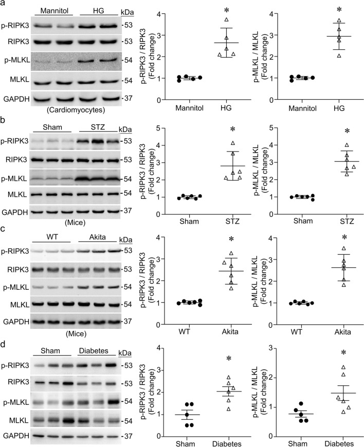
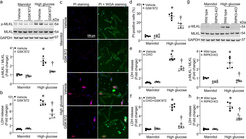
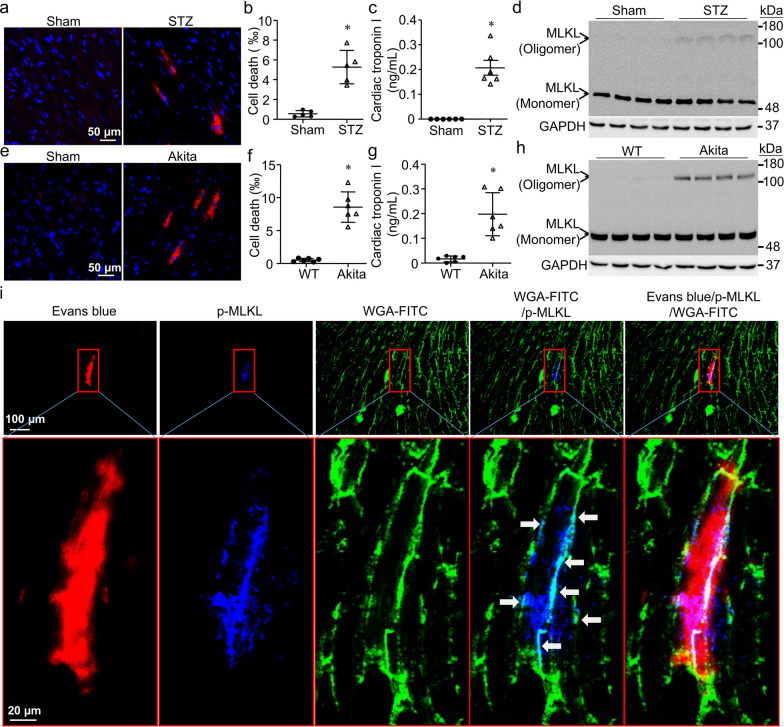
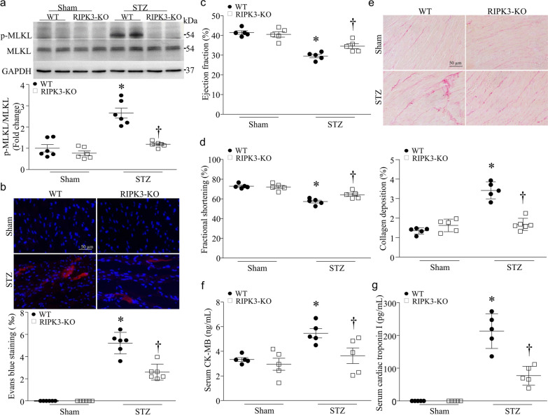
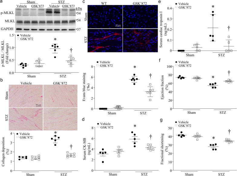
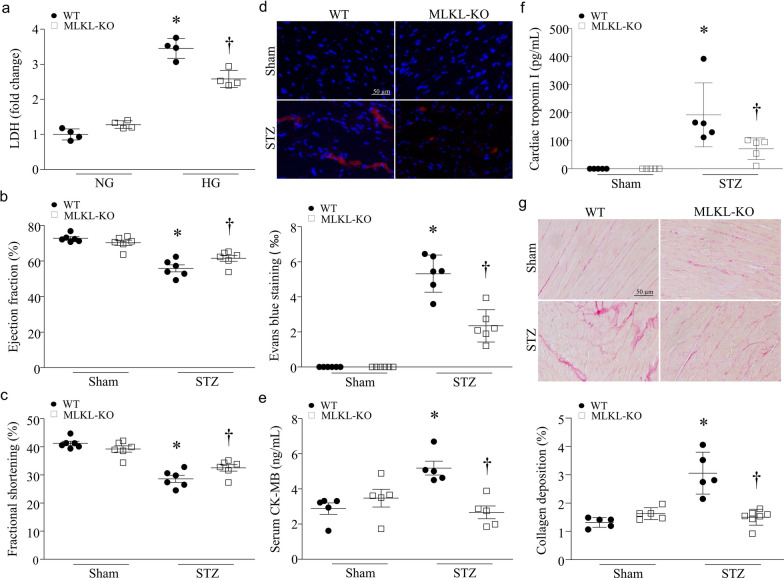
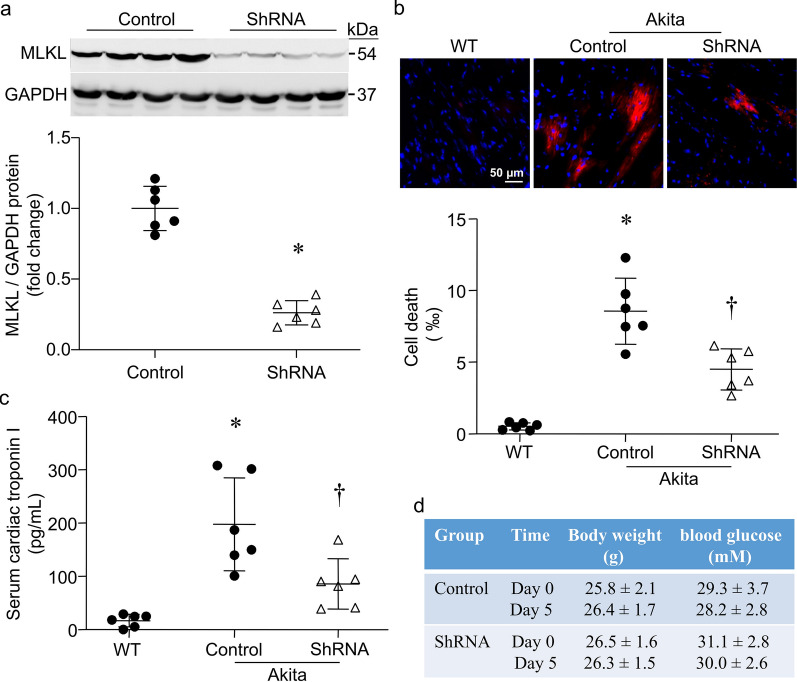
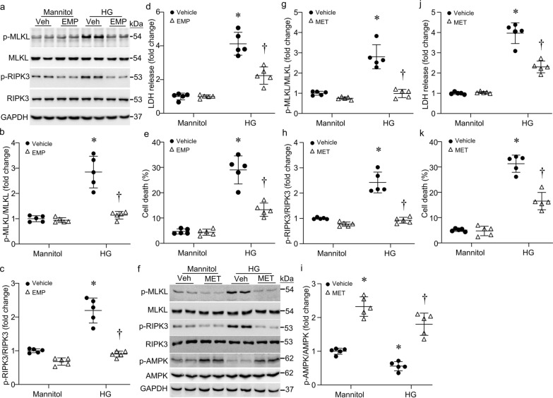
Results: We showed that the levels of phosphorylated RIPK3 and MLKL were higher in high glucose-stimulated cardiomyocytes and hearts of STZ-induced type-1 diabetic mice, akita mice and type-1 diabetic monkeys when compared to non-diabetic controls. Inhibition of RIPK3 by its pharmacological inhibitor or gene deletion, or MLKL deletion prevented high glucose-induced MLKL phosphorylation and attenuated necroptosis in cardiomyocytes. In STZ-induced type-1 diabetic mice, cardiomyocyte necroptosis was present along with elevated cardiac troponin I in serum and MLKL oligomerization, and co-localized with phosphorylated MLKL. Deletion of RIPK3 or MLKL prevented MLKL phosphorylation and cardiac necroptosis, attenuated serum cardiac troponin I levels, reduced myocardial collagen deposition and improved myocardial function in STZ-injected mice. Additionally, shRNA-mediated down-regulation of MLKL reduced cardiomyocyte necroptosis in akita mice. Interestingly, incubation with anti-diabetic drugs (empagliflozin and metformin) prevented phosphorylation of RIPK3 and MLKL, and reduced cell death in high glucose-induced cardiomyocytes.

Conclusions: We have provided evidence that cardiomyocyte necroptosis is present in diabetic hearts and that MLKL-mediated cardiomyocyte necroptosis contributes to diabetic cardiomyopathy. These findings highlight MLKL-mediated necroptosis as a target for cardiac protection in diabetes.

Keywords: Cardiomyocytes; Diabetes; Heart; MLKL; Necroptosis; RIPK3.


Conflict of interest statement

The authors declare that they have no competing interests.

Figures

Fig. 1
Fig. 1
Phosphorylation of RIPK3 and MLKL. a Adult mouse cardiomyocytes were incubated with high glucose or mannitol (25 mmol/L) for 24 h. The levels of phosphorylated RIPK3 (p-RIPK3) and MLKL (p-MLKL) were determined by western blot analysis. Left panel: representative western blots for p-RIPK3, p-MLKL and their total proteins as well as GAPDH; Right two panels: quantification of p-RIPK3 and p-MLKL relative to their total proteins, respectively. bd The levels of p-RIPK3 and p-MLKL in hearts of streptozotocin (STZ)-injected mice (b), Akita mice (c) and monkeys (d) were analyzed by western blot. Left panel: representative western blots from 3 out of 5–6 different hearts in each group for p-RIPK3 and p-MLKL, total RIPK3, total MLKL and GAPDH; Right two panels: quantification of p-RIPK3 and p-MLKL (WT: wild-type). Data are mean ± SD, n = 5–6. * P < 0.05 versus Mannitol, Sham or WT (t test)
Fig. 2
Fig. 2
Effects of RIPK3 inhibition on cell death in cardiomyocytes. Adult mouse cardiomyocytes isolated from RIPK3 knockout (RIPK3-KO) and wild-type mice (WT) were incubated with high glucose or mannitol (25 mmol/L) in the presence of GSK’872 and CHO alone or in combination for 24 h. a The levels of phosphorylated MLKL were determined by western blot analysis. Upper panel: a representative western blot for phosphorylated MLKL (p-MLKL), total MLKL and GAPDH; Bottom panel: quantification of p-MLKL relative to total MLKL. b LDH was measured in culture medium. c Necrotic cell death was assessed using PI staining. A representative micro-photograph for PI staining positive cells (red), nucleus Hoechst33342 staining (blue) and FITC-WGA staining for cell membrane (green). d Quantification of necrotic cell death. e and f LDH was measured in culture medium. g The phosphorylated levels of MLKL were determined in WT and RIPK3-KO cardiomyocytes. Upper panel: a representative western blot for p-MLKL, total MLKL and GAPDH; Bottom panel: quantification of p-MLKL relative to total MLKL. h LDH was measured in culture medium. Data are mean ± SD, n = 5 different cultures. *P < 0.05 versus Mannitol + Vehicle or Mannitol + WT and P < 0.05 versus HG + Vehicle or HG + WT (ANOVA)
Fig. 3
Fig. 3
Characterization of cardiomyocyte necroptosis in the heart. a and e Necrotic cell death was assessed by Evans blue staining assay. Representative micro-photographs show Evans blue stained necrotic cell death (red) in hearts of STZ-injected mice and Akita mice but not in Sham animals. Nuclei were stained blue by Hoechst33342. b and f Quantification of Evans blue stained necrotic cell death in the heart. c and g Serum cardiac troponin I. d and h MLKL oligomerization. Representative western blot from 4 different hearts in each group shows the presence of MLKL oligomerization in STZ-injected and Akita but not Sham mouse hearts. i Confocal microscopy. Representative micro-photograph indicates that Evans blue stained necrotic cells (red) are presented with phosphorylated MLKL (p-MLKL, blue), and that punctate staining of p-MLKL co-localizes with WGA-FITC (cyan). Data are mean ± SD, n = 5–6. * P < 0.05 versus Sham or WT (t test)
Fig. 4
Fig. 4
Effects of RIPK3 knockout in streptozocin (STZ)-induced diabetic hearts. RIPK3 knockout (RIPK3-KO) and wild-type mice (WT) were rendered diabetic by STZ injection. a The levels of phosphorylated MLKL (p-MLKL) were analyzed by western blot in the heart. Upper panel: Upper panel: a representative western blot from 2 out of 6 different hearts in each group for p-MLKL and total MLKL; Bottom panel: quantification of p-MLKL relative to total MLKL. b Necrotic cell death in the heart was determined by Evans blue staining assay. Upper panel: representative micro-photograph for necrotic cells in the heart; Bottom panel: quantification of necrotic cell death. c and d Echocardiographic analysis for myocardial function. e Myocardial fibrosis was assessed by collagen deposition in the heart. Upper panel: representative micro-photograph for collagen deposition in the heart (red); Bottom panel: quantification of collagen deposition. f and g Biomarkers of myocardial injury: serum CK-MB (f) and cardiac troponin I (g). Data are mean ± SD, n = 5–6. *P < 0.05 versus Sham + WT and P < 0.05 versus STZ + WT (ANOVA)
Fig. 5
Fig. 5
Effects of GSK’872 in streptozocin (STZ)-induced diabetic hearts. Wild-type mice were rendered diabetic by STZ injection and after the induction of diabetes, GSK’872 was administrated for a total of 2 months. a The levels of phosphorylated MLKL (p-MLKL) were analyzed by western blot in the heart. Upper panel: a representative western blot from 2 out of 6 different hearts in each group for p-MLKL and total MLKL; Bottom panel: quantification of p-MLKL relative to total MLKL. b Myocardial fibrosis was assessed by collagen deposition in the heart. Upper panel: representative micro-photograph for collagen deposition in the heart (red); Bottom panel: quantification of collagen deposition. c Necrotic cell death in the heart was determined by Evans blue staining assay. Upper panel: representative micro-photograph for necrotic cells in the heart; Bottom panel: quantification of necrotic cell death. d and e Biomarkers of myocardial injury: serum cardiac troponin I (e) and CK-MB (d) and. f and g Echocardiographic analysis for myocardial function. Data are mean ± SD, n = 5–6. * P < 0.05 versus Sham + Vehicle and P < 0.05 versus STZ + Vehicle (ANOVA)
Fig. 6
Fig. 6
Effects of MLKKL knockout in streptozocin (STZ)-induced diabetic hearts. a Adult cardiomyocytes isolated from MLKL knockout (MLKL-KO) and wild-type mice (WT) were incubated with high glucose or Mannitol (25 mmol/L) for 24 h. LDH was measured in culture medium. bg MLKL-KO and WT mice were rendered diabetic by STZ injection. b and c Echocardiographic analysis for myocardial function. d Necrotic cell death in the heart was determined by Evans blue staining assay. Upper panel: representative micro-photograph for necrotic cells in the heart; Bottom panel: quantification of necrotic cell death. e and f Biomarkers of myocardial injury: serum CK-MB (e) and cardiac troponin I (f). g Myocardial fibrosis was assessed by collagen deposition in the heart. Upper panel: representative micro-photograph for collagen deposition in the heart (red); Bottom panel: quantification of collagen deposition. Data are mean ± SD, n = 5–6. *P < 0.05 versus Sham + WT and P < 0.05 versus STZ + WT (ANOVA)
Fig. 7
Fig. 7
Effects of MLKL knockdown in streptozocin (STZ)-induced diabetic hearts. Akita mice received two separate doses of shRNA for MLKL on day 0 and day 3, respectively. Wild-type mice (WT) served as Sham. Control Akita mice received same amounts of an empty plasmid. On day 5, mice were sacrificed. a The levels of MLKL protein were analyzed by western blot in the heart. Upper panel: a representative western blot from 4 out of 6 different hearts in each group for MLKL and GAPDH; Bottom panel: quantification of MLKL relative to GAPDH protein. b Necrotic cell death in the heart was determined by Evans blue staining assay. Upper panel: representative micro-photograph for necrotic cells in the heart; Bottom panel: quantification of necrotic cell death. c Biomarker of myocardial injury: serum cardiac troponin I. d Body weight and blood glucose levels in Akita mice on day 0 and day 5. Data are mean ± SD, n = 6. *P < 0.05 versus Control and P < 0.05 versus Control + Akita (t test and ANOVA)
Fig. 8
Fig. 8
Effects of anti-diabetic drugs on necroptosis in cardiomyocytes. Adult mouse cardiomyocytes were incubated with high glucose (HG) or mannitol (25 mmol/L) in the presence of empagliflozin (EMP, 1 µmol/L), metformin (MET, 500 µmol/L) or Vehicle (Veh) for 24 h. a Representative western blots for phosphorylated RIPK3 (p-RIPK3) and MLKL (p-MLKL) and their total proteins as well as GAPDH. b and c; Quantification of p-RIPK3 and p-MLKL relative to their total proteins, respectively. d and j LDH was measured in culture medium. (e and k) Cell death was assessed by annexin V staining. f Representative western blots for p-RIPK3, p-MLKKL, phosphorylated AMPK (p-AMPK) and their total proteins as well as GAPDH. gi Quantification of p-MLKL, p-RIPK3 and p-AMPK relative to their total proteins, respectively. Data are mean ± SD, n = 5 different cultures. *P < 0.05 versus Mannitol + Veh and P < 0.05 versus HG + Veh (ANOVA)

References

    1. Kannel WB, McGee DL. Diabetes and cardiovascular disease. The Framingham study. JAMA. 1979;241(19):2035–2038. doi: 10.1001/jama.1979.03290450033020. - DOI - PubMed
    1. Garcia MJ, McNamara PM, Gordon T, Kannel WB. Morbidity and mortality in diabetics in the Framingham population. Sixteen year follow-up study. Diabetes. 1974;23(2):105–111. doi: 10.2337/diab.23.2.105. - DOI - PubMed
    1. Nichols GA, Gullion CM, Koro CE, Ephross SA, Brown JB. The incidence of congestive heart failure in type 2 diabetes: an update. Diabetes Care. 2004;27(8):1879–1884. doi: 10.2337/diacare.27.8.1879. - DOI - PubMed
    1. Kannel WB. Framingham study insights on diabetes and cardiovascular disease. Clin Chem. 2011;57(2):338–339. doi: 10.1373/clinchem.2010.149740. - DOI - PubMed
    1. Boudina S, Abel ED. Diabetic cardiomyopathy revisited. Circulation. 2007;115(25):3213–3223. doi: 10.1161/CIRCULATIONAHA.106.679597. - DOI - PubMed

Publication types