

A dopamine-methacrylated hyaluronic acid hydrogel as an effective carrier for stem cells in skin regeneration therapy
- PMID: 36030275
- PMCID: PMC9420120
- DOI: 10.1038/s41419-022-05060-9
A dopamine-methacrylated hyaluronic acid hydrogel as an effective carrier for stem cells in skin regeneration therapy
Abstract
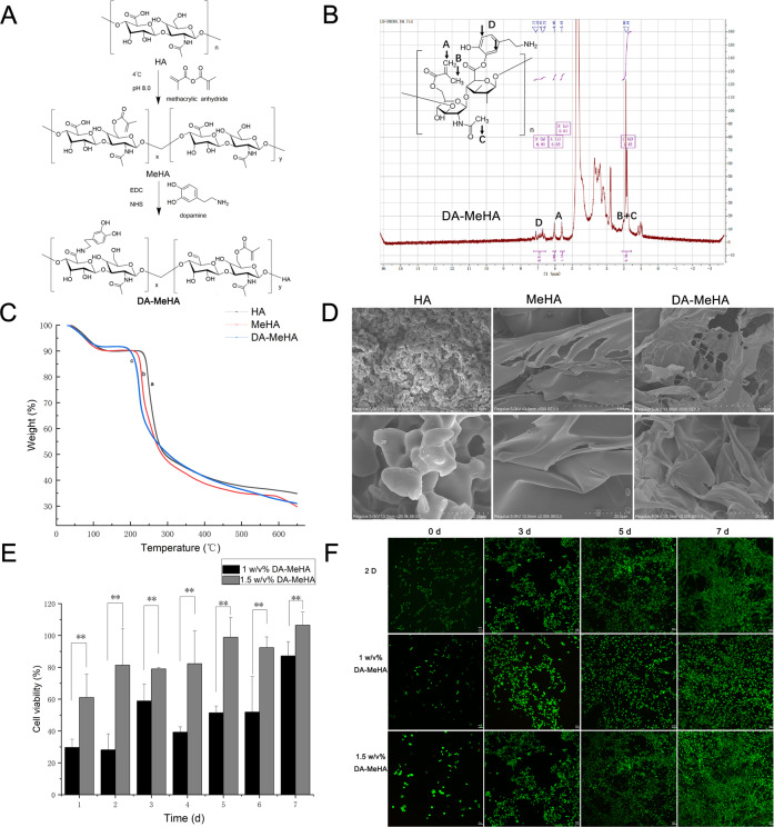
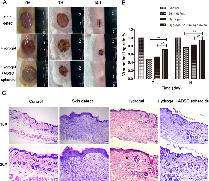
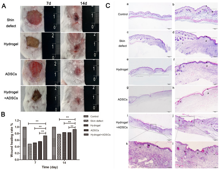
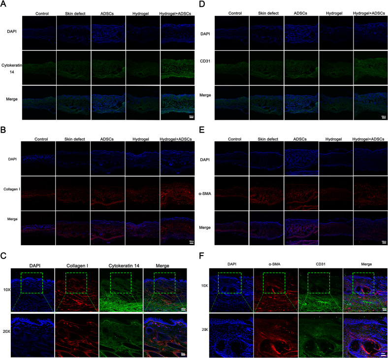
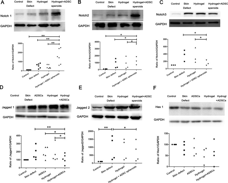
Adipose-derived stem cells (ADSCs) show potential in skin regeneration research. A previous study reported the failure of full-thickness skin self-repair in an injury area exceeding 4 cm in diameter. Stem cell therapies have shown promise in accelerating skin regeneration; however, the low survival rate of transplanted cells due to the lack of protection during and after transplantation leads to low efficacy. Hence, effective biomaterials for the delivery and retention of ADSCs are urgently needed for skin regeneration purposes. Here, we covalently crosslinked hyaluronic acid with methacrylic anhydride and then covalently crosslinked the product with dopamine to engineer dopamine-methacrylated hyaluronic acid (DA-MeHA). Our experiments suggested that the DA-MeHA hydrogel firmly adhered to the skin wound defect and promoted cell proliferation in vitro and skin defect regeneration in vivo. Mechanistic analyses revealed that the beneficial effect of the DA-MeHA hydrogel combined with ADSCs on skin defect repair may be closely related to the Notch signaling pathway. The ADSCs from the DA-MeHA hydrogel secrete high levels of growth factors and are thus highly efficacious for promoting skin wound healing. This DA-MeHA hydrogel may be used as an effective potential carrier for stem cells as it enhances the efficacy of ADSCs in skin regeneration.
© 2022. The Author(s).
Conflict of interest statement
The authors declare no competing interests.
Figures

Similar articles
-
Heparin-hyaluronic acid hydrogel in support of cellular activities of 3D encapsulated adipose derived stem cells.Acta Biomater. 2017 Feb;49:284-295. doi: 10.1016/j.actbio.2016.12.001. Epub 2016 Dec 5. Acta Biomater. 2017. PMID: 27919839
-
Conformable hyaluronic acid hydrogel delivers adipose-derived stem cells and promotes regeneration of burn injury.Acta Biomater. 2020 May;108:56-66. doi: 10.1016/j.actbio.2020.03.040. Epub 2020 Apr 3. Acta Biomater. 2020. PMID: 32251786
-
Development of a UV crosslinked biodegradable hydrogel containing adipose derived stem cells to promote vascularization for skin wounds and tissue engineering.Biomaterials. 2017 Jun;129:188-198. doi: 10.1016/j.biomaterials.2017.03.021. Epub 2017 Mar 18. Biomaterials. 2017. PMID: 28343005
-
The wound-healing and antioxidant effects of adipose-derived stem cells.Expert Opin Biol Ther. 2009 Jul;9(7):879-87. doi: 10.1517/14712590903039684. Expert Opin Biol Ther. 2009. PMID: 19522555 Review.
-
Prospective application of exosomes derived from adipose-derived stem cells in skin wound healing: A review.J Cosmet Dermatol. 2020 Mar;19(3):574-581. doi: 10.1111/jocd.13215. Epub 2019 Nov 21. J Cosmet Dermatol. 2020. PMID: 31755172 Review.
Cited by
-
Harnessing the potential of hyaluronic acid methacrylate (HAMA) hydrogel for clinical applications in orthopaedic diseases.J Orthop Translat. 2025 Jan 8;50:111-128. doi: 10.1016/j.jot.2024.11.004. eCollection 2025 Jan. J Orthop Translat. 2025. PMID: 39886531 Free PMC article. Review.
-
Biological study of skin wound treated with Alginate/Carboxymethyl cellulose/chorion membrane, diopside nanoparticles, and Botox A.NPJ Regen Med. 2024 Feb 27;9(1):9. doi: 10.1038/s41536-024-00354-2. NPJ Regen Med. 2024. PMID: 38413625 Free PMC article.
-
Bioactive Materials That Promote the Homing of Endogenous Mesenchymal Stem Cells to Improve Wound Healing.Int J Nanomedicine. 2024 Jul 30;19:7751-7773. doi: 10.2147/IJN.S455469. eCollection 2024. Int J Nanomedicine. 2024. PMID: 39099796 Free PMC article. Review.
-
Bioadhesive and antioxidant hydrogel by MoS2-TA dual-catalytic system initiated free radical polymerization combined with phototherapy for cartilage regeneration.Mater Today Bio. 2025 Jul 8;33:102056. doi: 10.1016/j.mtbio.2025.102056. eCollection 2025 Aug. Mater Today Bio. 2025. PMID: 40688664 Free PMC article.
-
Enhancing regenerative medicine: the crucial role of stem cell therapy.Front Neurosci. 2024 Feb 8;18:1269577. doi: 10.3389/fnins.2024.1269577. eCollection 2024. Front Neurosci. 2024. PMID: 38389789 Free PMC article. Review.
References
Publication types
MeSH terms
Substances
LinkOut - more resources
Full Text Sources
