. 2022 Sep-Oct;15(5):1192-1205.
doi: 10.1016/j.brs.2022.08.013. Epub 2022 Aug 27.

Neuronavigation maximizes accuracy and precision in TMS positioning: Evidence from 11,230 distance, angle, and electric field modeling measurements

Affiliations

Neuronavigation maximizes accuracy and precision in TMS positioning: Evidence from 11,230 distance, angle, and electric field modeling measurements

Kevin A Caulfield et al. Brain Stimul. 2022 Sep-Oct.

Abstract

Background: Researchers and clinicians have traditionally relied on elastic caps with markings to reposition the transcranial magnetic stimulation (TMS) coil between trains and sessions. Newer neuronavigation technology co-registers the patient's head and structural magnetic resonance imaging (MRI) scan, providing the researcher with real-time feedback about how to adjust the coil to be on-target. However, there has been no head to head comparison of accuracy and precision across treatment sessions.

Objective: /Hypothesis: In this two-part study, we compared elastic cap and neuronavigation targeting methodologies on distance, angle, and electric field (E-field) magnitude values.

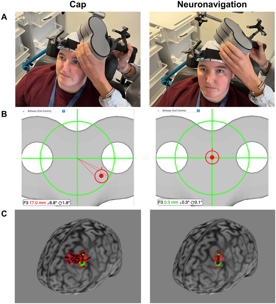
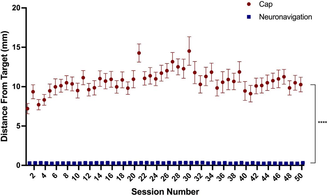
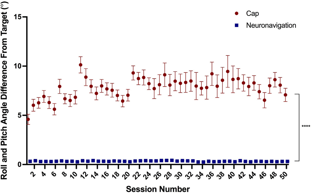
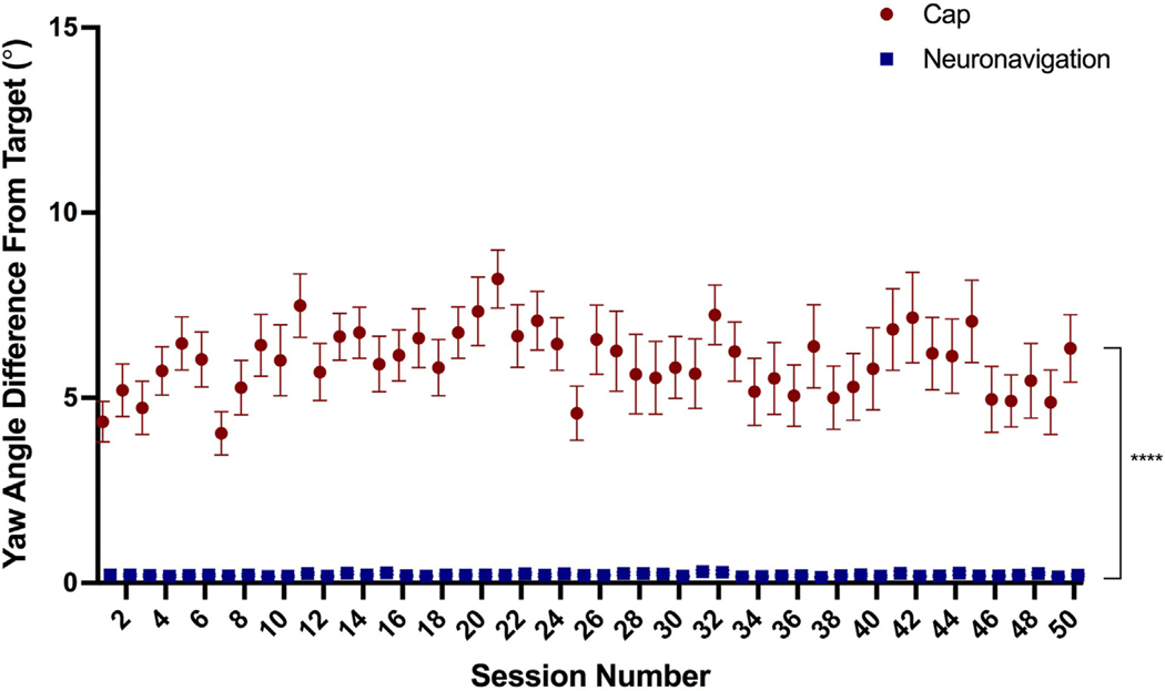
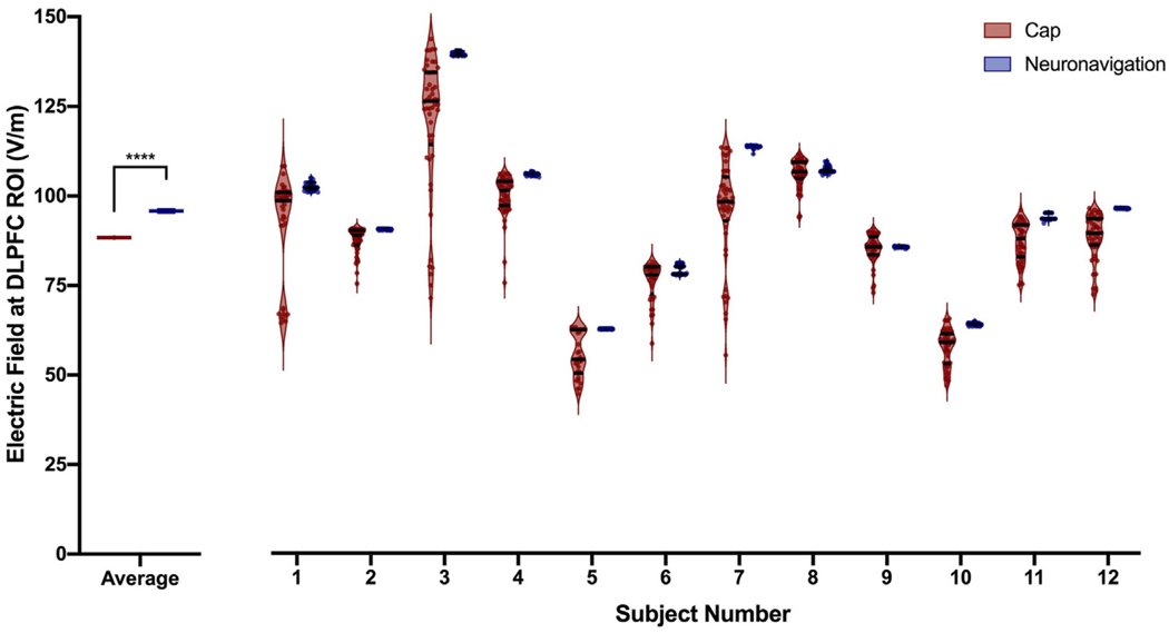
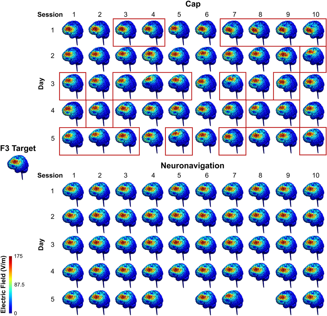
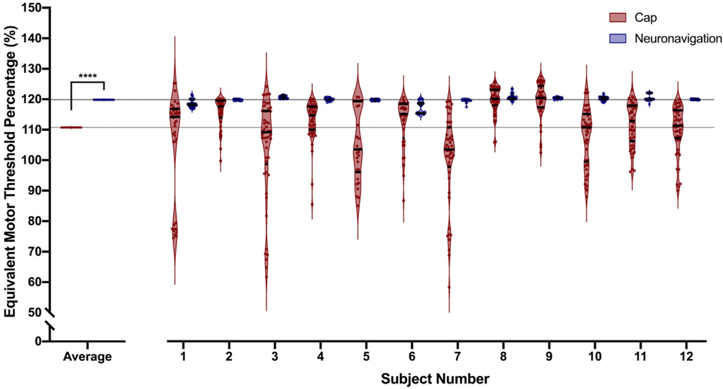
Methods: In 42 participants receiving up to 50 total accelerated rTMS sessions in 5 days, we compared cap and neuronavigation targeting approaches in 3408 distance and 6816 angle measurements. In Experiment 1, TMS administrators saved an on-target neuronavigation location at Beam F3, which served as the landmark for all other measurements. Next, the operators placed the TMS coil based on cap markings or neuronavigation software to measure the distance and angle differences from the on-target sample. In Experiment 2, we saved each XYZ coordinate of the TMS coil from cap and neuronavigation targeting in 12 participants to compare the E-field magnitude differences at the cortical prefrontal target in 1106 cap and neuronavigation models.

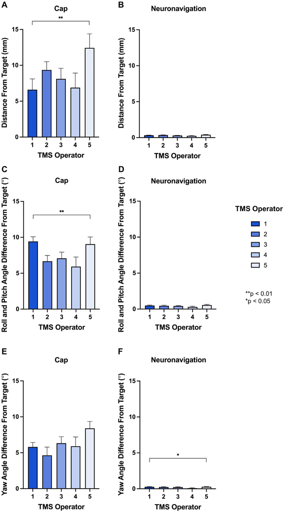
Results: Cap targeting was significantly off-target for distance, placing the coil an average of 10.66 mm off-target (Standard error of the mean; SEM = 0.19 mm) compared to 0.3 mm (SEM = 0.03 mm) for neuronavigation (p < 0.0001). Cap targeting also significantly deviated for angles off-target, averaging 7.79 roll/pitch degrees (SEM = 1.07°) off-target and 5.99 yaw degrees (SEM = 0.12°) off-target; in comparison, neuronavigation targeting positioned the coil 0.34 roll/pitch degrees (SEM = 0.01°) and 0.22 yaw (SEM = 0.004°) off-target (both p < 0.0001). Further analyses revealed that there were significant inter-operator differences on distance and angle positioning for F3 (all p < 0.05), but not neuronavigation. Lastly, cap targeting resulted in significantly lower E-fields at the intended prefrontal cortical target, with equivalent E-fields as 110.7% motor threshold (MT; range = 58.3-127.4%) stimulation vs. 119.9% MT (range = 115-123.3%) from neuronavigated targeting with 120% MT stimulation applied (p < 0.001).

Conclusions: Cap-based targeting is an inherent source of target variability compared to neuronavigation. Additionally, cap-based coil placement is more prone to differences across operators. Off-target coil placement secondary to cap-based measurements results in significantly lower amounts of stimulation reaching the cortical target, with some individuals receiving only 48.6% of the intended on-target E-field. Neuronavigation technology enables more precise and accurate TMS positioning, resulting in the intended stimulation intensities at the targeted cortical level.

Keywords: Dorsolateral prefrontal cortex (DLPFC); Elastic cap targeting; Electric field (E-field) modeling; Neuronavigated rTMS; Neuronavigation; Transcranial magnetic stimulation.


Conflict of interest statement

Declaration of competing interest We confirm that there are no known conflicts of interest associated with this publication and there was no financial support for this work that could have influenced its outcome.

Figures

Fig. 1.
Fig. 1.. Cap vs. Neuronavigation Coil Placement Procedure.
A. Photographs of Cap vs. Neuronavigation Targeting in a Representative Participant. Cap-based targeting uses the cap markings to place the TMS coil. In contrast, neuronavigation-based targeting enables the TMS operator to rely on the bullseye shown in Panel B. B. Neuronavigation Bullseye. Prior to the first stimulation session, the TMS operator used the Beam F3 method to place the TMS coil at the left prefrontal cortex and saved this location in the neuronavigation software. In this example, cap-based targeting placed the TMS coil 17.0 mm, 6.8° roll/pitch angle, and 1.8°yaw angle from the sampled F3 target; neuronavigation-based targeting placed the TMS coil 0.3 mm, 0.3° roll/pitch angle, and 0.1° yaw angle from the F3 target. Note that this example represents one stimulation session of the 3408 total stimulation sessions, and that there was variability of values in both the cap-based and neuronavigation-based TMS coil placements. C. TMS Coil XYZ Coordinate Locations. We saved the XYZ coordinates for up to 50 cap and neuronavigation locations in 12 healthy participants. In this example participant, we show the cortical projection of the Beam F3 location (green), the cap XYZ coordinates (50x red projections), and the neuronavigation XYZ coordinates (50x blue projections). We used these XYZ coordinates for electric field modeling in Experiment 2. (For interpretation of the references to colour in this figure legend, the reader is referred to the Web version of this article.)
Fig. 2.
Fig. 2.. Distance from F3 Target By Targeting Technique.
Cap targeting placed the coil a significantly greater distance from the F3 target, averaging 10.66 mm off-target compared to 0.30 mm in neuronavigation, p < 0.0001; error bars+/−SEM. There was also a significant effect of session and interaction between targeting technique by session (both p < 0.0001).
Fig. 3.
Fig. 3.. Roll and Pitch Angle from F3 Target By Targeting Technique.
Cap targeting placed the coil significantly more off-target in terms of roll/pitch angle, averaging 7.79° off-target compared to 0.34° for neuronavigation targeting, p < 0.0001; error bars+/−SEM. There was also a significant main effect of session number and interaction between targeting technique by session (both p < 0.0001).
Fig. 4.
Fig. 4.. Yaw Angle from F3 Target By Targeting Technique.
Similarly, yaw angle was significantly more off-target using cap-based targeting (average = 5.99°) compared to neuronavigation targeting (average = 0.22°), p < 0.0001; error bars+/−SEM. However, there was no significant effect of session or targeting method by session interaction (both p > 0.05).
Fig. 5.
Fig. 5.. Inter-Operator Accuracy of Cap Placement on Distance, Roll/Pitch Angle, and Yaw Angle.
In Experiment 1C, we tested the effects of operator cap placement on the distance, roll/pitch angle, and yaw angle deviation from the F3 target. Examining the first session of each day (i.e., immediately after the cap was repositioned), we analyzed each of the 5 TMS operators who placed the cap on at least one day. There were significant inter-operator differences on distance and roll/pitch angle from the F3 target (**p < 0.01; Fig. 5A and C). These inter-operator differences were not present when using neuronavigation (Fig. 5B and D), suggesting that neuronavigation is not only useful for reducing distance and angle deviation from the intended target, but also for reducing inter-operator differences that are present in cap- but not neuronavigation-based targeting. For yaw angle, there was no significant inter-operator differences for cap-based positioning (due to wide variation for each operator), but there was a significant difference for neuronavigation-based positioning (*p < 0.05; Fig. 5E–F). However, the average neuronavigation-based coil placements were all under 0.3° off-target. In sum, neuronavigation significantly reduces the error in coil placement for distance, roll/pitch angle, and yaw angle (Figs. 2–4), and additionally reduces the inter-operator differences in coil placement deviation (Fig. 5).
Fig. 6.
Fig. 6.. Raw Electric Field Magnitude Values at the Prefrontal Cortical Target.
In Experiment 2, we calculated 1106 electric field models across 12 healthy adult participants to compare the impact of off-target TMS coil positioning. Left: At a group level, neuronavigation-based targeting produced significantly higher electric fields at the dorsolateral prefrontal cortex (DLPFC) target region of interest (ROI), with an average value of 95.83 V/m compared to 88.36 V/m from cap-based targeting (****p < 0.0001; error bars+/−SEM). Right: Electric field magnitudes widely varied by stimulation technique and per participant. In each participant, neuronavigation targeting produced more closely grouped electric fields than cap targeting. The violin plots show the average and inter-quartile range per participant and targeting technique.
Fig. 7.
Fig. 7.. Visualization of Electric Fields in a Representative Participant.
In Experiment 2, we collected up to 50 cap and 50 neuro-navigation locations in 12 healthy control participants receiving 50 accelerated rTMS sessions over 5 days (10 sessions/day). Here we visually show the electric field distribution from cap vs. neuronavigation-based targeting in a representative participant with 50 cap and 48 neuronavigation E-field models, in comparison to the electric field produced form the coil at the targeted Beam F3 location. Red rectangles indicate the sessions in which the E-field magnitude was ≥10% lower than the E-field produced with the coil positioned on-target at the F3 target location. The E-field magnitude was ≥10% lower than on-target in 22/50 cap-based coil placements but 0/48 neuronavigation-based coil placements. There is a noticeably wider distribution of where is stimulated with cap targeting, compared to that of neuronavigation targeting. See Supplementary Video for an animated depiction of coil drift in cap-based vs. neuronavigated TMS. (For interpretation of the references to colour in this figure legend, the reader is referred to the Web version of this article.)
Fig. 8.
Fig. 8.. Intra- and Inter-Subject Variability in Electric Field Magnitude as an Equivalent Motor Threshold Percentage.
To put the raw electric field magnitude differences into more relatable terms, we converted the stimulation intensity from 120% motor threshold (MT) stimulation into the MT percentage that would produce the experienced electric field at the cortical target. The dotted horizontal lines represent the intended MT percentage of stimulation (120%, black line), the MT percentage of neuronavigated TMS (blue line), and the MT percentage of cap-based TMS (red line). Left: Cap-based targeting produced a significantly lower group average MT equivalent value of 110.7% MT, compared to 119.9% MT from neuronavigation (****p < 0.0001; error bars+/−SEM). On an individual level, cap-based targeting produced cortical target electric fields with a wide range (range = 58.3%–126.4%), compared to a much more tightly grouped electric field range from neuronavigated rTMS (range = 115–123.3%). Off-target coil placements due to cap targeting significantly affect the electric field delivered to the cortical prefrontal target. (For interpretation of the references to colour in this figure legend, the reader is referred to the Web version of this article.)

References

    1. O’Reardon JP, Solvason HB, Janicak PG, Sampson S, Isenberg KE, Nahas Z, et al. Efficacy and safety of transcranial magnetic stimulation in the acute treatment of major depression: a multisite randomized controlled trial. Biol Psychiatr 2007;62(11):1208–16. - PubMed
    1. George MS, Lisanby SH, Avery D, McDonald WM, Durkalski V, Pavlicova M, et al. Daily left prefrontal transcranial magnetic stimulation therapy for major depressive disorder: a sham-controlled randomized trial. Arch Gen Psychiatr 2010;67(5):507–16. - PubMed
    1. Carmi L, Tendler A, Bystritsky A, Hollander E, Blumberger DM, Daskalakis J, et al. Efficacy and safety of deep transcranial magnetic stimulation for obsessive-compulsive disorder: a prospective multicenter randomized double-blind placebo-controlled trial. Am J Psychiatr 2019;176(11):931–8. - PubMed
    1. Zangen A, Moshe H, Martinez D, Barnea-Ygael N, Vapnik T, Bystritsky A, et al. Repetitive transcranial magnetic stimulation for smoking cessation: a pivotal multicenter double-blind randomized controlled trial. World Psychiatr 2021;20(3):397–404. - PMC - PubMed
    1. Sparing R, Buelte D, Meister IG, Paus T, Fink GR. Transcranial magnetic stimulation and the challenge of coil placement: a comparison of conventional and stereotaxic neuronavigational strategies. Hum Brain Mapp 2008;29(1):82–96. - PMC - PubMed

Publication types

MeSH terms