Review
. 2022:189:153-176.
doi: 10.1016/B978-0-323-91532-8.00012-4.

Sudden unexpected death in epilepsy: Respiratory mechanisms

Affiliations
Review

Sudden unexpected death in epilepsy: Respiratory mechanisms

Frida A Teran et al. Handb Clin Neurol. 2022.

Abstract

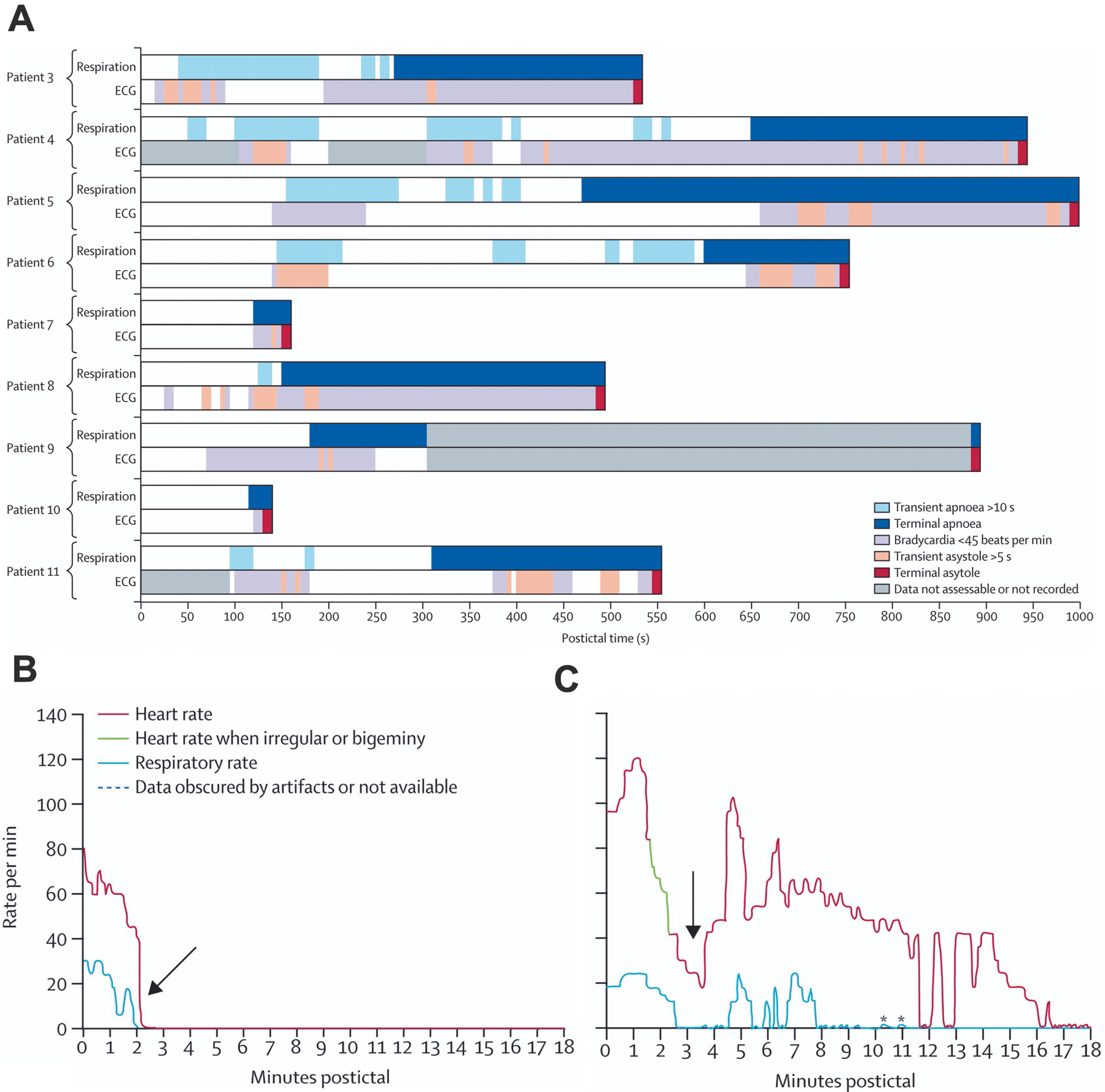
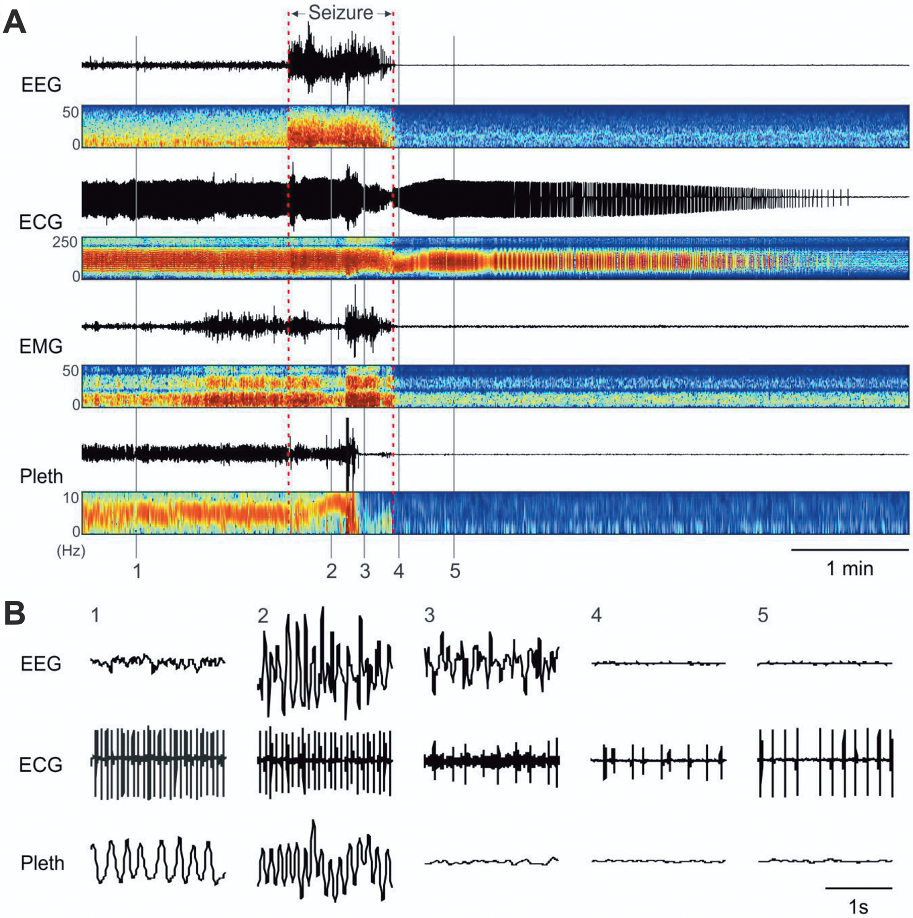
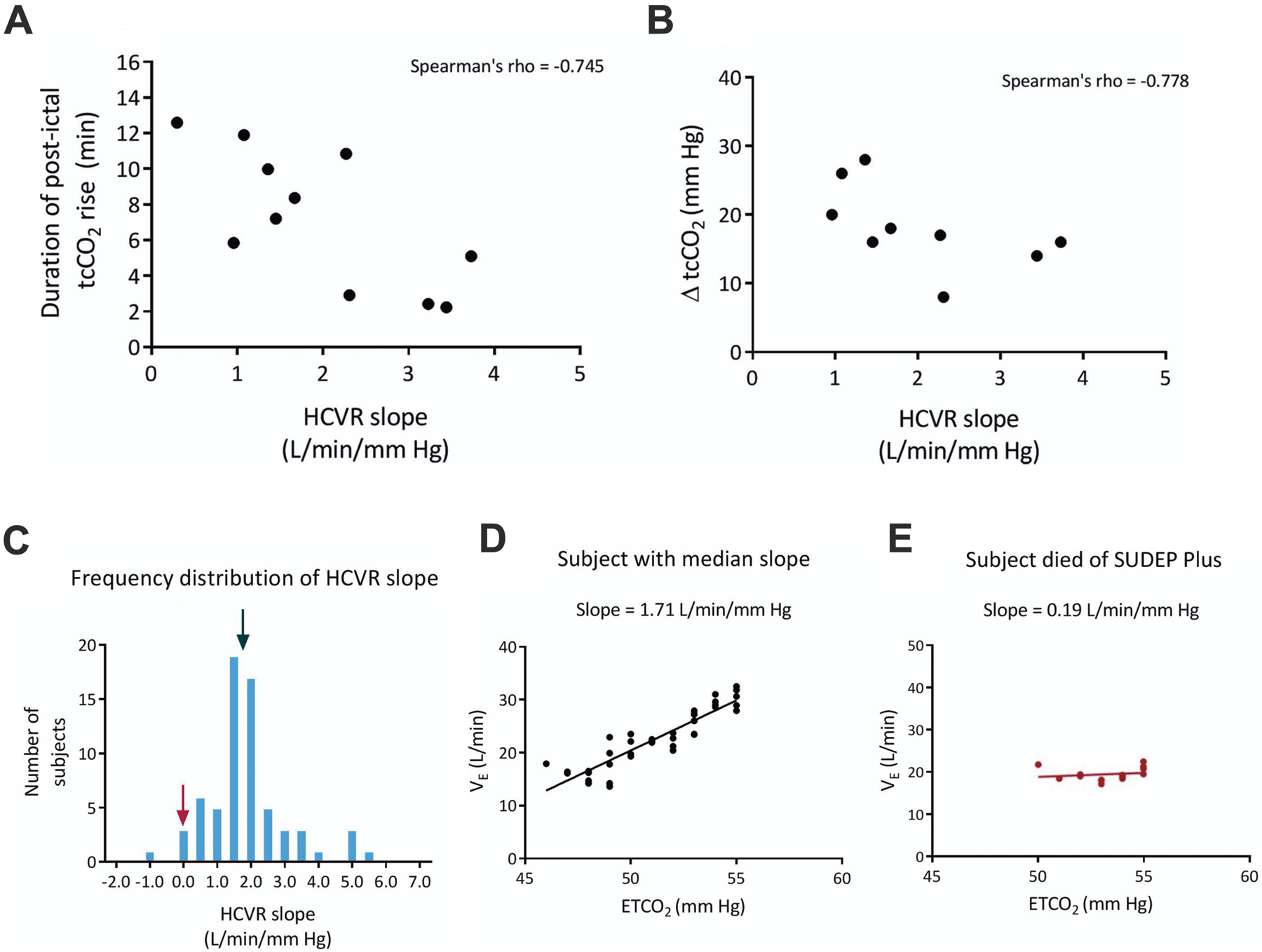
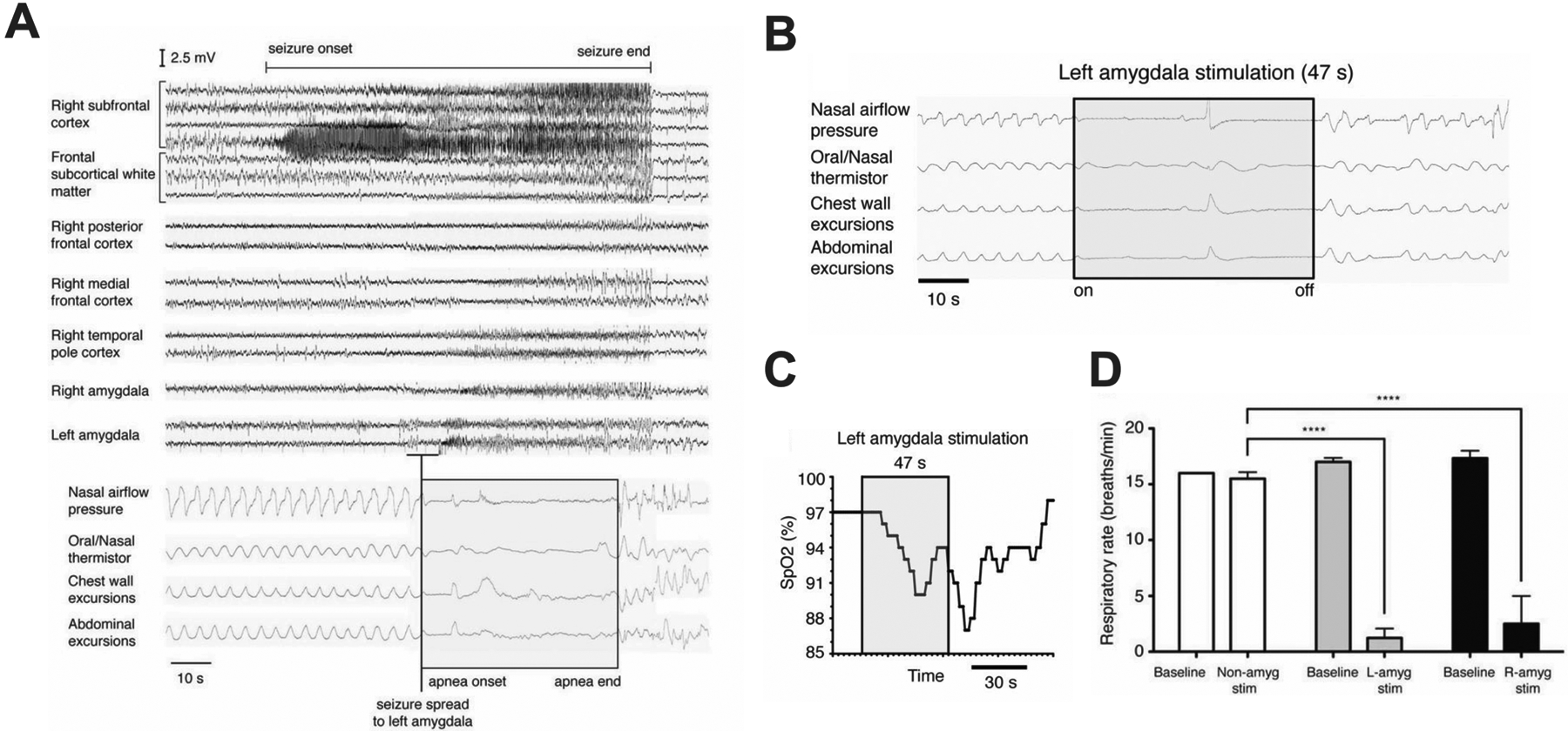
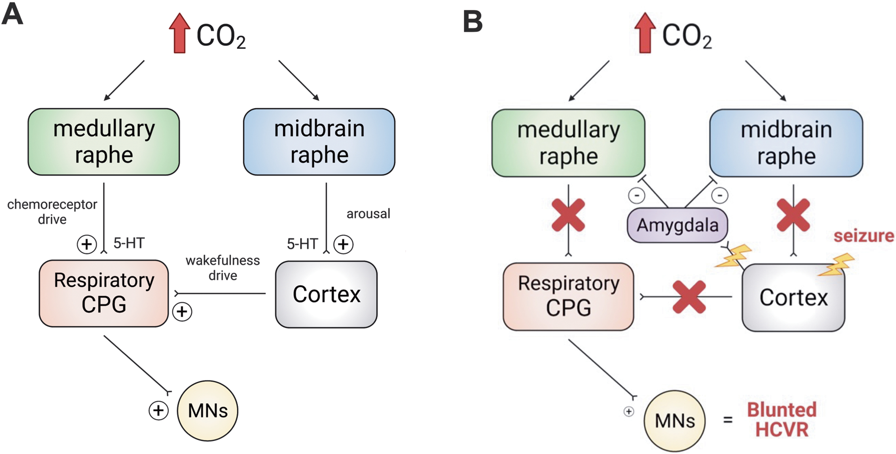
Epilepsy is one of the most common chronic neurologic diseases, with a prevalence of 1% in the US population. Many people with epilepsy live normal lives, but are at risk of sudden unexpected death in epilepsy (SUDEP). This mysterious comorbidity of epilepsy causes premature death in 17%-50% of those with epilepsy. Most SUDEP occurs after a generalized seizure, and patients are typically found in bed in the prone position. Until recently, it was thought that SUDEP was due to cardiovascular failure, but patients who died while being monitored in hospital epilepsy units revealed that most SUDEP is due to postictal central apnea. Some cases may occur when seizures invade the amygdala and activate projections to the brainstem. Evidence suggests that the pathophysiology is linked to defects in the serotonin system and central CO2 chemoreception, and that there is considerable overlap with mechanisms thought to be involved in sudden infant death syndrome (SIDS). Future work is needed to identify biomarkers for patients at highest risk, improve ascertainment, develop methods to alert caregivers when SUDEP is imminent, and find effective approaches to prevent these fatal events.

Keywords: Apnea; Breathing; CO(2); Chemoreception; SIDS; SUDEP; Serotonin; Ventilation.


Figures

Figure 1.
Figure 1.
Age and sex distribution of definite and probable sudden unexpected death occurring in epilepsy cases in Sweden in 2008. Reproduced with permission from Sveinsson et al (2017).
Figure 2.
Figure 2.
Pronounced oxygen desaturation with a complex partial left temporal onset seizure without secondary generalization. Patient was a 19-year-old male with a body mass index of 19.9. Seizure onset occurred with the patient awake and sitting in bed. He became unresponsive with lip smacking, a slight head turn to the left followed by forceful head turning to the right. He remained sitting for the duration of the seizure. The heart rate (beats per minute.) at various times is shown. Two other complex partial seizures in this patient (one left and one right temporal onset) were accompanied by oxygen desaturations below 50%. Oxygen saturation percent is shown on the ordinate. Reproduced with permission from Bateman et al (2008).
Figure 3.
Figure 3.
SUDEP cases monitored in epilepsy units at the time of death always occurred after a generalized tonic-clonic seizure and had terminal apnea that preceded terminal asystole. A) Shown are data from nine separate patients with bar graphs colored to show time of breathing patterns plotted above those of cardiac patterns. Time = 0 marks the end of each electrographic seizure. B) Pattern of postictal cardiorespiratory function from an individual patient (Patient 10) with near-simultaneous cessation of respiratory and cardiac activity. C) Postictal cardiorespiratory activity from an individual patient (Patient 5) with severe disruption of respiratory activity 14 minutes before cessation of cardiac activity. Breathing was measured by observation of respiratory movements on video. Breathing could not be determined during seizures due to movement artifact. Reproduced with permission from Ryvlin et al, (2013).
Figure 4.
Figure 4.
Death after spontaneous seizures in Scn1aR1407X/+ mice is initiated by respiratory arrest. A) Events leading to death in an Scn1aR1407X/+ mouse. Traces from top down are EEG, ECG, EMG, and whole-animal plethysmography (Pleth), in each case with paired power spectrum heatmaps shown below. A spontaneous seizure occurred at the time indicated, and this was followed by electrocerebral silence in the EEG. Shortly after the onset of the seizure, breathing became disrupted. Respiratory activity ceased halfway through the seizure. In contrast, the ECG did not change until near the end of the seizure, after which the frequency and amplitude slowly decreased over the next 4 minutes. B) Expanded traces of EEG, ECG, and plethysmography at the times labeled in (A) as 1–5. Reproduced with permission from Kim et al (2018).
Figure 5.
Figure 5.
The 5-HT system is involved in the ventilatory response to hypercapnia. A) Medullary raphe neurons are located close to the basilar artery and its large penetrating branches. Shown is a transverse section of the midline rostral medulla (dorsal to the basilar artery). 5-HT neurons in the raphe are immunostained with an antibody against tryptophan hydroxylase (green). Blood vessels are filled with fluorescently-tagged albumin (red). Scale bar: 50 μm. Reproduced with permission from Fiske (2002). B). 5-HT neurons project to neurons in the major respiratory nuclei. Reconstruction of two biocytin-filled raphe obscurus 5-HT neurons (red, blue) and two pre-Bötzinger complex neurons (green, black) in a brain slice illustrating dendrites and axonal projections. Reproduced from Ptak et al (2009). C) Awake mice with genetic deletion of 5-HT neurons (Lmx1bf/f/p) have a smaller ventilatory response to an increase in ambient CO2 compared to WT littermates. Adapted from Hodges et al (2008).
Figure 6.
Figure 6.
Decreased multiunit activity (MUA) in medullary raphe and reduced breathing during a seizure. A) Seizure induced by 2 s stimulation of hippocampus (HC). After the stimulus, fast polyspike activity is seen in the HC local field potential (LFP). MUA from the medullary raphe shows marked suppression of neuronal firing during the seizure with slow recovery in the post-ictal period. Respiratory trace shows airflow amplitude (proportional to volume) and respiratory rate both decreased markedly during the ictal and post-ictal periods. B) Expanded segments of data from baseline, seizure, post-ictal, and recovery periods from the boxed regions in (A). Reproduced from Zhan et al (2016).
Figure 7.
Figure 7.
Serum 5-HT levels and postconvulsive central apnea (PCCA) after generalized convulsive seizures. Elevated levels of postictal 5-HT in generalized convulsive seizures (GCS). The mean serum interictal 5-HT levels are shown in light green bars and postictal 5-HT levels (ng/mL) are shown in dark green bars for the 2 seizure groups: PCCA (n = 8) and non-PCCA (n = 19). The levels of postictal serum 5-HT in the absence of PCCA were higher when compared to interictal levels (p < 0.001), but not when PCCA occurred (p = 0.22), suggesting the elevated 5-HT may be protective against PCCA. Reproduced with permission from Murugesan et al (2019).
Figure 8.
Figure 8.
Patients with DS have abnormal breathing after seizures. A) Schematic of events while recording cardiorespiratory activity during and after a seizure in a 9-year-old girl with DS. Arrows labeled “b-e” denote the time of recordings shown in parts B–E, respectively. Area within red bars denotes convulsive seizures. B–E) Respiratory impedance plethysmography (Pleth) and ECG during normal breathing when tcCO2 was 43 mmHg (B), between convulsive seizures when there was apnea (C), after the seizures when apnea continued (D), and 44 minutes after the seizures when tcCO2 had risen to 68 mmHg (E). At 2 hours, tcCO2 decreased when the patient was stimulated to arouse, but did not return to baseline until 4 hours. Reproduced with permission from Kim et al (2018).
Figure 9.
Figure 9.
A low interictal HCVR slope is associated with more severe postictal apnea, and may increase the risk of SUDEP in epilepsy patients. A) Scatterplot depicting the correlation between HCVR slope and duration of postictal tcCO2 rise and, B) HCVR and magnitude of postictal tcCO2 rise (ΔtcCO2). C) Frequency distribution of HCVR slopes in epilepsy patients. The black arrow points to the median slope for all patients and the maroon arrow indicates a patient who later died of SUDEP plus. D) Linear regression of VE and ETCO2 for a patient with the median response indicated in (C). E) Linear regression for the patient in (C) shown with the maroon arrow. Adapted with permission from Sainju et al (2019).
Figure 10.
Figure 10.
Central apnea occurs in patients when seizures spread to the left amygdala, and can be induced by electrical stimulation. A) EEG of complex partial seizure with onset in the right subfrontal cortex. Breathing pattern is normal before and after seizure initiation. Apnea occurred when the seizure spread to the left amygdala and before it spread to the right amygdala, right temporal pole, right medial frontal cortex, and right posterior frontal cortex. B) Apnea could be induced by electrical stimulation at the electrode contact in (A) coinciding with apnea. C) Apnea was severe enough to cause O2 desaturation below 90%. D) Apnea was induced by stimulation of either the left or right amygdala. Adapted from Dlouhy et al (2015).
Figure 11.
Figure 11.
Working model for seizure-induced respiratory depression. A) There are two main sources of respiratory drive. While awake, breathing is maintained by drive from chemoreceptors (including medullary raphe neurons) and cortical “wakefulness drive.” When CO2 increases it causes arousal from sleep and stimulates chemoreceptor drive. B) When a seizure occurs and propagates from the cortex to the brainstem (including via the amygdala), both chemoreceptor and wakefulness drives are lost. This leads to decreased baseline ventilation and a blunted HCVR. This postictal HCVR depression and hypoventilation could increase the risk of respiratory arrest and SUDEP. MNs = motor neurons, CPG = central pattern generator.
Figure 12.
Figure 12.
The number of published studies on SUDEP has increased dramatically from 1971 to 2021. PubMed was used to search for articles using ‘sudden unexpected death in epilepsy’ or ‘SUDEP’.

References

    1. Ahmad S, Fowler LJ & Whitton PS (2005). Effects of combined lamotrigine and valproate on basal and stimulated extracellular amino acids and monoamines in the hippocampus of freely moving rats. Naunyn Schmiedebergs Arch Pharmacol, 371, 1–8. - PubMed
    1. Aiba I & Noebels JL (2015). Spreading depolarization in the brainstem mediates sudden cardiorespiratory arrest in mouse SUDEP models. Sci Transl Med, 7, 282ra46. - PMC - PubMed
    1. Albano C, Cupello A, Mainardi P, et al. (2006). Successful treatment of epilepsy with serotonin reuptake inhibitors: proposed mechanism. Neurochem Res, 31, 509–14. - PubMed
    1. Alper K, Schwartz KA, Kolts RL, et al. (2007). Seizure incidence in psychopharmacological clinical trials: an analysis of Food and Drug Administration (FDA) summary basis of approval reports. Biol Psychiatry, 62, 345–54. - PubMed
    1. Anderson LL, Thompson CH, Hawkins NA, et al. (2014). Antiepileptic activity of preferential inhibitors of persistent sodium current. Epilepsia, 55, 1274–83. - PMC - PubMed