. 2022 Dec;22(12):3061-3068.
doi: 10.1111/ajt.17181. Epub 2022 Sep 14.

IL-2 receptor engineering enhances regulatory T cell function suppressed by calcineurin inhibitor

Affiliations

IL-2 receptor engineering enhances regulatory T cell function suppressed by calcineurin inhibitor

Toshihito Hirai et al. Am J Transplant. 2022 Dec.

Abstract

Clinical trials utilizing regulatory T cell (Treg) therapy in organ transplantation have shown promising results, however, the choice of a standard immunosuppressive regimen is still controversial. Calcineurin inhibitors (CNIs) are one of the most common immunosuppressants for organ transplantation, although they may negatively affect Tregs by inhibiting IL-2 production by conventional T cells. As a strategy to replace IL-2 signaling selectively in Tregs, we have introduced an engineered orthogonal IL-2 (ortho IL-2) cytokine/cytokine receptor (R) pair that specifically binds with each other but does not bind with their wild-type counterparts. Murine Tregs were isolated from recipients and retrovirally transduced with ortho IL-2Rβ during ex vivo expansion. Transduced Tregs (ortho Tregs) were transferred into recipient mice in a mixed hematopoietic chimerism model with tacrolimus administration. Ortho IL-2 treatment significantly increased the ortho IL-2Rβ(+) Treg population in the presence of tacrolimus without stimulating other T cell subsets. All the mice treated with tacrolimus plus ortho IL-2 achieved heart allograft tolerance, even after tacrolimus cessation, whereas those receiving tacrolimus treatment alone did not. These data demonstrate that Treg therapy can be adopted into a CNI-based regimen by utilizing cytokine receptor engineering.

Keywords: animal models: murine; basic (laboratory) research/science; bioengineering; cellular biology; cytokines/cytokine receptors; immunobiology; immunosuppressant - calcineurin inhibitor (CNI); immunosuppression/immune modulation; tolerance: chimerism.


Conflict of interest statement

DISCLOSURE

The authors of this manuscript have conflicts of interest to disclose as described by the American Journal of Transplantation. Ortho IL-2 is the subject of a pending patent filing with LLS, LKP, and KCG as inventors. KCG is a founder of Synthekine. The other authors declare that they have no competing interests.

Figures

FIGURE 1
FIGURE 1
Ortho IL-2 and tacrolimus synergistically increase %ortho Tregs. Foxp3GFP+ Tregs transduced with ortho IL-2Rβ (ortho Tregs) and naïve Tcons were incubated with CD3/CD28 beads. Wt IL-2 (1000 IU/ml), ortho IL-2 (100 000 IU/ml), and tacrolimus (100 ng/ml) were added as indicated and replenished every 2 days. (A) Representative pseudocolor plots of co-cultured cells. (B-D) Box plots indicating the number of Tcons (B), ortho Tregs (C), and the proportion of ortho Tregs among total cells (D) on day 4. Quantification of triplicate wells in 1 representative experiment of 2 independent experiments. *<0.05, **<0.01, ***<0.001; p-values calculated by Dunnett test comparing each column to PBS-control (gray bar) among tacrolimus (−) or tacrolimus (+) group. ns, not significant.
FIGURE 2
FIGURE 2
Ortho IL-2 increase ortho IL-2Rβ hi Tregs. (A) Representative pseudo color plot for tRFP (ortho IL-2Rβ) hi and low gate. The absolute number of ortho IL-2Rβ hi (B) and low (C) fractions in each treatment group are shown. Quantification of triplicate wells in 1 representative experiment of 2 independent experiments. *<0.05, **<0.01, ***<0.001; p-values calculated by Dunnett test comparing each column to PBS-control (gray bar) among tacrolimus (−) or tacrolimus (+) group.
FIGURE 3
FIGURE 3
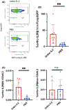
Ortho IL-2 injection together with tacrolimus facilitates ortho Treg expansion and the establishment of heart transplant tolerance. (A) Schema of in vivo experiment. Anti-CD40L (MR1); 0.5 mg i.p. on d0. ortho IL-2; 25 000 IU, tacrolimus; 5 mg/kg, both i.p. from d0 to d14. (B) Left; representative pseudocolor plots for CD4+ gated cells on d14. Right; mean ± SD of %Foxp3GFP+/CD4+ cells at indicated time points. (C) Mean ± SD of %donor (CD45.1+ cells/CD45+ PBMCs) at indicated time points, dashed line: 1%. Pooled data from two independent experiments with at least three mice per group. *p-values <0.05 calculated by unpaired t-test between indicated two groups.
FIGURE 4
FIGURE 4
Ortho IL-2 increased the expression of ortho IL-2Rβ. (A) Representative pseudocolor plot of CD4+Foxp3GFP+ gated cells in the peripheral blood on d14 showing gating strategy for ortho IL-2Rβ (tRFP) hi and low fraction. Mean ± SD with individual plots showing the proportion of ortho IL-2Rβ hi in Foxp3GFP+ cells (B) and the proportion of ortho IL-2Rβ hi and low cells among CD45.2 (C). Pooled data from two independent experiments with at least three mice per group. *p-values <0.05 calculated by unpaired t-test between indicated two groups.
FIGURE 5
FIGURE 5
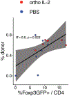
The proportion of ortho Treg and donor cell chimerism shows positive correlation. Pearson's correlation for %Foxp3GFP+/CD4+ cells (x-axis) and %donor (y-axis) on d14. Each dots represent individual mouse in ortho IL-2-treated (red) and PBS-treated mouse (blue). R = 0.6, p = 0.023.
FIGURE 6
FIGURE 6
Combination of ortho IL-2 plus tacrolimus treatment promotes heart allograft tolerance. Heart allograft survival curve. **p-value <0.01 calculated by Log-rank test. Red, ortho IL-2-treated, N = 7; blue, PBS-treated, N = 7. Pooled data from two independent experiments.
FIGURE 7
FIGURE 7
Ortho IL-2 replaces IL-2 signaling in ortho IL-2Rβ+ Tregs that was deprived by calcineurin inhibitor treatment. (Left) TCR stimulation activates NFAT and promotes transcription of IL-2 in Teffs. Wt IL-2 secreted from activated Teffs stimulate both Teffs and Tregs. (Mid) CNIs inhibit NFAT activation and result in IL-2 deprivation for both Teffs and Tregs. (Right) ortho IL-2 restores IL-2 signaling through transduced ortho IL-2R in Tregs but does not affect wt IL-2R on Teffs. Created with BioRender.com.

References

    1. Tang Q, Vincenti F. Transplant trials with Tregs: perils and promises. J Clin Invest. 2017;127(7):2505–2512. doi:10.1172/JCI90598 - DOI - PMC - PubMed
    1. Chandran S, Tang Q, Sarwal M, et al. Polyclonal regulatory T cell therapy for control of inflammation in kidney transplants. Am J Transplant. 2017;17(11):2945–2954. doi:10.1111/ajt.14415 - DOI - PMC - PubMed
    1. Todo S, Yamashita K, Goto R, et al. A pilot study of operational tolerance with a regulatory T-cell-based cell therapy in living donor liver transplantation. Hepatology. 2016;64(2):632–643. doi:10.1002/hep.28459 - DOI - PubMed
    1. Sawitzki B, Harden PN, Reinke P, et al. Regulatory cell therapy in kidney transplantation (the ONE study): a harmonised design and analysis of seven non-randomised, single-arm, phase 1/2A trials. Lancet. 2020;395(10237):1627–1639. doi:10.1016/S0140-6736(20)30167-7 - DOI - PMC - PubMed
    1. Akimova T, Kamath BM, Goebel JW, et al. Differing effects of rapamycin or calcineurin inhibitor on T-regulatory cells in pediatric liver and kidney transplant recipients. Am J Transplant. 2012;12(12):3449–3461. doi:10.1111/j.1600-6143.2012.04269.x - DOI - PMC - PubMed

Publication types