

Chiropractic Management of Symptomatic Pedicle Hemangioma: a Case Report
- PMID: 36032593
- PMCID: PMC9375871
- DOI: 10.26574/maedica.2022.17.2.528
Chiropractic Management of Symptomatic Pedicle Hemangioma: a Case Report
Abstract
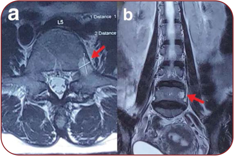
Spinal hemangiomas are the most commonly encountered primary vertebral tumors, which are benign and asymptomatic. They usually occur in the vertebral body and rarely extend into or originate from the posterior column. Thus, hemangiomas of the pedicle are extremely rare. Only one case of pedicle hemangioma has been documented in the literature. Disc herniations and annular tears are not always symptomatic, and they have always been observed in asymptomatic patients. Therefore, even with a thorough medical history and physical examination, patients with comorbid hemangioma and annular tears present a formidable challenge to the most experienced clinicians. This report describes a rare case of pedicle hemangioma and disc herniation without spinal cord compression in a 47-year-old woman complaining of lower back pain and inability to walk.
Figures

References
-
- Patel S, Ansari D, Patil SN, et al. High-grade spinal hemangioma: a national cancer database analysis. World Neurosurg. 2021;148:e527–e535. - PubMed
-
- Tang C, Liao YH, Tang Q, et al. What is the difference in pedicle morphology of the fifth lumbar vertebra between isthmic and degenerative L5–S1 spondylolisthesis? An anatomic study of 328 patients via multi-slice spiral computed tomography. Eur Spine J. 2021;30:2301–2310. - PubMed
-
- Ekin EE, Altunrende ME. Pedicle stress injury in children and adolescents with low back pain. Spine. 2019;44:E1038–E1044. - PubMed
Publication types
LinkOut - more resources
Full Text Sources