

An Anatomical Evaluation of Normal and Aberrant Foramen Ovale in Skull Base with Its Clinical Significance
- PMID: 36032599
- PMCID: PMC9375881
- DOI: 10.26574/maedica.2022.17.2.357
An Anatomical Evaluation of Normal and Aberrant Foramen Ovale in Skull Base with Its Clinical Significance
Abstract
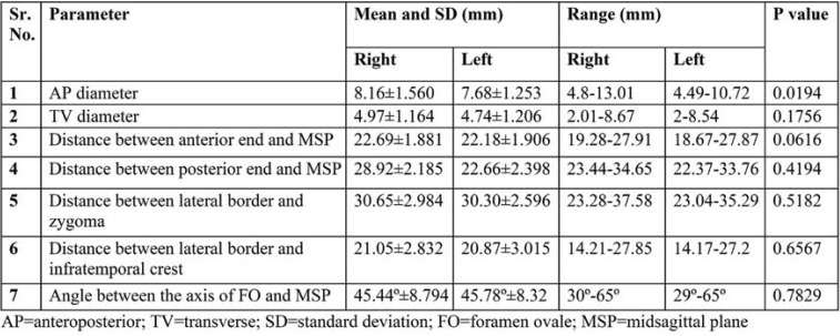
Introduction: Foramen ovale is one of the most significant foramina of skull base and transmits mandibular nerve. Its detailed knowledge is crucial in treatment of trigeminal neuralgia and various diagnostic practices. Aim: Aim of the study was to provide anatomical data of foramen ovale regarding number, shape, diameters and its relation to nearby bony landmarks. Material and method: The present study was ethically approved and 100 dry adult human skulls were included in the study to evaluate 200 foramina ovale. Non-metric parameters were observed and metric parameters were measured with Vernier calliper and goniometer. Results:Different kinds of shapes were found in foramen ovale. Variant features in the form of bony spine, ridge, foramen or bar were identified. The means of anteroposterior and transverse diameter of foramen ovale were found to be 8.16 and 4.97 mm on the right side, and 7.68 and 4.74 mm on the left side. The mean distance of its anterior and posterior ends from the midsagittal plane were 22.69 and 28.92 mm on the right side, and 22.18 and 22.66 mm on the left side. Distance from the lateral border of foramen ovale to the posterior end of zygoma and midpoint of infratemporal crest was found to be 30.65 and 21.05 mm on the right, and 30.30 and 20.87 mm on the left side. The mean of angle of foramen ovale with midsagittal was 45.44º on the right side and 45.78º on the left side. Conclusion:Variations found in foramen ovale are key points to keep in mind while operating in this region. Measured metric parameters were found to a higher extent on the right side than the left one. The present study will be helpful for both further research and neurosurgeons operating in this region.
Figures


References
-
- Teul I, Czerwiński F, Gawlikowska A, et al. Asymmetry of the ovale and spinous foramina in mediaeval and contemporary skulls in radiological examinations. Folia Morphol. 2002;61:147–152. - PubMed
-
- Wieser HG, Siegel AM. Analysis of foramen ovale electrode recorded seizures and correlation with outcome following amygdalohippocamectomy. Epilepsia. 1991;32:838–850. - PubMed
-
- Ray B, Gupta N, Ghose S. Anatomic variations of foramen ovale. Kathmandu Univ Med. 2005;3:64–68. - PubMed
-
- Skrzat J, Walocha J, Środek R. An anatomical study of the pterygoalar bar and the pterygoalar foramen. Folia Morphol. 2005;64:92–96. - PubMed
-
- Freire AR, Rossi AC, de Oliveira VCS, et al. Emissary foramens of the human skull: anatomical characteristics & its relations with clinical neurosurgery. Int J Morphol. 2013;31:287–292.
Publication types
LinkOut - more resources
Full Text Sources