. 2022 Aug 12:9:958586.
doi: 10.3389/fmolb.2022.958586. eCollection 2022.

Biochemical and Structural Insights on the Poplar Tau Glutathione Transferase GSTU19 and 20 Paralogs Binding Flavonoids

Affiliations

Biochemical and Structural Insights on the Poplar Tau Glutathione Transferase GSTU19 and 20 Paralogs Binding Flavonoids

Elodie Sylvestre-Gonon et al. Front Mol Biosci. .

Abstract

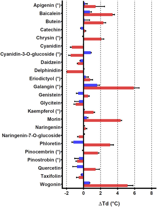
Glutathione transferases (GSTs) constitute a widespread superfamily of enzymes notably involved in xenobiotic detoxification and/or in specialized metabolism. Populus trichocarpa genome (V4.1 assembly, Phytozome 13) consists of 74 genes coding for full-length GSTs and ten likely pseudogenes. These GSTs are divided into 11 classes, in which the tau class (GSTU) is the most abundant with 54 isoforms. PtGSTU19 and 20, two paralogs sharing more than 91% sequence identity (95% of sequence similarity), would have diverged from a common ancestor of P. trichocarpa and P. yatungensis species. These enzymes display the distinctive glutathione (GSH)-conjugation and peroxidase activities against model substrates. The resolution of the crystal structures of these proteins revealed significant structural differences despite their high sequence identity. PtGSTU20 has a well-defined deep pocket in the active site whereas the bottom of this pocket is disordered in PtGSTU19. In a screen of potential ligands, we were able to identify an interaction with flavonoids. Some of them, previously identified in poplar (chrysin, galangin, and pinocembrin), inhibited GSH-conjugation activity of both enzymes with a more pronounced effect on PtGSTU20. The crystal structures of PtGSTU20 complexed with these molecules provide evidence for their potential involvement in flavonoid transport in P. trichocarpa.

Keywords: Populus trichocarpa; flavonoids; glutathione transferase (GST); ligandin property; photosynthetic organisms; poplar; specialized metabolism; structure.


Conflict of interest statement

The authors declare that the research was conducted in the absence of any commercial or financial relationships that could be construed as a potential conflict of interest.

Figures

FIGURE 1
FIGURE 1
Crystal structures of PtGSTU20. (A) View of the dimer of PtGSTU20 that highlights its putative H-site. The backbone atoms (cartoon) are colored by monomer. The morin flavonoid is represented as gray sticks with the non-carbon atoms colored according to their types (red, oxygen; blue, nitrogen; yellow, sulfur). (B) View of PtGSTU20 monomer that shows both G- and H-sites. The G-site is occupied by the glutathionyl moiety of glutathionyl-phenylacetophenone (GS-PAP). The phenylacetophenone moiety, disordered over two positions, defines the boundaries of the putative H-site. The backbone atoms (cartoon) of PtGSTU20 are colored according to their secondary structure (cyan, helix; red, strand; magenta, loop). GS-PAP is represented as gray sticks with the non-carbon atoms colored according to their types (red, oxygen; blue, nitrogen; yellow, sulfur).
FIGURE 2
FIGURE 2
Structure-based sequence alignment of GSTUs highlighting common regions. The sequence alignment was generated with mTM-align. Sequences were retrieved from the RCSB PDB: SbGSTU6 (GSTU6 from Salix babylonica, PDB ID 7DW2), MiGSTU1 (GSTU1 from Mangifera indica, 5G5E), TaGSTU4 (GSTU4 from Aegilops tauschii, 1GWC), AtGSTU25 (GSTU25 from Arabidopsis thaliana, 5G5A), AtGSTU20 (GSTU20 from A. thaliana, 5ECS), PtGSTU30 (GSTU30 from P. trichocarpa, 5J4U), GmGSTU10 (GSTU10 from Glycine max, 4CHS), GmGSTU4 (GSTU4 from G. max, 2VO4), OsGSTU1 (GSTU1 from Oryza sativa, 1OYJ), AtGSTU23 (GSTU23 from A. thaliana, 6EP7), RcGSTU1 (GSTU1 from Ricinus communis, 4J2F), PtGSTU19 (this study), SbGSTU7 (GSTU7 from S. babylonica, PDB ID 7DWD), and PtGSTU20 (this study). Secondary structures are labeled and shown using arrows (β-strands) and squiggles (helices). Common regions, i.e., regions with no gap and with pairwise residue distances less than 4Å are highlighted in blue. Residues participating in dimer stabilization via polar interactions are marked with ■. Residues involved in glutathione binding (G-site) are marked with ▲. Residues involved in the putative H-site of PtGSTU20 are marked with ●.
FIGURE 3
FIGURE 3
Binding of GS-PAP (A) and morin (B) in the putative H-site of PtGSTU20. Putative H-site of PtGSTU20 is a well-delineated cavity deeply inserted between the α4 and α6 helices of the C-terminal domain. Both conformations of GS-PAP are shown. The GS-PAP (A) and morin (B) ligands are represented in sticks as their surrounding residues. Intermolecular contacts are materialized as dashed lines. The N- and C-terminal domains are colored orange and magenta, respectively, and ligands are colored green. Non-carbon atoms are colored according to their types (red, oxygen; blue, nitrogen; yellow, sulfur).
FIGURE 4
FIGURE 4
Effects of 23 flavonoids on the thermostability of PtGSTU19 and 20 isoforms. Thermostability of PtGSTU19 (blue bars) and 20 (red bars) isoforms has been analyzed by using 20 μM of protein with or without 100 µM of chemical compounds diluted in 8% DMSO (Supplementary Table S3). The denaturation temperature difference (ΔTd) corresponds to the difference between the denaturation temperature of the protein in the presence of a potential ligand and a reference assay in which the potential ligand is replaced by an equivalent DMSO concentration.

Similar articles

Cited by

References

    1. Ahmad L., Rylott E. L., Bruce N. C., Edwards R., Grogan G. (2017). Structural Evidence for Arabidopsis Glutathione Transferase AtGSTF2 Functioning as a Transporter of small Organic Ligands. FEBS Open Bio 7, 122–132. 10.1002/2211-5463.12168 - DOI - PMC - PubMed
    1. Allocati N., Masulli M., Pietracupa M., Federici L., Di Ilio C. (2006). Evolutionarily Conserved Structural Motifs in Bacterial GST (Glutathione S-Transferase) Are Involved in Protein Folding and Stability. Biochem. J. 394, 11–17. 10.1042/BJ20051367 - DOI - PMC - PubMed
    1. Axarli I., Dhavala P., Papageorgiou A. C., Labrou N. E. (2009a). Crystal Structure of Glycine max Glutathione Transferase in Complex with Glutathione: Investigation of the Mechanism Operating by the Tau Class Glutathione Transferases. Biochem. J. 422, 247–256. 10.1042/BJ20090224 - DOI - PubMed
    1. Axarli I., Dhavala P., Papageorgiou A. C., Labrou N. E. (2009b). Crystallographic and Functional Characterization of the Fluorodifen-Inducible Glutathione Transferase from Glycine max Reveals an Active Site Topography Suited for Diphenylether Herbicides and a Novel L-Site. J. Mol. Biol. 385, 984–1002. 10.1016/j.jmb.2008.10.084 - DOI - PubMed
    1. Axarli I., Muleta A. W., Vlachakis D., Kossida S., Kotzia G., Maltezos A., et al. (2016). Directed Evolution of Tau Class Glutathione Transferases Reveals a Site that Regulates Catalytic Efficiency and Masks Co-operativity. Biochem. J. 473, 559–570. 10.1042/BJ20150930 - DOI - PubMed