. 2022 Aug 24;8(8):1091-1101.
doi: 10.1021/acscentsci.2c00057. Epub 2022 Apr 25.

Intersubunit Coupling Enables Fast CO2-Fixation by Reductive Carboxylases

Affiliations

Intersubunit Coupling Enables Fast CO2-Fixation by Reductive Carboxylases

Hasan DeMirci et al. ACS Cent Sci. .

Abstract

Enoyl-CoA carboxylases/reductases (ECRs) are some of the most efficient CO2-fixing enzymes described to date. However, the molecular mechanisms underlying the extraordinary catalytic activity of ECRs on the level of the protein assembly remain elusive. Here we used a combination of ambient-temperature X-ray free electron laser (XFEL) and cryogenic synchrotron experiments to study the structural organization of the ECR from Kitasatospora setae. The K. setae ECR is a homotetramer that differentiates into a pair of dimers of open- and closed-form subunits in the catalytically active state. Using molecular dynamics simulations and structure-based mutagenesis, we show that catalysis is synchronized in the K. setae ECR across the pair of dimers. This conformational coupling of catalytic domains is conferred by individual amino acids to achieve high CO2-fixation rates. Our results provide unprecedented insights into the dynamic organization and synchronized inter- and intrasubunit communications of this remarkably efficient CO2-fixing enzyme during catalysis.


Conflict of interest statement

The authors declare no competing financial interest.

Figures

Figure 1
Figure 1
Reaction scheme and structural organization of the K. setae ECR complex. (a) Carboxylation reaction scheme of ECR. (b) Anisotropic B-factors of the tetramer of the different ECR complexes solved in this study are shown color coded according to the B factors (blue for low values and red for high values).
Figure 2
Figure 2
Binding of NADPH results in global and local conformational changes in K. setae ECR. (a) NADPH-bound tetramer complex that is organized as pair of dimers, a pair of closed (green) and open (orange) subunits, and another pair containing closed (gray) and open (blue) subunits. (b) The foreground dimer with open (orange)- and closed-form (green) subunits rotated by 30° from the view in (a). Each subunit is composed of a catalytic and an oligomerization domain. (c) Comparison of the putative substrate binding sites between the open- and closed-form subunits.
Figure 3
Figure 3
Structure of the ternary ECR complex. (a) ECR tetramer in a complex with NADPH and butyryl-CoA organized as a pair of dimers, the foreground dimer with one closed subunit (green) with NADPH and butyryl-CoA, an open (orange) subunit containing NADPH, and another pair in the background with one closed (gray) and one open (blue) subunit. (b) The foreground dimer with closed (green) and open (orange) subunits, rotated by 30° from the view in (a). Butyryl-CoA and NADPH atoms are represented as spheres. (c) Comparison of the product binding sites between the open- and closed-form subunits. (d) Cartoon and stick representation of the closed-form subunit active site. In (d–g), simple 2FoFc density contoured at the 1.5 σ level is shown for butyryl-CoA, or pa ortion thereof, and NADPH within 3 Å from the molecules. (e) Cartoon and stick representation of the open-form subunit active site. (f) Superposition of the open-form subunit onto the closed-form subunit with a stick representation of the residues surrounding butyryl-CoA. (g) Butyryl-CoA binding in both open- and closed-form subunits with the electron density of the bound butyryl-CoA and NADPH at the active site of the closed subunit (green) and the adenine ring of butyryl-CoA at the active site of the open subunit (only the adenine ring electron density is visible). Left inset: the adenine binding pocket of the open-form subunit stabilizing the adenine ring of butyryl-CoA that stretches into the adjacent closed-form subunit. Right inset: the adenine binding pocket of the closed-form subunit holding the adenine ring of butyryl-CoA. Note that only the adenine ring of butyryl-CoA has visible electron density, while the rest of the molecule is disordered, and hence is indicated by a transparent stick model. In both cases, three residues of the adjacent subunits, K296, R303, and Y328, together hold the adenine ring.
Figure 4
Figure 4
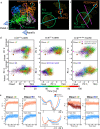
Molecular dynamics simulations and principal component analyses reveal that the absence of the substrate induces a transition between the open and closed forms of the catalytic domain in the ECR tetramer and a twist in the subunits, which would move a bound substrate toward the NADPH cofactor. (a) Representation of the swing motion of the catalytic domain in dimer AC described by the first principal component (PC1) and the twist motion of one subunit to the other by PC2. This twist motion would bring a bound substrate closer to the NADPH cofactor. PC1 and PC2 axes of A (closed) and C (open) subunits are superimposed on the ternary tetramer structure in the same orientation as in Figure 3a, showing the functional relevance of PC1 as opening–closing and PC2 as twisting of the catalytic domains. (b, c) Sets of vectors between initial and final Cα positions of the catalytic domains are shown viewed along the PC1 (b) and PC2 (c) axes of subunit A as shown on the bottom left of (a). (d) Projection of 10 100 ns trajectories on the first two principal components of the wild type binary system with the NADPH cofactor ECRwt·NADPH (crosses and diamonds represent the open- and closed-form subunits from the X-ray structure, respectively). The middle panels show the projection on the same PCs for the ECRwt ternary complex bearing only one substrate in the B subunit of the BD dimer ([B·Crot-CoA]D). The right panels represent the dynamics of the E151D/N157E/N218E triple variant with the NADPH cofactor (ECRTriple.Mut·NADPH) on the same eigenvectors. Colors of each points represent the time frame according to the scale bar at the bottom. (e) Mean values and 1.5 times the standard deviation of the principal components of 10 trajectories as a function of simulation time for each dimer in the WT (orange) and E151D/N157E/N218E triple variant (blue) ECR·NADPH complex. Time traces of PC1 are shown on the left and those of PC2 on the right. In the middle, free energy profiles as a function of PC1 and PC2 were derived for the wild type (orange) and variant (blue) on the basis of the observed kinetics. The triple variant with slower opening kinetics (PC1) is consistent with a barrier in the transition to the open form, and its larger standard deviation suggests a shallower minimum in the closed form. The free energy profile of PC2 corresponds to a broad minimum for the triple variant because of the larger standard deviation observed in the kinetics and a large barrier to reach the twisted conformation observed in the WT dynamics (brown trajectory shown in dimer AC for PC2 on the right).
Figure 5
Figure 5
Inter- and intradimer communications drive fast CO2 fixation by K. setae ECR. (a) Two distinct sets of communications: interdimer interactions between the catalytic domains from two dimers (purple arrows) and intradimer communication between the open and closed subunits within each dimer (brown arrows). (b) Interdimer catalytic domain interface and positions of selected amino acids that were mutated in this study to affect the interface between the two catalytic domains (open-form subunit in orange and closed form subunit in gray). The right panel shows the mutual H-bonding interaction between N218 and N157 from open- and closed-form subunits and H-bonding between E151 and the N atom from the protein backbone. (c) Alignment of ECR protein sequences from the primary (upper row) and the secondary (lower row) metabolisms represented as sequence logos. The numbering of residues, above the first row, is according to their position in the K. setae ECR. (d) Communication between the closed (green) and open (orange) subunits across the two dimers of the K. setae ECR. In the closed conformation the contacts between NADPH-H365-E165 and K332 of the adjacent open subunit allow for the correct intradimer communication. In the open conformation the communication network is compromised, as indicated by the increased distances between the amino acid side chains that cause the incorrect positioning of the nicotinamide ring of NADPH.

References

    1. Schwander T.; Schada von Borzyskowski L.; Burgener S.; Cortina N. S.; Erb T. J. A synthetic pathway for the fixation of carbon dioxide in vitro. Science (80-.). 2016, 354, 900–904. 10.1126/science.aah5237. - DOI - PMC - PubMed
    1. Erb T. J.; et al. Synthesis of C5-dicarboxylic acids from C2-units involving crotonyl-CoA carboxylase/reductase: the ethylmalonyl-CoA pathway. Proc. Natl. Acad. Sci. U. S. A. 2007, 104, 10631–6. 10.1073/pnas.0702791104. - DOI - PMC - PubMed
    1. Schada von Borzyskowski L.; Rosenthal R. G.; Erb T. J. Evolutionary history and biotechnological future of carboxylases. J. Biotechnol. 2013, 168, 243–251. 10.1016/j.jbiotec.2013.05.007. - DOI - PubMed
    1. Amao Y. Formate dehydrogenase for CO2 utilization and its application. J. of CO22 Utilization 2018, 26, 623–641. 10.1016/j.jcou.2018.06.022. - DOI
    1. Erb T. J.; Brecht V.; Fuchs G.; Muller M.; Alber B. E. Carboxylation mechanism and stereochemistry of crotonyl-CoA carboxylase/reductase, a carboxylating enoyl-thioester reductase. Proc. Natl. Acad. Sci. U. S. A. 2009, 106, 8871–8876. 10.1073/pnas.0903939106. - DOI - PMC - PubMed