. 2022 Aug;74(Suppl 1):442-448.
doi: 10.1007/s12070-020-02207-y. Epub 2020 Oct 23.

Primary Endoscopic Stapes Surgery: A Comparison of Adipose Tissue and Gelfoam Seal

Affiliations

Primary Endoscopic Stapes Surgery: A Comparison of Adipose Tissue and Gelfoam Seal

Pradeep Pradhan et al. Indian J Otolaryngol Head Neck Surg. 2022 Aug.

Abstract

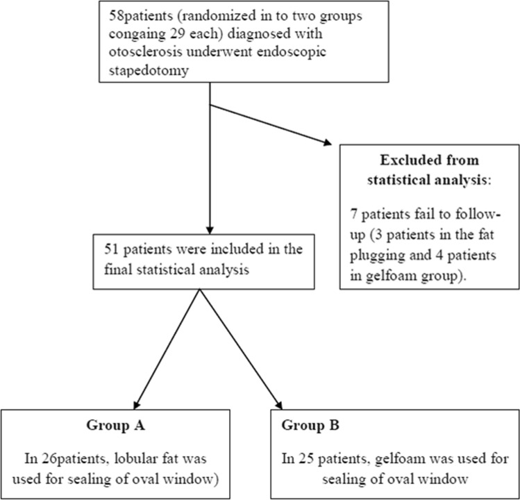
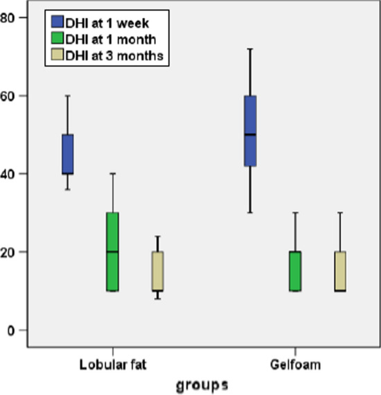
To compare the efficacy between the commonly used sealing materials, i.e., adipose tissue and the gelfoam in primary endoscopic stapedotomy. Lobular fat and gelfoam have been used in patients who underwent endoscopic stapedotomy between two groups, each containing 29 patients. The hearing outcomes and postoperative complications were compared at the end of 12 weeks between two groups. The ABG of ≤ 10 dB was achieved in 69% of cases in group A and 76% of cases in group B. There was a significant short-term (1 week) improvement in the Dizziness Handicap Inventory score (p = 00) with patients of adipose tissue seal compared to the gelfoam. Although the audiological outcomes were comparable between the two groups, the use of the adipose tissue can be a better alternative than gelfoam to control vertigo in the early postoperative period without causing any significant morbidity to the patient.

Keywords: Adipose tissue seal; Comparison; Endoscopic stapes surgery; Gelfoam seal.


Conflict of interest statement

Conflict of interestThere are no conflicts of interest among the authors.

Figures

Fig. 1
Fig. 1
Transcanal incision given (Right ear) from 6 o’clock to 12 o’clock poison
Fig. 2
Fig. 2
Complete exposure of stapes suprastructure
Fig. 3
Fig. 3
Fenestra was made over the stapes footplate with 0.8 mm perforator
Fig. 4
Fig. 4
Teflon piston (0.6 mm diameter, 4.25 mm length) was inserted through the stapedial window, anchored over the long process of incus, and a piece of lobular fat was used to seal the stapedial window after the insertion of the piston
Fig. 5
Fig. 5
Study design
Fig. 6
Fig. 6
Diagram shows the comparative DHI scores between fat and gelfoam plugging at the end of 1,4 and 12 weeks in the postoperative period

References

    1. Hunter JB, Rivas A. Outcomes following endoscopic stapes surgery. Otolaryngol Clin North Am. 2016;49:1215–1225. doi: 10.1016/j.otc.2016.05.012. - DOI - PubMed
    1. Kojima H, Komori M, Chikazawa S, et al. Comparison between endoscopic and microscopic stapes surgery. Laryngoscope. 2014;124:266–271. doi: 10.1002/lary.24144. - DOI - PubMed
    1. Iannella G, Marcotullio D, Re M, et al. Endoscopic vs microscopic approach in stapes surgery: advantages in the middle ear structures visualization and trainee’s point of view. J Int Adv Otol. 2017;13:14–20. doi: 10.5152/iao.2017.3322. - DOI - PubMed
    1. Tarabichi M. Endoscopic middle ear surgery. Ann Otol Rhinol Laryngol. 1999;108:39–46. doi: 10.1177/000348949910800106. - DOI - PubMed
    1. Bottrill ID, Poe DS. Endoscope-assisted ear surgery. Am J Otol. 1995;16:158–163. doi: 10.1016/0196-0709(95)90095-0. - DOI - PubMed

LinkOut - more resources