

doi: 10.1007/s12070-020-02145-9.
Epub 2020 Sep 17.
Cochlear Implant in a Child with a Large Arachnoid Cyst and Cysto-peritoneal Shunt
Affiliations
- PMID: 36032920
- PMCID: PMC9411317
- DOI: 10.1007/s12070-020-02145-9
Item in Clipboard
Cochlear Implant in a Child with a Large Arachnoid Cyst and Cysto-peritoneal Shunt
Indian J Otolaryngol Head Neck Surg.
2022 Aug.
Abstract
Profound hearing loss requiring cochlear implantation and arachnoid cyst requiring placement of Cysto-Peritoneal Shunt (CPS) are two commonly seen entities. However, there are very few published cases of patients requiring both of them. The present report describes the importance of multidisciplinary surgical planning in one such patient.
Keywords: Arachnoid cyst; Cochlear implantation; Cysto-peritoneal shunt; Trans-Tympanic Electrically-evoked Auditory Brainstem Response (TTEABR).
© Association of Otolaryngologists of India 2020.
Conflict of interest statement
Conflict of interestNo conflict of interest to be declared.
Figures
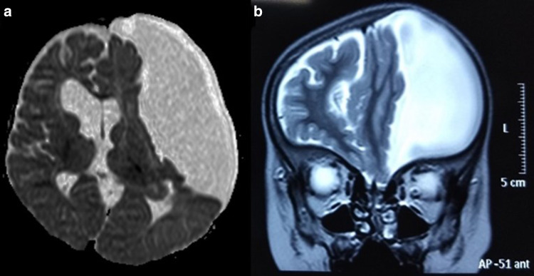
a MRI brain, axial image showing large arachnoid cyst in the left middle cranial fossa with significant mass effect on the left cerebral hemisphere. b T2-weighted MRI, coronal section, showing the large arachnoid cyst
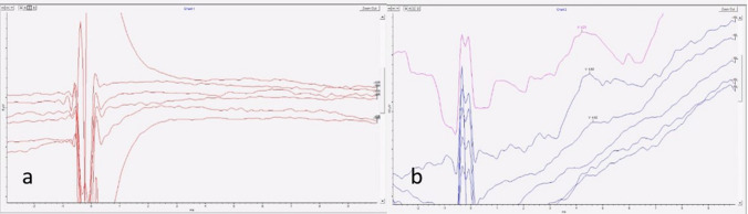
a Recording of TTEABR of right ear showing no responses. b Recording of TTEABR of left ear showing moderate responses
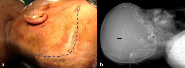
a Intra operative picture of the position and trajectory of the CPS (blue dotted line). b X-ray skull lateral view showing the position of CPS (black arrow)
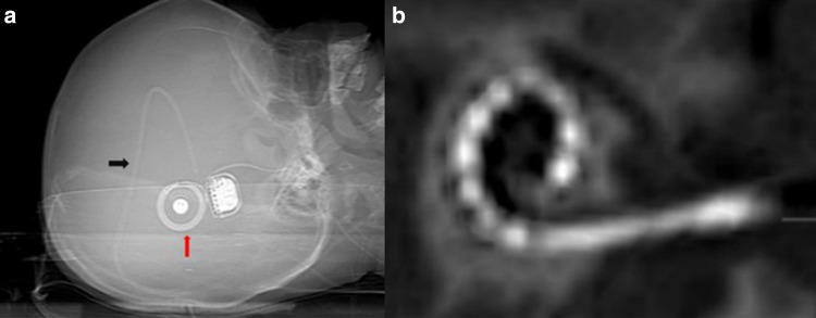
a X-ray skull lateral view (post cochlear implantation) showing the relative placement of the CPS (black arrow) and the RS package (red arrow) b HRCT temporal bones (post cochlear implantation) showing the electrode within the cochlea
References
LinkOut - more resources
Full Text Sources