[Preprint]. 2022 Aug 15:2022.08.14.503921.
doi: 10.1101/2022.08.14.503921.

Evasion of Neutralizing Antibody Response by the SARS-CoV-2 BA.2.75 Variant

Affiliations

Evasion of Neutralizing Antibody Response by the SARS-CoV-2 BA.2.75 Variant

Panke Qu et al. bioRxiv. .

Update in

Abstract

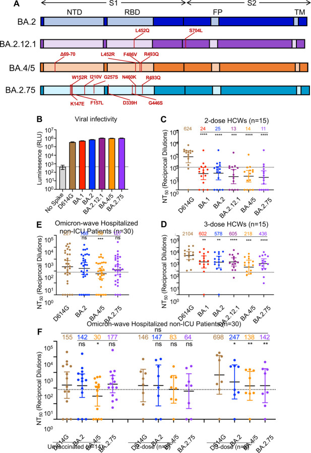
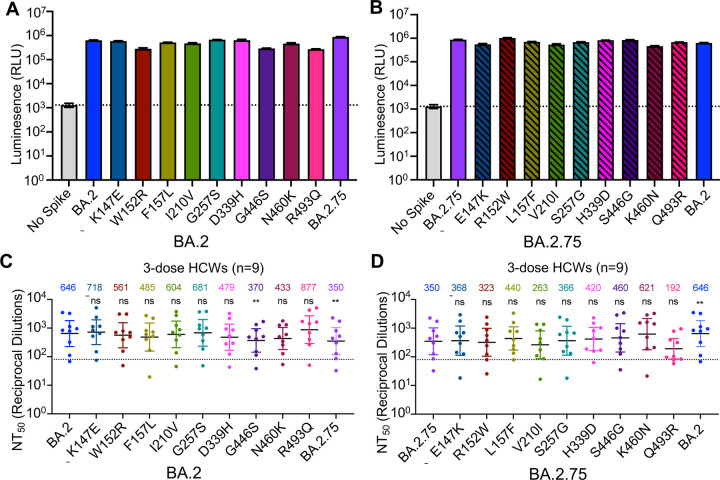
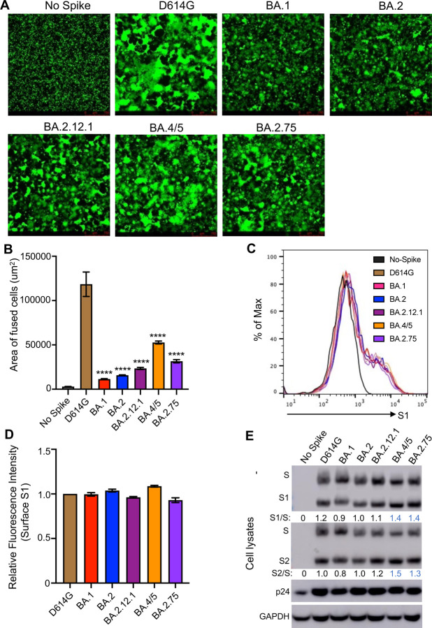
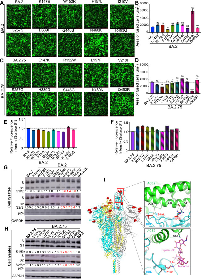
The newly emerged BA.2.75 SARS-CoV-2 variant exhibits an alarming 9 additional mutations in its spike (S) protein compared to the ancestral BA.2 variant. Here we examine the neutralizing antibody escape of BA.2.75 in mRNA-vaccinated and BA.1-infected individuals, as well as the molecular basis underlying functional changes in the S protein. Notably, BA.2.75 exhibits enhanced neutralization resistance over BA.2, but less than the BA.4/5 variant. The G446S and N460K mutations of BA.2.75 are primarily responsible for its enhanced resistance to neutralizing antibodies. The R493Q mutation, a reversion to the prototype sequence, reduces BA.2.75 neutralization resistance. The mutational impact is consistent with their locations in common neutralizing antibody epitopes. Further, the BA.2.75 variant shows enhanced cell-cell fusion over BA.2, driven largely by the N460K mutation, which enhances S processing. Structural modeling revealed a new receptor contact introduced by N460K, supporting a mechanism of potentiated receptor utilization and syncytia formation.


Conflict of interest statement

Declaration of Interests

The authors declare no competing interests.

Figures

Figure 1:
Figure 1:. BA.2.75 exhibits strong neutralization resistance to 2-dose and 3-dose mRNA vaccinee sera and Omicron wave patient sera.
(A) Schematic of BA.2-derived SARS-CoV-2 variants with mutations relative to the BA.2 background indicated. Highlighted are the S1 and S2 subunits, N-terminal domain (NTD), receptor binding domain (RBD), fusion peptide (FP), and transmembrane domain (TM). (B) Infectivity of pseudotyped lentivirus bearing S protein from SARS-CoV-2 variants of study; bars represent means ± standard error. (C-D) Neutralizing antibody titers against lentivirus pseudotyped with S from individual SARS-CoV-2 variants for 15 health care workers for sera collected 3–4 weeks after second mRNA vaccination (C) or 1–12 weeks after homologous mRNA booster vaccination (D). (E) Neutralizing antibody titers for sera collected from 30 COVID-19 patients hospitalized during the BA.1 pandemic wave. (F) Neutralizing antibody titers against hospitalized BA.1 wave patients are divided by vaccination status. (C-F) Dots indicate individual patient samples; bars represent geometric means with 95% confidence intervals; significance relative to D614G was determined by one-way repeated measures ANOVA with Bonferroni multiplicity correction. P-values are displayed as *p < 0.05, **p < 0.01, ***p < 0.001, ****p < 0.0001, and ns for not significant.
Figure 2:
Figure 2:. The G446S, N460K, and R493Q mutations modulate BA.2.75 neutralization sensitivity.
(A) Relative infectivity of lentivirus pseudotyped with BA.2 S with single mutations from BA.2.75 lineage defining mutations; bars represent means ± standard error. (B) Relative infectivity of lentivirus pseudotyped with BA.2.75 S with single reversion mutations to remove BA.2.75 lineage defining mutations; bars represent means ± standard error. (C-D) Neutralizing antibody titers against lentivirus pseudotyped with S from BA.2 with single mutations from BA.2.75 lineage-defining mutations (C) or BA.2.75 with single reversion mutations from BA.2.75 lineage-defining mutations (D) for sera collected from 9 health care workers 1–12 weeks after homologous mRNA booster vaccination. Dots indicate individual patient samples; bars represent geometric means with 95% confidence intervals; significance relative to D614G was determined by one-way repeated measures ANOVA with Bonferroni multiplicity correction. P-values are displayed as **p < 0.01, and ns for not significant.
Figure 3:
Figure 3:. BA.2.75 exhibits enhanced cell-cell fusion and S processing.
(A) Fluorescence images displaying syncytia formation are presented for HEK293T-ACE2 cells 48 hr after co-transfection with a GFP expression construct and SARS-CoV-2 variant S proteins. (B) Quantification of syncytia formation in panel (A) displays the mean syncytia size; bars represent means ± standard error, with significance relative to D614G determined by one-way ANOVA with Bonferroni multiplicity correction. (C) Histogram displays of the surface staining of HEK293T cells expressing S proteins, which were detected by an anti-S1 antibody (T62). (D) Quantification of relative surface expression as shown in (C); bars represent means ± standard error. (E) Pseudotyped lentivirus producer cell lysate was assessed for processing of S by probing with anti-S1 (T62), anti-S2, anti-HIV-1 Gag (anti-p24), and anti-GAPDH. Band intensities were quantified in ImageJ and the ratio of S1/S or S2/S is displayed relative to the S1/S or S2/S ratio of BA.2. P-values are displayed as ****p < 0.0001.
Figure 4:
Figure 4:. The N460K mutation determines enhanced cell-cell fusion and S processing of BA.2.75.
(A) Fluorescence images displaying syncytia formation are presented for HEK293T-ACE2 cells 48 hr after co-transfection with a GFP expression construct and BA.2 single mutant S proteins. (B) Quantification of syncytia formation in panel (A) displays the mean syncytia size; bars represent means ± standard error, with significance relative to D614G determined by one-way ANOVA with Bonferroni multiplicity correction. (C) Fluorescence images displaying syncytia formation are presented for HEK293T-ACE2 cells 48-hrs after co-transfection with a GFP expression construct and BA.2.75 single reversion mutant S proteins. (D) Quantification of syncytia formation in panel (C) displays the mean syncytia size; bars represent means ± standard error, with significance relative to D614G determined by one-way ANOVA with Bonferroni multiplicity correction. (E-F) Quantification of relative S surface expression in transfected HEK293T cells for BA.2 single mutants (E) or BA.2.75 reversion mutants (F), as examined by flow cytometry; bars represent means ± standard error. (G) Pseudotyped lentivirus producer cell lysate was assessed for processing of S from BA.2 single mutants by probing with anti-S1 (T62), anti-S2, anti-HIV-1 p24, and anti-GAPDH. Band intensities were quantified in ImageJ and the ratios of S1/S and S2/S are displayed relative to the S1/S and S2/S ratios of BA.2. (H) Pseudotyped lentivirus producer cell lysate was assessed for processing of S from BA.2.75 reversion mutants by probing with anti-S1, anti-S2, anti-HIV-1 p24, and anti-GAPDH. Band intensities were quantified in ImageJ and the ratios of S1/S and S2/S are displayed relative to the S1/S and S2/S ratios of BA.2.75. (I) Structural modelling of Omicron BA.2.75 spike protein viewed as a ribbon. Mutations of BA.2.75 specific mutants are highlighted by red spheres. The RBD of the cyan spike protomer is in an “up” conformation. Upper inset: The mutation G446S reduces the backbone flexibility and possibly stabilizes the hydrogen bond between its carbonyl group and the residue Q42 on ACE2 receptor (green); the mutation R493Q abolishes the salt-bridge interaction with the E35 on ACE2 receptor and potentially forms two hydrogen bonds with E35 and K31. Lower inset: the mutation N460K enables formation of a hydrogen bond with the glycan-N90 on ACE2 receptor (green). In all cases, p-values are displayed as *p < 0.05, **p < 0.01, ****p < 0.0001, and ns for not significant.

References

    1. Callaway E. 2022. Will ‘Centaurus’ be the next global coronavirus variant? Indian cases offer clues. Nature. - PubMed
    1. Cao Y., Song W., Wang L., Liu P., Yue C., Jian F., Yu Y., Yisimayi A., Wang P., Wang Y., Zhu Q., Deng J., Fu W., Yu L., Zhang N., Wang J., Xiao T., An R., Wang J., Liu L., Yang S., Niu X., Gu Q., Shao F., Hao X., Jin R., Wang Y., Xie X.S., and Wang X.. 2022. Characterizations of enhanced infectivity and antibody evasion of Omicron BA.2.75. bioRxiv:2022.2007.2018.500332. - PMC - PubMed
    1. Centers for Disease Control and Prevention. 2022. COVID Data Tracker. Atlanta, GA: US Department of Health and Human Services, CDC. 2022, August 10. https://covid.cdc.gov/covid-data-tracker
    1. Evans J.P., Zeng C., Qu P., Faraone J., Zheng Y.M., Carlin C., Bednash J.S., Zhou T., Lozanski G., Mallampalli R., Saif L.J., Oltz E.M., Mohler P.J., Xu K., Gumina R.J., and Liu S.L.. 2022. Neutralization of SARS-CoV-2 Omicron sub-lineages BA.1, BA.1.1, and BA.2. Cell Host Microbe. 30:1093–1102.e1093. - PMC - PubMed
    1. Goerke A.R., Loening A.M., Gambhir S.S., and Swartz J.R.. 2008. Cell-free metabolic engineering promotes high-level production of bioactive Gaussia princeps luciferase. Metab Eng. 10:187–200. - PubMed

Publication types