Review
. 2022 Aug 10:12:964265.
doi: 10.3389/fcimb.2022.964265. eCollection 2022.

Where do T cell subsets stand in SARS-CoV-2 infection: an update

Affiliations
Review

Where do T cell subsets stand in SARS-CoV-2 infection: an update

Mohammad Tarique et al. Front Cell Infect Microbiol. .

Abstract

An outbreak of coronavirus disease 2019 (COVID-19) emerged in China in December 2019 and spread so rapidly all around the globe. It's continued and spreading more dangerously in India and Brazil with higher mortality rate. Understanding of the pathophysiology of COVID-19 depends on unraveling of interactional mechanism of SARS-CoV-2 and human immune response. The immune response is a complex process, which can be better understood by understanding the immunological response and pathological mechanisms of COVID-19, which will provide new treatments, increase treatment efficacy, and decrease mortality associated with the disease. In this review we present a amalgamate viewpoint based on the current available knowledge on COVID-19 which includes entry of the virus and multiplication of virus, its pathological effects on the cellular level, immunological reaction, systemic and organ presentation. T cells play a crucial role in controlling and clearing viral infections. Several studies have now shown that the severity of the COVID-19 disease is inversely correlated with the magnitude of the T cell response. Understanding SARS-CoV-2 T cell responses is of high interest because T cells are attractive vaccine targets and could help reduce COVID-19 severity. Even though there is a significant amount of literature regarding SARS-CoV-2, there are still very few studies focused on understanding the T cell response to this novel virus. Nevertheless, a majority of these studies focused on peripheral blood CD4+ and CD8+ T cells that were specific for viruses. The focus of this review is on different subtypes of T cell responses in COVID-19 patients, Th17, follicular helper T (TFH), regulatory T (Treg) cells, and less classical, invariant T cell populations, such as δγ T cells and mucosal-associated invariant T (MAIT) cells etc that could influence disease outcome.

Keywords: COVID-19; T cell; immune response; immunological reaction; pathophysiology.


Conflict of interest statement

The authors declare that the research was conducted in the absence of any commercial or financial relationships that could be construed as a potential conflict of interest.

Figures

Figure 1
Figure 1
The following schematic diagram illustrates the steps taking place during the infection and replication of SARS-CoV-2 into host cells: (A) SARS-CoV-2 enter into the cell by binding to the ACE2 receptor present on the host cell membrane. As the virus replicates in the lung cells, it induces pyroptosis and the release of DAMPs (disease associated molecular patterns). Several pro-inflammatory cytokines and chemokines, including IL-6, IL-10, MIP1-α, MIP1-1β, and MCP-1, are secreted by adjoining epithelial cells, alveolar macrophages, and endothelial cells in response to contact with DAMPs. As a result of these cytokines and chemokines, monocytes, macrophages, and neutrophils are recruited to the lung, thereby releasing more inflammatory cytokines in the process, which cause hypoxia, fluid accumulation in lung, fever and acute lung injury (B) SARS-CoV-2 enters the cytoplasm and releases viral RNA, which is translated into polyproteins after entering the cytoplasm. The (+) stand genomic RNA is used as a template to begin replication transcription. Nucleoprotein complexes are formed from sub genomic RNA synthesized by transcription and the (+) strand genomic RNA. Virions are released from host cells following assembly of nucleoprotein complexes and proteins.
Figure 2
Figure 2
Recruitments of immune cells and damage of Lung epithelium and cytokine storm: SARS-CoV-2 enter into the cell by binding to the ACE2 receptor present on the host cell membrane. As the virus replicates in the lung cells and damage lung epithelium. Damaged lung epithelium cell secretes inflammatory signals and immune cells recruited in lung blood vessels which secrete pro-inflammatory cytokines and chemokines, including IL-6, IL-10, MIP1-α, MIP1-1β, and MCP-1, are secreted by adjoining epithelial cells, alveolar macrophages, and endothelial cells. As a result of these cytokines storm happens and increased blood vessels permeability, accumulation of fluid in the lung, hypoxia and ultimately acute respiratory distress syndrome (ARDS) and multiple organ failure.
Figure 3
Figure 3
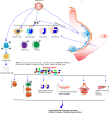
T cells in severe COVID-19: presence and activation.: Numbers of CD4 and CD8 decreased in peripheral blood (lymphopenia) while activated CD4 and CD8 cells are increased in lung secernated excessive amount of TNF-α and IFN-γ which damaged lung and inhibits function of Tregs. IL-17 secreting Th17 cells and follicular helper T (Tfh) cells increased in peripheral blood of COVID-19 patients, however there are conflicting finding in case of Tregs. The number of NKT, MAIT, and γδ T cells decreases in peripheral blood and is detected in lung aspirates with heightened levels of activation, as evaluated by CD69 and PD-1. Activated conventional and unconventional T cells in the lung may secrete multiple inflammatory cytokines such as IL-17A, IL-6, TNF-α, and IFN-γ that contribute to the cytokine milieu and/or disease pathology.

References

    1. Anghelina D., Zhao J., Trandem K., Perlman S. (2009). Role of regulatory T cells in coronavirus-induced acute encephalitis. Virology 385, 358–367. doi: 10.1016/j.virol.2008.12.014 - DOI - PMC - PubMed
    1. Balachandran H., Phetsouphanh C., Agapiou D., Adhikari A., Rodrigo C., Hammoud M., et al. . (2022). Maintenance of broad neutralizing antibodies and memory b cells 1 year post-infection is predicted by SARS-CoV-2-specific CD4+ T cell responses. Cell Rep. 38, 110345. doi: 10.1016/j.celrep.2022.110345 - DOI - PMC - PubMed
    1. Barathan M., Mohamed R., Vadivelu J., Chang L. Y., Saeidi A., Yong Y. K., et al. . (2016). Peripheral loss of CD8(+) CD161(++) TCRValpha7.2(+) mucosal-associated invariant T cells in chronic hepatitis c virus-infected patients. Eur. J. Clin. Invest. 46, 170–180. doi: 10.1111/eci.12581 - DOI - PubMed
    1. Battistini L., Caccamo N., Borsellino G., Meraviglia S., Angelini D. F., Dieli F., et al. . (2005). Homing and memory patterns of human gammadelta T cells in physiopathological situations. Microbes Infect. 7, 510–517. doi: 10.1016/j.micinf.2004.12.008 - DOI - PubMed
    1. Bhardwaj A., Sapra L., Saini C., Azam Z., Mishra P. K., Verma B., et al. . (2022). COVID-19: Immunology, immunopathogenesis and potential therapies. Int. Rev. Immunol. 41, 171–206. doi: 10.1080/08830185.2021.1883600 - DOI - PMC - PubMed

Publication types