Review
. 2022 Aug 3:9:952147.
doi: 10.3389/fnut.2022.952147. eCollection 2022.

Research progress on extraction technology and biomedical function of natural sugar substitutes

Affiliations
Review

Research progress on extraction technology and biomedical function of natural sugar substitutes

Pengyu Lei et al. Front Nutr. .

Abstract

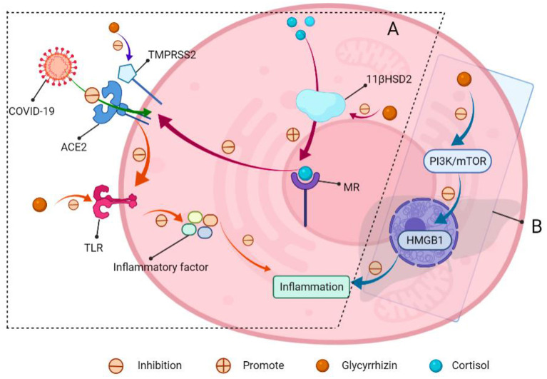
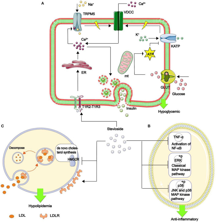
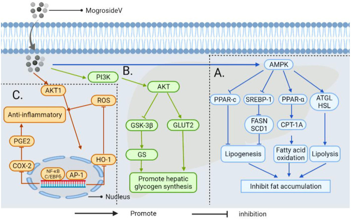
Improved human material living standards have resulted in a continuous increase in the rate of obesity caused by excessive sugar intake. Consequently, the number of diabetic patients has skyrocketed, not only resulting in a global health problem but also causing huge medical pressure on the government. Limiting sugar intake is a serious problem in many countries worldwide. To this end, the market for sugar substitute products, such as artificial sweeteners and natural sugar substitutes (NSS), has begun to rapidly grow. In contrast to controversial artificial sweeteners, NSS, which are linked to health concepts, have received particular attention. This review focuses on the extraction technology and biomedical function of NSS, with a view of generating insights to improve extraction for its large-scale application. Further, we highlight research progress in the use of NSS as food for special medical purpose (FSMP) for patients.

Keywords: diabetes; extraction technology; inflammation; natural sugar substitutes; obesity.


Conflict of interest statement

The authors declare that the research was conducted in the absence of any commercial or financial relationships that could be construed as a potential conflict of interest.

Figures

Figure 1
Figure 1
Mechanism through which the human body feels sweetness. The front end of the tongue is the main area for sweet taste. Taste pores at the front of taste buds are mainly composed of type II taste receptor cells (TRCs), while the calcium homeostasis regulatory protein 1 (CALHM1) gene is specifically expressed only in type II taste sensing cells. The sweet substance binds to the sweet receptor T1R2–T1R3 dimer on TRCs, thereby activating the coupled G protein, then the phospholipase C β 2 (PLCβ2) is deactivated. PLCβ2 acts as a catalyst, participating in the decomposition of phosphatidylinositol diphosphate (PIP2) to produce inositol triphosphate (IP3), diacylglycerol (DAG) and H +. IP3 combines with inositol triphosphate receptor (IP3R) in the endoplasmic reticulum to promote release of Ca2+ in the endoplasmic reticulum. Ca2+ binds to TRPM5. The channel is opened to allow Na + to flow in, depolarize the membrane potential, promote the opening of CALHM1 channel, and the adenosine-triphosphate (ATP) outflow as a neurotransmitter transmits information to the synapse of taste afferent nerve. Finally, sweetness is produced in the cerebral cortex.
Figure 2
Figure 2
Artificial sweeteners are harmful to human health and the environment. (A) Artificial sweeteners mediate a reduction in glucose tolerance and insulin resistance by altering the composition of human gut microbes. (B) Environmental hazards caused by artificial sweeteners. (C) Artificial sweeteners can increase biofilm formation in Enterococcus faecalis and E. coli, adhere to and invade the epithelial cells on the intestinal wall, and kill these cells. The sweeteners can easily pass through the intestinal wall, into the blood, and accumulate in the lymph nodes, liver and spleen, thereby causing secretion of some toxins, and predisposing the body to infections, sepsis and multiple organ failure. (D) Artificial sweeteners increase the risk of cancer.
Figure 3
Figure 3
The popularity of sugar substitutes. Range from very negative (left) to positive (right), Red for artificial sweeteners, green for NSS, and purple for sucrose. Adapted with permission from (15). Copyright 2020, WILEY.
Figure 4
Figure 4
Classification, structure and source of NSS.
Figure 5
Figure 5
Improved schematic diagram showing the extraction method.
Figure 6
Figure 6
Schematic representation of the biomedical function of NSS.
Figure 7
Figure 7
Schematic presentation of the mechanisms of low GI and low calorie levels of maltitol, psicose, stevia glycoside and erythritol. (A) Psicose and erythritol enter the bloodstream and are subsequently eliminated via urine from the body through the kidney. They do not stimulate pancreatic β-cells to produce insulin and have limited effects on blood glucose levels. (B) These NSS are difficult to be broken down and be absorbed by digestive enzymes in the human gastrointestinal tract. (C) Indigestible NSS are excreted from the intestines.
Figure 8
Figure 8
Mechanisms of psicose in decreasing blood glucose levels and inhibiting fat accumulation. (A) Psicose inhibits fatty acid synthase and promotes lipoxygenase activities, down-regulates the expressions of fat synthesis-related PPAR-7 and C/EBPα genes. In addition, it promotes the use of glucose in the liver and synthesis of liver glycogen. (B) Psicose promotes muscle glycogen synthesis. (C) It promotes the secretion of GLP-1 and activates the GLP-1 receptor to stimulate the vagal afferent nerve, allowing people to develop satiety. (D) Moreover, it stimulates glucokinase to maintain a normal GI and insulin receptor sensitivity. It scavenges for reactive oxygen species, thereby exerting antioxidant effects and plays a role in protecting pancreatic β-cells.
Figure 9
Figure 9
Schematic of glycyrrhizin for COVID-19 resistance and acute hepatitis therapy. (A) COVID-19 entry into the cells through ACE2. (I) The mineralocorticoid receptor (MR) regulates ACE2 expression, and MR activation decreases ACE2 expression. Meanwhile, glycyrrhizin promotes cortisol activation of MR by inhibiting 11-β-hydroxy steroid dehydrogenase 2 (11β-HSD2), lowering ACE2 expression and preventing virus entry into cells; (II) transmembrane protease serines TMPRSS2 (which is a cofactor that promotes viral entry into cells through ACE2). Glycyrrhizin suppressed the expression of TMPRSS2; (III) ACE2 can produce angiotensin, which inhibits TLR4 receptor expression. Meanwhile, glycyrrhizin can directly block TLR4 receptors, preventing the production of inflammatory factors and lowering lung inflammation. (B) Glycyrrhizin relieves acute hepatitis.
Figure 10
Figure 10
The mechanism of stevia glycoside in hypoglycemic, anti-inflammatory, and hypolipidemic. (A) Stevia glycoside binds to sweet receptors (T1R2–T1R3 dimer), prompting the endoplasmic reticulum to release Ca2+. Ca2+ opens the transient receptor potential ion channel protein 5 (TRPM5), allowing Na+ to enter and generate action potentials. At the same time, glucose is transferred to the mitochondria by enhanced diffusion of glucose transporter to generate ATP, which inhibits the opening of the ATP-sensitive potassium (KATP) and increases action potential production. Finally, the action potential opens the voltage-dependent calcium channel (VDCC), which allows Ca2+ to flow in. Thus, an increase in Ca2+ in β cells stimulates insulin secretion and hypoglycemia. (B) Stevia glycoside reduces the inflammatory factors IL-1β And IL-6 production, as well as the phosphorylation of two critical MAPK pathway proteins, p38 and ERK, as well as decreased TNF-α expression and hence NF-κB activation, all of which contribute to inflammation resistance. (C) Stevia glycoside decreases intracellular cholesterol by inhibiting HMGCR expression, the rate-limiting enzyme in the de novo cholesterol synthesis pathway, while increasing the cell surface LDL binding receptor (LDLR), promoting cholesterol breakdown in the cell, and the LDLR returns to the cell surface, resulting in hypolipidemia.
Figure 11
Figure 11
The mechanism by which androgynous mogroside V inhibits fat accumulation lowers blood glucose and exerts anti-inflammatory effects. (A) In LPS induced inflammatory cells, mogrosideV inhibited the phosphorylation of AKT1(protein kinase B, regulating cell proliferation and growth) and blocked AKT1 mediated NF-κB and C/EBP δ Pathway, resulting in cyclooxygenase-2 (COX-2) expression, and inhibit prostaglandin E2 (PGE2) production. In addition, dephosphorylation of AKT1 can inhibit transcriptional activator protein-1 (AP-1) to restore hemoglobin oxygenase-1 (HO-1) overexpression to the normal level and reduce reactive oxygen species (ROS) level. Thereby decreasing the occurrence of inflammation. (B) It upregulates phosphatidylinositol-3-kinase (PI3K) and activates downstream AKT, thereby promoting GLUT2 transport and inhibiting the activity of GSK-3β, thereby enhancing the activity of GS and accelerating glucose uptake by hepatocytes. (C) Mogroside V inhibits fat accumulation by activating AMP-activated protein kinase (AMPK): (I) It can down-regulate the transcription factor sterol-regulatory element binding proteins-1 (SREBP-1) and inhibit fatty acid synthase (FASN) and stearoyl-CoA desaturase 1(SCD1); (II) It can down-regulate peroxisome proliferator-activated receptor-c (PPAR-c) (involved in regulating triacylglycerol synthesis in the liver); (III) It regulates the activation of carnitine Palmitoyltransferase-1A (CPT-1A) by PPAR-α, which is involved in mitochondrial transport and oxidation of fatty acids. Thus improving liver steatosis and inhibiting fat accumulation; (IV) It enhances the expression of genes for adipose triglyceride lipase (ATGL) and hormone-sensitive lipase (HSL) in the liver to promote lipolysis.
Figure 12
Figure 12
Side effects of some NSS and solutions. (A) Sugar alcohols are difficult for pepsin and gastric acid to digest. (B) Sugar alcohols and some carbohydrates are fermented together in the gut by microbes to produce gases (CO2, H2, and CH4). (C) Sugar alcohol sweeteners enter the intestine rapidly due to their inability to be digested, increased transport into the small intestine may lead to increased permeability in the lumen of the small intestine, resulting in water retention. (D) Glycyrrhizin inhibits the 11β-hydroxy steroid dehydrogenase activity, preventing cortisol from being converted to corticosterone, and it also increases the level of aldosterone in the human body when combined with cortisol, aldosterone, and MR, resulting in mineralocorticoid excess, increases the level of water and sodium ions in the blood, resulting in water sodium retention. As a consequence, increased blood volume results in hypertension and hypokalemia. (I) The administration to regulate food labeling to indicate the recommended intake range of NSS and the consumption group. (II) Contraindicated in patients with hypertension.

Similar articles

Cited by

References

    1. Bradley D. The intriguing intersection of type 2 diabetes, obesity-related insulin resistance, and osteoarthritis. J Clin Endocrinol Metab. (2021) 106:e2370–2. 10.1210/clinem/dgab009 - DOI - PMC - PubMed
    1. Almourani R, Chinnakotla B, Patel R, Kurukulasuriya LR, Sowers J. Diabetes and cardiovascular disease: an update. Curr Diab Rep. (2019) 19. 10.1007/s11892-019-1239-x - DOI - PubMed
    1. World Health Organization . Guideline: Sugars Intake for Adults and Children. Geneva: World Health Organization; (2015). - PubMed
    1. Daher MI, Matta JM, Abdel Nour AM. Non-nutritive sweeteners and type 2 diabetes: Should we ring the bell? Diab Res Clin Pract. (2019) 155:107786. 10.1016/j.diabres.2019.107786 - DOI - PubMed
    1. Lee A, Owyang C. Sugars, sweet taste receptors, and brain responses. Nutrients. (2017) 9:653. 10.3390/nu9070653 - DOI - PMC - PubMed

LinkOut - more resources