. 2022 Aug 10:6:100071.
doi: 10.1016/j.yjsbx.2022.100071. eCollection 2022.

The role of C-terminal helix in the conformational transition of an arginine binding protein

Affiliations

The role of C-terminal helix in the conformational transition of an arginine binding protein

Vinothini Santhakumar et al. J Struct Biol X. .

Abstract

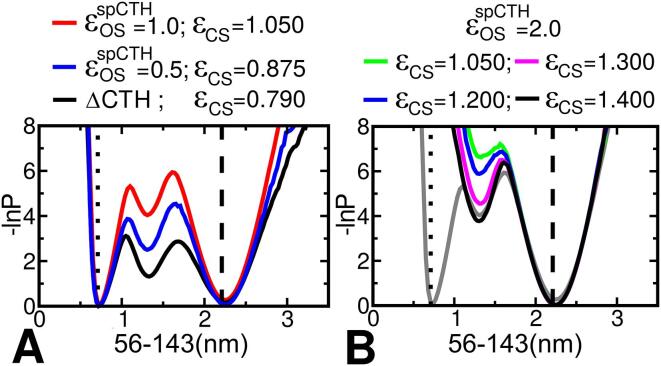
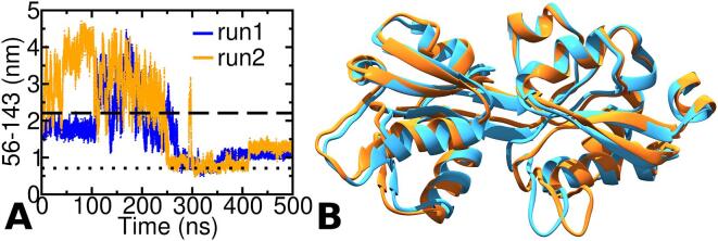
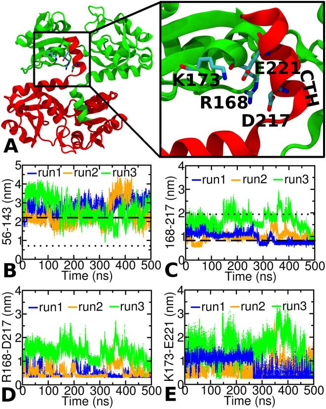
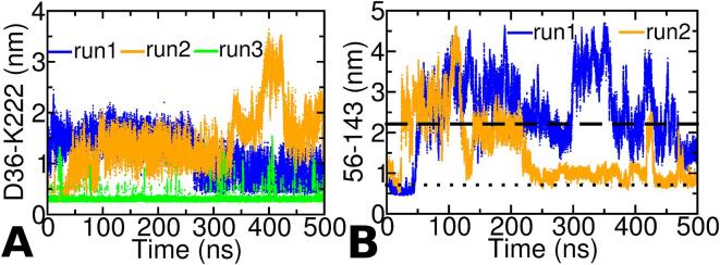
The thermotoga maritima arginine binding protein (TmArgBP) is a periplasmic binding protein that has a short helix at the C-terminal end (CTH), which is swapped between the two chains. We apply a coarse-grained structure-based model (SBM) and all-atom MD simulation on this protein to understand the mechanism and the role of CTH in the conformational transition. When the results of SBM simulations of TmArgBP in the presence and absence of CTH are compared, we find that CTH is strategically located at the back of the binding pocket restraining the open-state conformation thereby disengaging access to the closed-state. We also ran all-atom MD simulations of open-state TmArgBP with and without CTH and discovered that in the absence of CTH the protein could reach the closed-state within 250 ns, while in its presence, the protein remained predominantly in its open-state conformation. In the simulation started from unliganded closed-state conformation without CTH, the protein exhibited multiple transitions between the two states, suggesting CTH as an essential structural element to stabilize the open-state conformation. In another simulation that began with an unliganded closed-state conformation with CTH, the protein was able to access the open-state. In this simulation the CTH was observed to reorient itself to interact with the protein emphasizing its role in assisting the conformational change. Based on our findings, we believe that CTH not only acts as a structural element that constraints the protein in its open-state but it may also guide the protein back to its open-state conformation upon ligand unbinding.

Keywords: Arginine binding protein; C-terminal helix; CTH, C-terminal helix; Conformational transition; MD simulation; SBM, structure-based model; Structure-based model; TmArgBP, thermotoga maritima arginine binding protein.


Conflict of interest statement

The authors declare that they have no known competing financial interests or personal relationships that could have appeared to influence the work reported in this paper.

Figures

Fig. 1
Fig. 1
A. A cartoon representation of the domain-swapped open-state and closed-state conformation of TmArgBP. The protein consists of two lobes, L1 and L2, with arginine (ligand) binding at their interface. The two protein chains are colored in green and red. In the open-state, the C-terminal helix (CTH) docks itself at the back of the binding pocket, while in the closed-state conformation the CTH looses many of its interactions with the neighboring protein chain. B. Crystal structure of apo TmArgBP in the open-state highlighting the swapping of CTH (PDB: 4PRS). The two chains of the protein are colored in green and red. Note how the short helix at the C-terminal end (CTH) projects onto the neighbouring chain. C. Comparison of the structures of open- and closed-state of TmArgBPΔCTH colored in cyan and orange, respectively. Here the two structures are aligned with respect to the L2 domain to demonstrate the huge variation (RMSD ∼ 0.58 nm) between the open- and closed-states. The spheres pertains to Cα atoms of residues 56 and 143 in open- (green) and closed-state (blue). The distance between the Cα atoms of 56–143 is used as the reaction coordinate to monitor the conformational change.
Fig. 2
Fig. 2
A. The free energy plots as a function of the distance between the Cα atoms of residues 56–143 for os-TmArgBP. The black curve corresponds to the simulation started with os-TmArgBPΔCTH (the open-state in the absence of CTH) with strength of open-state specific contacts fixed at εOS = 1.0. The blue and the red curves correspond to simulation started with the open-state in the presence of CTH (os-TmArgBPCTH) with a value of εOSspCTH (specific CTH contacts in the open-state) set to 0.5 and 1.0, respectively. In the simulation of os-TmArgBPCTH the strength of open-state specific contacts evaluated from os-TmArgBPΔCTH are still present (εOS = 1.0). B. Simulation of os-TmArgBPCTH with εOSspCTH = 2.0. The different curves correspond to simulation performed with increasing value of εCS. The grey curve is indicated for comparison, which corresponds to the red curve in A.
Fig. 3
Fig. 3
MD simulation results of os-TmArgBPΔCTH. A. The distance between the Cα atom of residue 56–143 plotted as a function of time for the two independent simulations of monomeric version of os-TmArgBPΔCTH. In both these simulations the distance between 56 and 143 decreases to a value that is indicative of a conformational transition to the closed-state. B. A snapshot from r1-TmArgBPΔCTH (orange) ∼ 256 ns, the snapshot with the lowest RMSD of 0.18 nm with the closed-state) compared with X-ray structure of the closed-state (cyan).
Fig. 4
Fig. 4
A. The overall structure of domain-swapped os-TmArgBP. The two chains are colored in green and red. The boxed region highlights the ionic interactions of CTH with its neighboring chain. The two acidic residues in CTH, namely D217 and E221, form salt-bridge like interaction with K173 and R168, respectively. Simulation results for os-TmArgBPCTH. The distance between the Cα atoms of residue 56–143 (B) and 168–217 (C) plotted as a function of time for three independent simulations of monomeric version of os-TmArgBPCTH. While the Cα distance between 56 and 143 monitors the open to close transition, that between 168 and 217 is a good proxy to monitor the closeness of CTH to the protein. To monitor the salt-bridge interactions across the three independent runs of os-TmArgBPCTH, the distance between the charged side-chain centers of residues R168-D217 (D) and K173-E221 (E) is plotted. The plot is the closest distance between any of the oxygen atoms of acidic residues and the nitrogen atoms of basic residues. In this study we set a cut-off of 0.5 nm for measuring the salt-bridge. Please note that in 168–217, we refer to the distance between the Cα-atoms of residues 168 and 217 while in R168-D217, we refer to the distance between the charged side-chain centers of the two residues.
Fig. 5
Fig. 5
A. Distance between the charged centers of D36-K222 in r1/r2/r3-TmArgBPCTH as a function of simulation time. The distance in case of r3-TmArgBPCTH alone is quite stable ∼ 0.4 nm indicative of a strong salt-bridge. In both r1/r2-TmArgBPCTH the distances are not as stable as that observed for r3-TmArgBPCTH. B. Distance between the Cα-residues of 56–143 as a function of time for the two independent simulation runs starting from the closed-state structure of TmArgBP without CTH (r1/r2-cs-TmArgBPΔCTH). In both simulations, the protein escapes to the open-state within ∼ 75 ns and in r2-TmArgBPΔCTH one can see the protein reverting back and forth between the two states.
Fig. 6
Fig. 6
A. Initial snapshots from 0 to 125 ns at intervals of 25 ns highlighting the conformational transition from closed-state (CS) to open-state (OS). The first (CS) and last (125 ns) snapshots are coloured red and blue, respectively, with intermediate (25 ns) snapshots colored according to the color bar. In the figure movement of the CTH and its insertion between the two lobes is clearly depicted. The location of 56–143 (Cα) atoms in the closed-state is indicated as green sphere. B. The distance between the Cα atom of residues 56–143 plotted as a function of time used to monitor the conformational change. C. Distance between the charged-centers of K173 (in L2 that is oriented towards CTH) and acidic residues (D218 and E222) in CTH. D. Distance between the charged-centers of R168 (in L2 that is oriented towards CTH) and acidic residues (D218 and E222) in CTH. E. Distance between the charged-centers of R168 and D93 (at the hinge).

References

    1. Abraham M.J., Murtola T., Schulz R., Páll S., Smith J.C., Hess B., Lindahl E. GROMACS: High performance molecular simulations through multi-level parallelism from laptops to supercomputers. SoftwareX. 2015;1–2:19–25. doi: 10.1016/j.softx.2015.06.001. - DOI
    1. Ames G.F.L. Bacterial periplasmic transport systems: structure, mechanism, and evolution. Annu. Rev. Biochem. 1986;55:397–425. doi: 10.1146/annurev.bi.55.070186.002145. - DOI - PubMed
    1. Ausili A., Pennacchio A., Staiano M., Dattelbaum J.D., Fessas D., Schiraldi A., D’Auria S. Amino acid transport in thermophiles: characterization of an arginine-binding protein from Thermotoga maritima. 3. Conformational dynamics and stability. J. Photochem. Photobiol. B. 2013;118:66–73. doi: 10.1016/j.jphotobiol.2012.11.004. - DOI - PubMed
    1. Bahar I., Lezon T.R., Yang L.-W., Eyal E. Global dynamics of proteins: bridging between structure and function. Annu. Rev. Biophys. 2010;39:23–42. doi: 10.1146/annurev.biophys.093008.131258. - DOI - PMC - PubMed
    1. Balasco N., Smaldone G., Vigorita M., Del Vecchio P., Graziano G., Ruggiero A., Vitagliano L. The characterization of thermotoga maritima arginine binding protein variants demonstrates that minimal local strains have an important impact on protein stability. Sci. Rep. 2019;9:6617. doi: 10.1038/s41598-019-43157-y. - DOI - PMC - PubMed

LinkOut - more resources