Review
. 2022 Aug 11:10:944016.
doi: 10.3389/fcell.2022.944016. eCollection 2022.

The vertebrate Embryo Clock: Common players dancing to a different beat

Affiliations
Review

The vertebrate Embryo Clock: Common players dancing to a different beat

Gil Carraco et al. Front Cell Dev Biol. .

Abstract

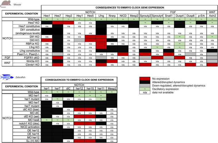
Vertebrate embryo somitogenesis is the earliest morphological manifestation of the characteristic patterned structure of the adult axial skeleton. Pairs of somites flanking the neural tube are formed periodically during early development, and the molecular mechanisms in temporal control of this early patterning event have been thoroughly studied. The discovery of a molecular Embryo Clock (EC) underlying the periodicity of somite formation shed light on the importance of gene expression dynamics for pattern formation. The EC is now known to be present in all vertebrate organisms studied and this mechanism was also described in limb development and stem cell differentiation. An outstanding question, however, remains unanswered: what sets the different EC paces observed in different organisms and tissues? This review aims to summarize the available knowledge regarding the pace of the EC, its regulation and experimental manipulation and to expose new questions that might help shed light on what is still to unveil.

Keywords: HES; embryo clock; negative feedback regulation; notch signalling; somitogenesis; temporal control.


Conflict of interest statement

The authors declare that the research was conducted in the absence of any commercial or financial relationships that could be construed as a potential conflict of interest.

Figures

FIGURE 1
FIGURE 1
Embryo Clock (EC) gene expression oscillations. (A) The EC encompasses oscillatory genes belonging to the Fgf, Wnt and Notch signalling pathways (representative genes are indicated); (B) Negative feedback regulation of hairy-enhancer-of-split (HES) oscillations. In PSM cells, hes transcription is induced by pulses of intercellular Notch-Delta signalling, leading to HES protein production. HES protein enters the nucleus and represses its own promoter. HES protein and mRNA are rapidly degraded allowing for a new cycle of expression. HES also inhibits delta and lfng expression ensuring coupled oscillations in neighbour cells of the tissue. Dashed line represents a delay imposed on Delta integration in the cell membrane (Yoshioka-Kobayashi et al., 2020). NICD: Notch intracellular domain.
FIGURE 2
FIGURE 2
The somitogenesis clock ticks with different paces among vertebrates. The periodicity of somitogenesis clock gene expression in different organisms correlates with somite formation time. Danio rerio: 30 min (blue); Xenopus laevis: 40 min (green); Gallus gallus: 90 min (orange); Mus musculus: 120 min (brown); Homo sapiens: ∼5 h (pink).
FIGURE 3
FIGURE 3
The pace of Gallus gallus Embryo Clock (EC) oscillations in different tissues. During somitogenesis (red and orange), the EC pace ranges from 1.5 to 2.5 h, while during forelimb development (green) a cycle lasts 6 h. EC dynamics in the early stages of somitogenesis (blue) and in neural development (purple) remain unknown.
FIGURE 4
FIGURE 4
Summary of the effects of genetic manipulation on mouse and zebrafish EC gene expression. EC genes were grouped by the main signalling pathways they are associated with.

Similar articles

Cited by

References

    1. Aulehla A., Johnson R. L. (1999). Dynamic expression of lunatic fringe suggests a link between notch signaling and an autonomous cellular oscillator driving somite segmentation. Dev. Biol. 207, 49–61. 10.1006/dbio.1998.9164 - DOI - PubMed
    1. Aulehla A., Wehrle C., Brand-Saberi B., Kemler R., Gossler A., Kanzler B., et al. (2003). Wnt3a plays a major role in the segmentation clock controlling somitogenesis. Dev. Cell 4, 395–406. 10.1016/s1534-5807(03)00055-8 - DOI - PubMed
    1. Aulehla A., Wiegraebe W., Baubet V., Wahl M. B., Deng C., Taketo M., et al. (2008). A β-catenin gradient links the clock and wavefront systems in mouse embryo segmentation. Nat. Cell Biol. 10, 186–193. 10.1038/ncb1679 - DOI - PMC - PubMed
    1. Ay A., Knierer S., Sperlea A., Holland J., Özbudak E. M. (2013). Short-lived her proteins drive robust synchronized oscillations in the Zebrafish segmentation clock. Development 140, 3244–3253. 10.1242/dev.093278 - DOI - PubMed
    1. Barrantes I. D. B., Elia A. J., Wünsch K., De Angelis M. H., Mak T. W., Rossant J., et al. (1999). Interaction between Notch signalling and Lunatic fringe during somite boundary formation in the mouse. Curr. Biol. 9, 470–480. 10.1016/S0960-9822(99)80212-7 - DOI - PubMed

LinkOut - more resources