. 2022 Jan-Dec;14(1):2110639.
doi: 10.1080/19490976.2022.2110639.

Akkermansia muciniphila upregulates genes involved in maintaining the intestinal barrier function via ADP-heptose-dependent activation of the ALPK1/TIFA pathway

Affiliations

Akkermansia muciniphila upregulates genes involved in maintaining the intestinal barrier function via ADP-heptose-dependent activation of the ALPK1/TIFA pathway

Camille Martin-Gallausiaux et al. Gut Microbes. 2022 Jan-Dec.

Abstract

The commensal bacteria that make up the gut microbiota impact the health of their host on multiple levels. In particular, the interactions taking place between the microbe-associated molecule patterns (MAMPs) and pattern recognition receptors (PRRs), expressed by intestinal epithelial cells (IECs), are crucial for maintaining intestinal homeostasis. While numerous studies showed that TLRs and NLRs are involved in the control of gut homeostasis by commensal bacteria, the role of additional innate immune receptors remains unclear. Here, we seek for novel MAMP-PRR interactions involved in the beneficial effect of the commensal bacterium Akkermansia muciniphila on intestinal homeostasis. We show that A. muciniphila strongly activates NF-κB in IECs by releasing one or more potent activating metabolites into the microenvironment. By using drugs, chemical and gene-editing tools, we found that the released metabolite(s) enter(s) epithelial cells and activate(s) NF-κB via an ALPK1, TIFA and TRAF6-dependent pathway. Furthermore, we show that the released molecule has the biological characteristics of the ALPK1 ligand ADP-heptose. Finally, we show that A. muciniphila induces the expression of the MUC2, BIRC3 and TNFAIP3 genes involved in the maintenance of the intestinal barrier function and that this process is dependent on TIFA. Altogether, our data strongly suggest that the commensal A. muciniphila promotes intestinal homeostasis by activating the ALPK1/TIFA/TRAF6 axis, an innate immune pathway exclusively described so far in the context of Gram-negative bacterial infections.

Keywords: ALPK1; Microbiota; NF-κB; akkermansia muciniphila; intestinal epithelial cells.


Conflict of interest statement

No potential conflict of interest was reported by the author(s).

Figures
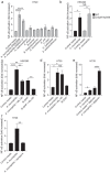
Figure 1.
Figure 1.
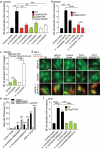
A. muciniphila supernatants activate NF-κB in IECs via a pathway that does not involve MYD88 and NOD1. A. Average NF-κB activity in HT29-NFκB reporter system induced by a wide range of supernatants derived from commensal Gram negative bacteria. B. HEK-NF-κB reporter (WT, black bars) and deleted for MYD88 (MYD88−/−, gray bars) cells were incubated with A. muciniphila DSM22959 supernatant or control media for 24 h. C-D Treatment of HEK-NF-κB (c) or HT29-NF-κB reporter cells with control media, A. muciniphila DSM22959 supernatant or the NOD1 ligand, IE-DAP for 24 h in presence or absence of a NOD1 inhibitor, ML130. E. NF-κB activity in HT29 cells treated with A. muciniphila DSM22959 supernatant in the presence or absence of the permeabilizing agent digitonin for 30 min. When incubated with digitonin, cells were washed with fresh media before measuring the NF-κB activity at 24 h. F. NF-κB activity in HT29 cells pretreated with DMSO or dynasore for 1 h prior to treatment with A. muciniphila supernatant. NF-κB activation was measured by SEAP secretion and expressed as mean ± SEM fold change toward unstimulated cells. Data represent ≥3 independent experiments performed in duplicate or triplicate. Data analysis: unpaired t test was used, ****P < 0,0001; ***P < 0,001; **P < 0,01; *P < 0,05; P > 0,05 was considered as not significant (ns).
Figure 2.
Figure 2.
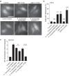
A. muciniphila supernatant activates the ALPK1-TIFA-TRAF6 axis. A. WT (black bars), ALPK1−/− (red bars), TIFA−/− (green bars) and TRAF6−/− (white bars) HEK-NF-κB reporter cells were stimulated with A. muciniphila DSM22959 supernatant or control media for 24 h. NF-κB activation was measured by SEAP secretion and expressed as mean ± SEM fold change toward unstimulated cells. B. WT (black bars) and ALPK1−/− (red bars) HEK-NF-κB reporter cells were incubated with control media, A. muciniphila DSM22959 supernatant or A. muciniphila supernatant in presence of a NOD1 inhibitor (ML130) for 24 h prior NF-κB activation measurement. C. NF-κB activity was measured in HEK293 TIFA−/− cells transfected (white bars) or not (green bars) with pTIFA and stimulated with A. muciniphila DSM22959 supernatant or control media for 24 h. NF-κB activation was measured by SEAP secretion and expressed as mean ± SEM fold change toward un-stimulated cells. D-E) After siRNA treatment with Control siRNA (black bars) or ALPK1 specific siRNA (white and gray bars), Hela cells were transfected with empty pCMV or pCMV-ALPK1-myc and were left unstimulated (NS) or stimulated with the control media, A. muciniphila supernatants or ADP-H (10−6M). D. Representative pictures of cells with TIFAsomes at 30 min in TIFA-GFP-expressing Hela cells, Scale bar: 20 μm. E. The graph shows the quantification of cells with TIFAsomes in each condition as shown in D. Data correspond to the mean ± SD from 3 independent experiments performed in triplicate wells. Statistical significance was assessed using one-way ANOVA followed by Tukey’s multiple comparisons test **P < 0,01; ***P < 0,001; ****P < 0,0001. F. WT (black bars) and TIFA−/− (green bars) HT29-NF-κB reporter cells were stimulated with A. muciniphila supernatants or control media for 24 h. NF-κB activation was measured by SEAP secretion and expressed as mean ± SEM fold change toward unstimulated cells. Data represent ≥3 independent experiments performed in triplicate. Data analysis: unpaired t test was used, ****P < 0,0001; ***P < 0,001; **P < 0,01; *P < 0,05; P > 0,05 was considered as not significant (NS).
Figure 3.
Figure 3.
HldE (Amuc_0415) from A. muciniphila from the heptose biosynthesis pathway is functional. A. Schematic view of the heptose biosynthesis pathway. The gene names annotated for A. muciniphila are in bold. B. HT29-NF-κB reporter WT or TIFA−/− cells were stimulated for 24 h with lysats from E. coli; E. coli ΔhldE; E. coli ΔhldE transformed with plasmid controls (pBAD), with hldE (Amuc_0415) from A. muciniphila (pBAD-hldE_AM) or ADP-H (10−6 M). NF-κB activation was measured by SEAP secretion and expressed as mean (%) ± SEM fold change toward supernatant-stimulated cells. Data represent ≥3 independent experiments performed in triplicate. Data analysis: unpaired t test was used, ****P < 0,0001; ***P < 0,001; **P < 0,01; *P < 0,05; P > 0,05 was considered as not significant (NS).
Figure 4.
Figure 4.
A. muciniphila derived molecule activating NF-κB has the biological properties of ADP-H. A. Representative pictures of cells with TIFAsomes at 30 min in TIFA-GFP-expressing Hela cells. Cells were treated with digitonin and A. muciniphila DSM22959 or DSM26127 supernatants, HBP (10−5M) or ADP-H (10−8M). Scale bar: 20 μm. B. Quantification of cells with TIFAsomes in each condition as shown in A. Statistical significance was assessed using one-way ANOVA followed by Tukey’s multiple comparisons test **P < 0,01; ****P < 0,0001; NS: non-significant. C. A. muciniphila supernatants were untreated, treated with calf intestine alkaline phosphatase (CIP), with Crotalus adamanteus phosphodiesterase (PDE) or their respective buffer (CIP buffer and PDE buffer) prior to stimulation of HT29-NF-κB reporter cells for 24 h. NF-κB activation was measured by SEAP secretion and expressed as mean (%) ± SEM fold change toward supernatant-stimulated cells. Data represent ≥3 independent experiments performed in triplicate. Data analysis: unpaired t test was used, ****P < 0,0001; ***P < 0,001; **P < 0,01; *P < 0,05; P > 0,05 was considered as not significant (NS).
Figure 5.
Figure 5.
ALPK1 and TIFA are expressed by IECs in mice gut.
Figure 6.
Figure 6.
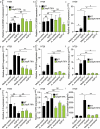
A. muciniphila activates the expression of TNFAIP3, BIRC3, MUC2 and CXCL8 via TIFA.
Figure 7.
Figure 7.
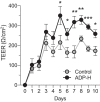
ADP-H improve the HT29 Cl.16E monolayer integrity.

References

    1. Ivanov II, Honda K.. Intestinal commensal microbes as immune modulators. Cell Host Microbe. 2012;12:496–18. doi:10.1016/j.chom.2012.09.009. - DOI - PMC - PubMed
    1. Naito T, Mulet C, De Castro C, Molinaro A, Saffarian A, Nigro G, Bérard M, Clerc M, Pedersen AB, Sansonetti PJ, Pédron T, et al. Lipopolysaccharide from crypt-specific core microbiota modulates the colonic epithelial proliferation-to-differentiation balance. mBio. 2017; 8(5):e01680–17. - PMC - PubMed
    1. Nigro G, Rossi R, Commere PH, Jay P, Sansonetti PJ. The cytosolic bacterial peptidoglycan sensor Nod2 affords stem cell protection and links microbes to gut epithelial regeneration. Cell Host Microbe. 2014;15(6):792–798. doi:10.1016/j.chom.2014.05.003. - DOI - PubMed
    1. Peterson LW, Artis D. Intestinal epithelial cells: regulators of barrier function and immune homeostasis. Nat Rev Immunol. 2014;14:141–153. doi:10.1038/nri3608. - DOI - PubMed
    1. Vlantis K, Polykratis A, Welz PS, van Loo G, Pasparakis M, Wullaert A. TLR-independent anti-inflammatory function of intestinal epithelial TRAF6 signalling prevents DSS-induced colitis in mice. Gut. 2016;65:935–943. doi:10.1136/gutjnl-2014-308323. - DOI - PMC - PubMed

Publication types

Supplementary concepts