. 2022 Oct 26;13(5):e0188622.
doi: 10.1128/mbio.01886-22. Epub 2022 Aug 29.

Dysfunctional Glymphatic System with Disrupted Aquaporin 4 Expression Pattern on Astrocytes Causes Bacterial Product Accumulation in the CSF during Pneumococcal Meningitis

Affiliations

Dysfunctional Glymphatic System with Disrupted Aquaporin 4 Expression Pattern on Astrocytes Causes Bacterial Product Accumulation in the CSF during Pneumococcal Meningitis

Jaqueline S Generoso et al. mBio. .

Abstract

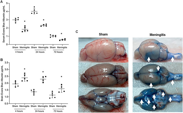
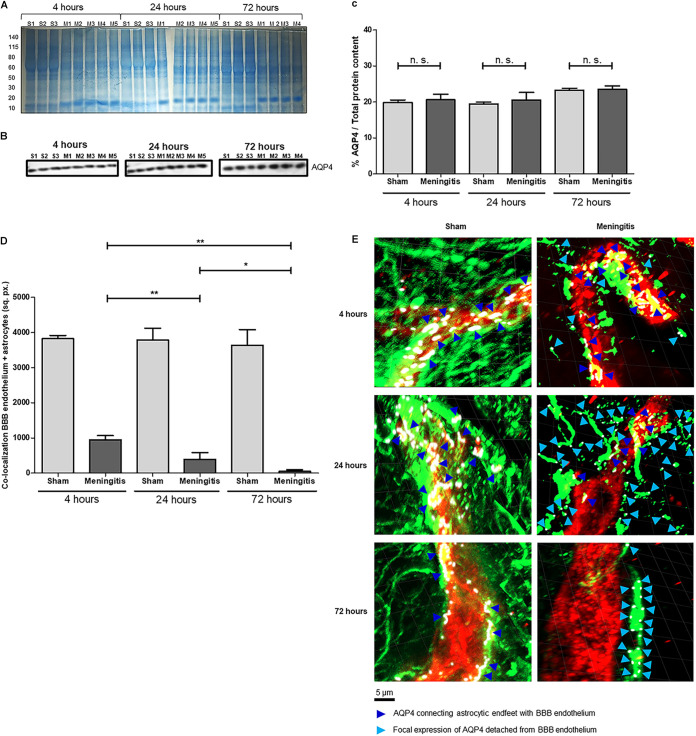
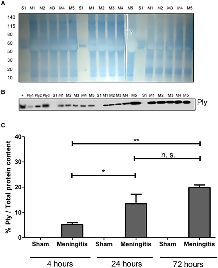
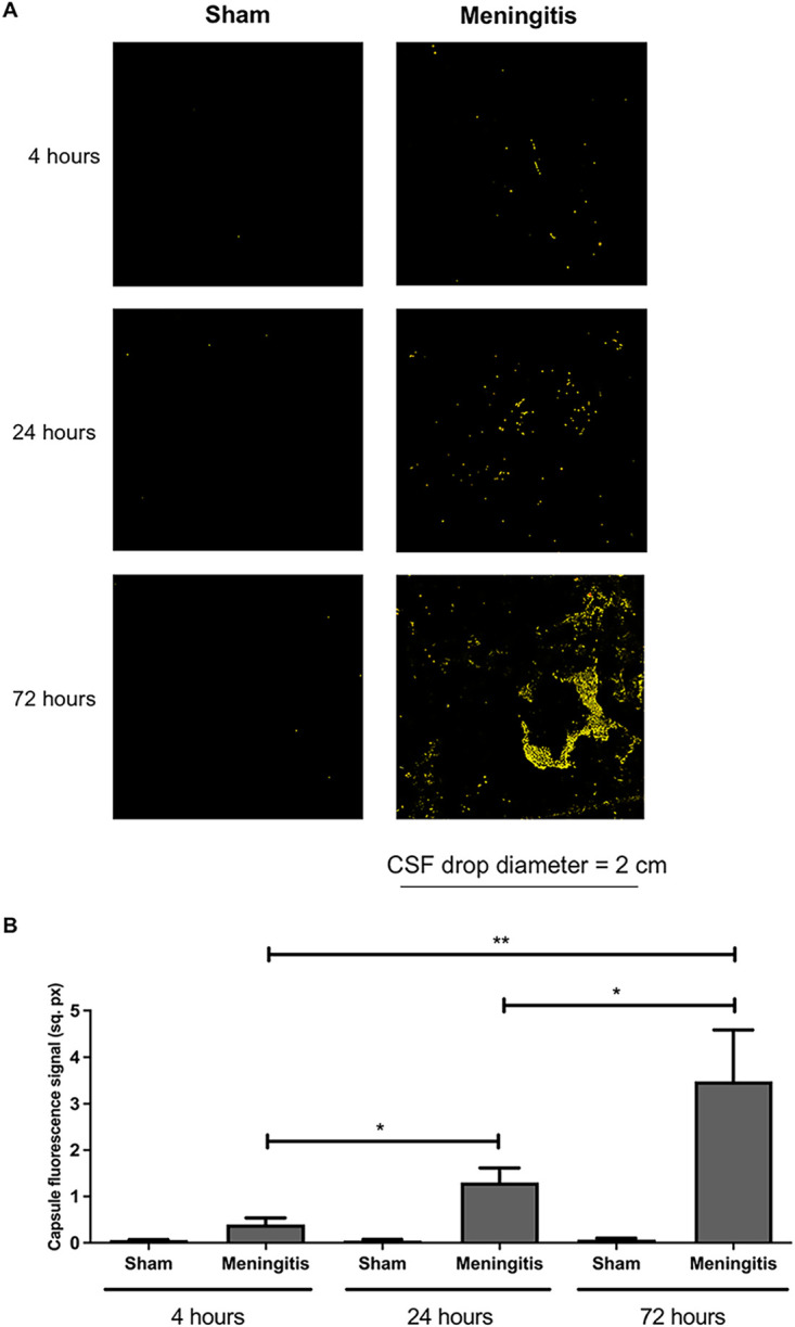
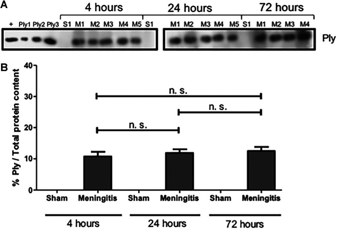
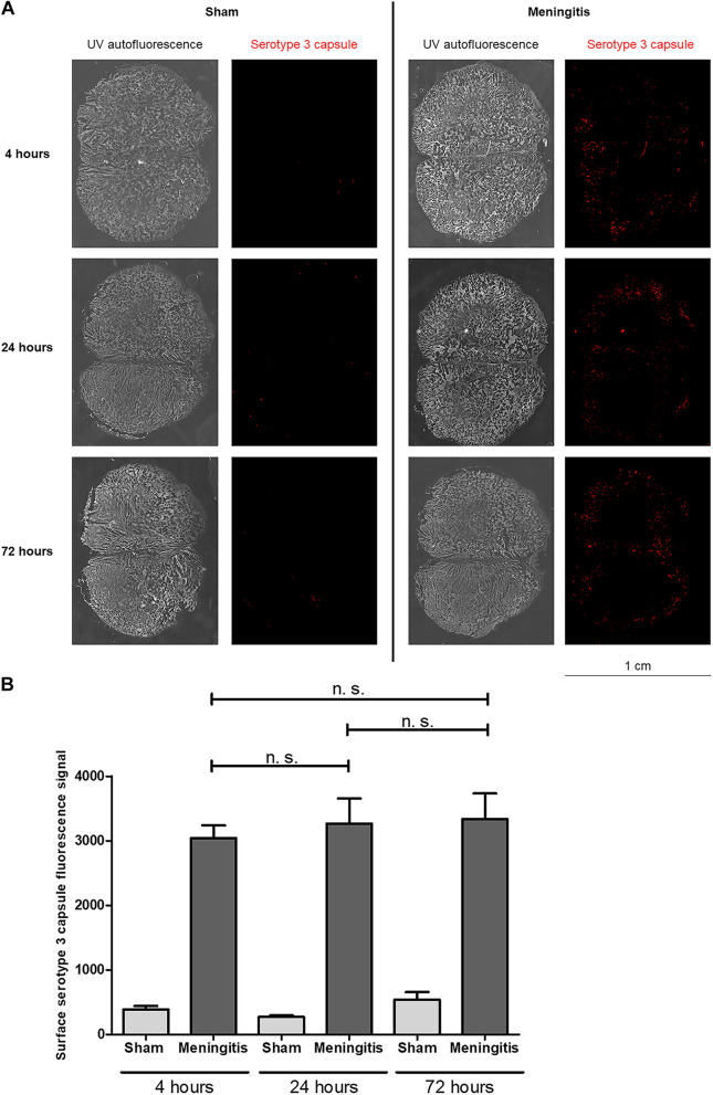
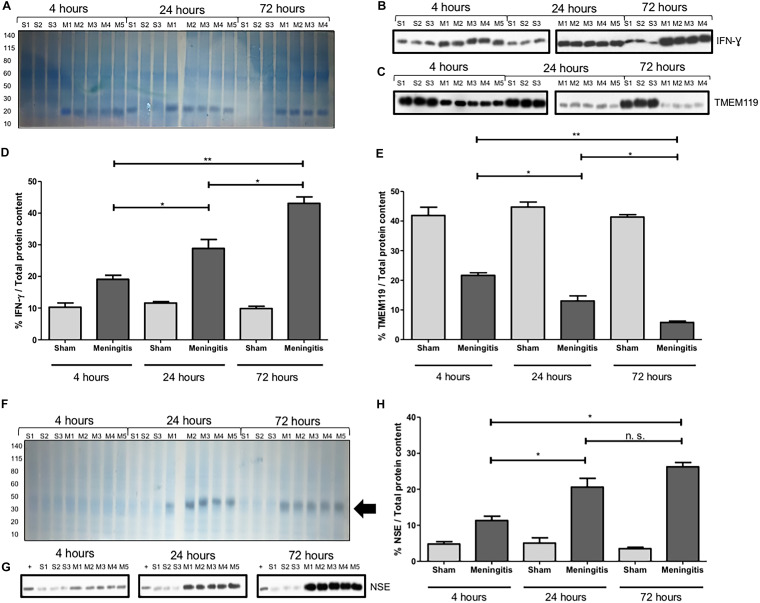
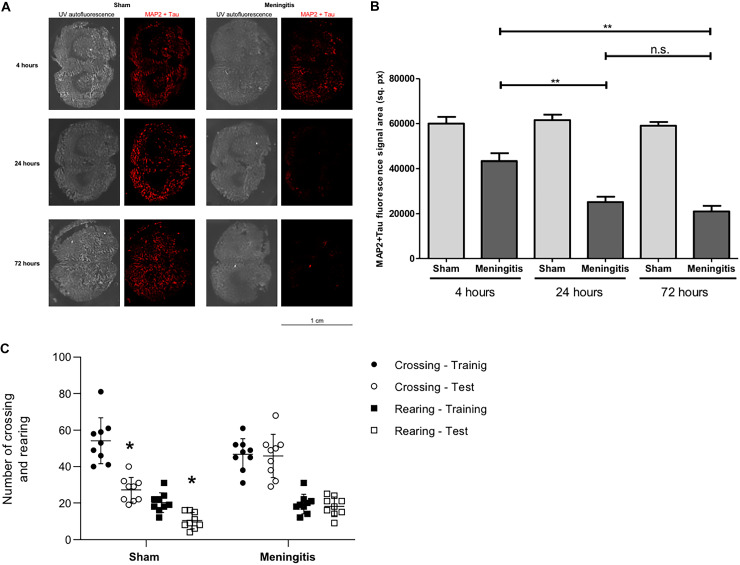
Pneumococcal meningitis, inflammation of the meninges due to an infection of the Central Nervous System caused by Streptococcus pneumoniae (the pneumococcus), is the most common form of community-acquired bacterial meningitis globally. Aquaporin 4 (AQP4) water channels on astrocytic end feet regulate the solute transport of the glymphatic system, facilitating the exchange of compounds between the brain parenchyma and the cerebrospinal fluid (CSF), which is important for the clearance of waste away from the brain. Wistar rats, subjected to either pneumococcal meningitis or artificial CSF (sham control), received Evans blue-albumin (EBA) intracisternally. Overall, the meningitis group presented a significant impairment of the glymphatic system by retaining the EBA in the CSF compartments compared to the uninfected sham group. Our results clearly showed that during pneumococcal meningitis, the glymphatic system does not function because of a detachment of the astrocytic end feet from the blood-brain barrier (BBB) vascular endothelium, which leads to misplacement of AQP4 with the consequent loss of the AQP4 water channel's functionality. IMPORTANCE The lack of solute drainage due to a dysfunctional glymphatic system leads to an increase of the neurotoxic bacterial material in the CSF compartments of the brain, ultimately leading to brain-wide neuroinflammation and neuronal damage with consequent impairment of neurological functions. The loss of function of the glymphatic system can therefore be a leading cause of the neurological sequelae developing post-bacterial meningitis.

Keywords: Streptococcus pneumoniae; aquaporin 4; astrocytic end feet; blood-brain barrier (BBB); glymphatic system; meningitis; neuroinflammation; neuronal damage.


Conflict of interest statement

The authors declare no conflict of interest.

Figures

FIG 1
FIG 1
Demonstration of impaired glymphatic system's functionality during experimental pneumococcal meningitis in vivo. (A and B) Transit of EBA from cisterna magna to serum (A) and from cisterna magna to the brain (B) of adult Wistar rats at 4, 24, and 72 h after induction of pneumococcal meningitis. Data are shown as mean and standard deviation. Statistical significance is shown compared to the control group (n = 5). *, P < 0.05. (C) Retained EBA in the brains of the sham (noninfected) control and meningitis groups at the 4-h time point (the time point with higher EBA retention for both groups). Images of one brain/group are shown as representatives for both groups, and images have been taken from three different perspectives (side, top, and backwards). White arrows point toward the CSF compartments of the brain with high EBA retention.
FIG 2
FIG 2
Loss of glymphatic system's functionality is not due to changes in AQP4 expression levels but to a misplacement of AQP4 water channels on astrocytic end feet. (A) The total protein content of brain homogenate samples was measured after Coomassie staining. S, sham (noninfected) control; M, meningitis. The numbers 1 to 5 refer to the rat number in the group (sham or meningitis) per time point; due to excessive bleeding, one brain (M5) from the 72-h time point was not used for this analysis. (B) Western blot detection of AQP4 in brain homogenate samples from sham and meningitis groups; since AQP4 is expressed by astrocytes independently from the infection, brain homogenates from three sham rats were analyzed; due to excessive bleeding; one brain (M5) from the 72-h time point was not used for this analysis. (C) The percentage of AQP4/total protein content was finally calculated using ImageJ, and data are shown as mean and standard deviation. n.s., nonsignificant. (D) Quantification of the area of colocalization (in square pixels [sq. px.]) between the BBB vascular endothelium (in red in panel E, as shown in Fig. S5) and the astrocytes (in green in panel E, as shown in Fig. S5) measured with ImageJ. Data are shown as mean and standard deviation. *, P < 0.05; **, P < 0.01. (E) Confocal microscopy analysis of BBB vascular endothelium (red), astrocytes (green), and AQP4 (purple, rendered in white using the imaging program ZEN lite). Dark blue arrows point toward AQP4 fluorescent signal connecting astrocytic end feet with BBB vascular endothelium, while light blue arrows point toward focal expression of AQP4 on astrocytes detached from the BBB endothelium. Four brain tissue sections from three sham control and four infected rats were analyzed per time point, and three images per section were taken. The displayed images are representative of each group. Plane images were angled 30° from the horizontal position on the z axis using Imaris.
FIG 3
FIG 3
Accumulation of pneumococcal pneumolysin in the cerebrospinal fluid as a consequence of an impaired functionality of the glymphatic system. (A) The total protein content of cerebrospinal fluid (CSF) samples was first measured by Coomassie staining. S, sham (noninfected) control; M, meningitis. The numbers 1 to 5 refer to the rat number in the group (sham or meningitis) per time point. (B) The same volume of the same samples was then used for Western blot analysis for the detection of Ply; a lysate of serotype 3 S. pneumoniae was used as a positive control (+) together with three serial amounts of purified Ply (Ply1 at 0.0005 μg, Ply2 at 0.005 μg, and Ply3 at 0.05 μg in loaded volumes of 10 μL). (C) The percentage of Ply/total protein content was finally calculated using ImageJ. Data are shown as mean and standard deviation. *, P < 0,05; **, P < 0.01; n.s., nonsignificant since sham control rats were not infected with pneumococci and therefore did not have any Ply. As a negative control, one CSF sample from the sham group for each time point was analyzed, for a total of three sham samples analyzed.
FIG 4
FIG 4
Accumulation of pneumococcal polysaccharide capsule in the CSF due to a malfunctioning glymphatic system during pneumococcal meningitis. (A) Ten-microliter CSF drops dried on microscope glass slides were stained for serotype 3 polysaccharide capsule (red). The surface area of the fluorescence images is within the yellow border (after the function “Edit selection” of ImageJ was applied to measure the surface area of the fluorescence signal) to enhance the contrast of the fluorescence signal. Per time point, CSF samples from three rats of the sham (noninfected) group were analyzed in order to have a broad assessment of the unspecific signal from noninfected CSF samples; one representative image is shown per group. (B) Quantification of the fluorescence signal of the pneumococcal capsule stained in Fig. 3A. Data are shown as mean and standard deviation. *, P < 0.05; **, P < 0.01.
FIG 5
FIG 5
Presence but not accumulation of Ply in the brain homogenates of rats affected by pneumococcal meningitis. (A) Western blot analysis for the detection of Ply on brain homogenate samples. A lysate of serotype 3 S. pneumoniae was used as a positive control (+) together with three serial amounts of purified Ply (Ply1 at 0.0005 μg, Ply2 at 0.005 μg, and Ply3 at 0.05 μg in loaded volumes of 10 μL). As a negative control, brain homogenate samples from three rats of the sham (noninfected) group per time point were analyzed. (B) The percentage of Ply/total protein content was finally calculated using ImageJ. Data are shown as mean and standard deviation. n.s., nonsignificant. (The total protein content was measured after the Coomassie staining shown in Fig. 2A.)
FIG 6
FIG 6
Presence but not accumulation of serotype 3 capsule in the brain parenchyma of rats with pneumococcal meningitis. (A) Brain tissue sections were stained for serotype 3 polysaccharide capsule (red), and the whole-tissue sections were imaged with autofluorescence under UV light. Per time point, tissue sections from one rat of the sham (noninfected) control group and three rats of the meningitis group with two sections per rat were analyzed in total. One representative image is shown per group (sham or meningitis) per time point. (B) Quantification of the fluorescence signal of the pneumococcal capsule stained in panel A. Data are shown as mean and standard deviation. n.s., nonsignificant.
FIG 7
FIG 7
Neuroinflammation and neuronal damage increase over time during pneumococcal infection with a malfunctioning glymphatic system. (A) The total protein content of brain homogenate samples was measured after Coomassie staining. S, sham (noninfected) control; M, meningitis. The numbers 1 to 5 refer to the rat number in the group (sham or meningitis) per time point. (B and C) Western blot detection of IFN-γ (B) and TMEM119 (C) was performed using brain homogenate samples of rats from sham and meningitis groups. Since both TMEM119 and IFN-γ are, respectively, microglial and microglial/macrophage markers present in the brain independently from the infection, brain homogenates from three sham rats were analyzed. Due to excessive bleeding, one brain (M5) from the 72-h time point was not used for this analysis. (D and E) The percentages of IFN-γ/total protein content (D) and TMEM119/total protein content (E) were finally calculated using ImageJ. Data are shown as mean and standard deviation. *, P < 0.05; **, P < 0.01; n.s., nonsignificant. As a negative control, brain homogenate samples from three rats of the sham group per time point were analyzed. The numbers 1 to 5 refer to the rat number in the group (sham or meningitis) per time point. (The total protein content was measured after the Coomassie staining shown in Fig. 6A.) (F) The total protein content of serum samples was first measured after Coomassie staining. S, sham control; M, meningitis. The numbers 1 to 5 refer to the rat number in the group (sham or meningitis) per time point, and the black arrow points toward an enhanced intensity of the Coomassie staining around the molecular weight of NSE (around 40 kDa), which is particularly evident for the meningitis group at 24 and 72 h. (G) The same volume of the same samples was then used for Western blot analysis for the detection of NSE. A lysate of differentiated neurons from SH-SY5Y cells was used as a positive control (+). (H) The percentage of NSE/total protein content was finally calculated using ImageJ, and data are shown as mean and standard deviation. *, P < 0.05; n.s., nonsignificant.
FIG 8
FIG 8
Loss of neuronal synaptic connections and consequent impairment of neurological functions during pneumococcal meningitis with a malfunctioning glymphatic system. (A) Brain tissue sections were stained for a combination of MAP2 and Tau proteins (red), and the whole-tissue sections were imaged with autofluorescence under UV light. Per time point, tissue sections from three rats of the sham (noninfected) group and three rats of the meningitis group with two sections per rat were analyzed in total. One representative image is shown per group (sham or meningitis) per time point. (B) Quantification of the fluorescence signal of MAP2+Tau stained in Fig. 5A. Data are shown as mean and standard deviation. **, P < 0.01; n.s., nonsignificant. (C) Open-field task 10 days after pneumococcal meningitis induction. Data from the open field task were reported as the mean and standard error of mean values and analyzed by paired Student's t tests (n = 9 animals per group). *, P < 0.05 versus training session.

Comment in

Similar articles

Cited by

References

    1. Iovino F, Seinen J, Henriques-Normark B, van Dijl JM. 2016. How does Streptococcus pneumoniae invade the brain? Trends Microbiol 24:307–515. doi:10.1016/j.tim.2015.12.012. - DOI - PubMed
    1. Oordt-Speets AM, Bolijn R, van Hoorn RC, Bhavsar A, Kyaw MH. 2018. Global etiology of bacterial meningitis: a systematic review and meta-analysis. PLoS One 13:e0198772. doi:10.1371/journal.pone.0198772. - DOI - PMC - PubMed
    1. Brouwer M, van de Beek D. 2018. Epidemiology of community-acquired bacterial meningitis. Curr Opin Infect Dis 31:78–84. doi:10.1097/QCO.0000000000000417. - DOI - PubMed
    1. Lucas MJ, Brouwer MC, van de Beek D. 2016. Neurological sequelae of bacterial meningitis. J Infect 73:18–27. doi:10.1016/j.jinf.2016.04.009. - DOI - PubMed
    1. Kloek AT, Brouwer MC, Schmand B, Tanck MWT, van de Beek D. 2020. Long-term neurologic and cognitive outcome and quality of life in adults after pneumococcal meningitis. Clin Microbiol Infect 26:1361–1367. doi:10.1016/j.cmi.2020.01.020. - DOI - PubMed

Publication types

LinkOut - more resources