. 2022 Oct 26;10(5):e0051822.
doi: 10.1128/spectrum.00518-22. Epub 2022 Aug 29.

FXR Signaling-Mediated Bile Acid Metabolism Is Critical for Alleviation of Cholesterol Gallstones by Lactobacillus Strains

Affiliations

FXR Signaling-Mediated Bile Acid Metabolism Is Critical for Alleviation of Cholesterol Gallstones by Lactobacillus Strains

Xin Ye et al. Microbiol Spectr. .

Abstract

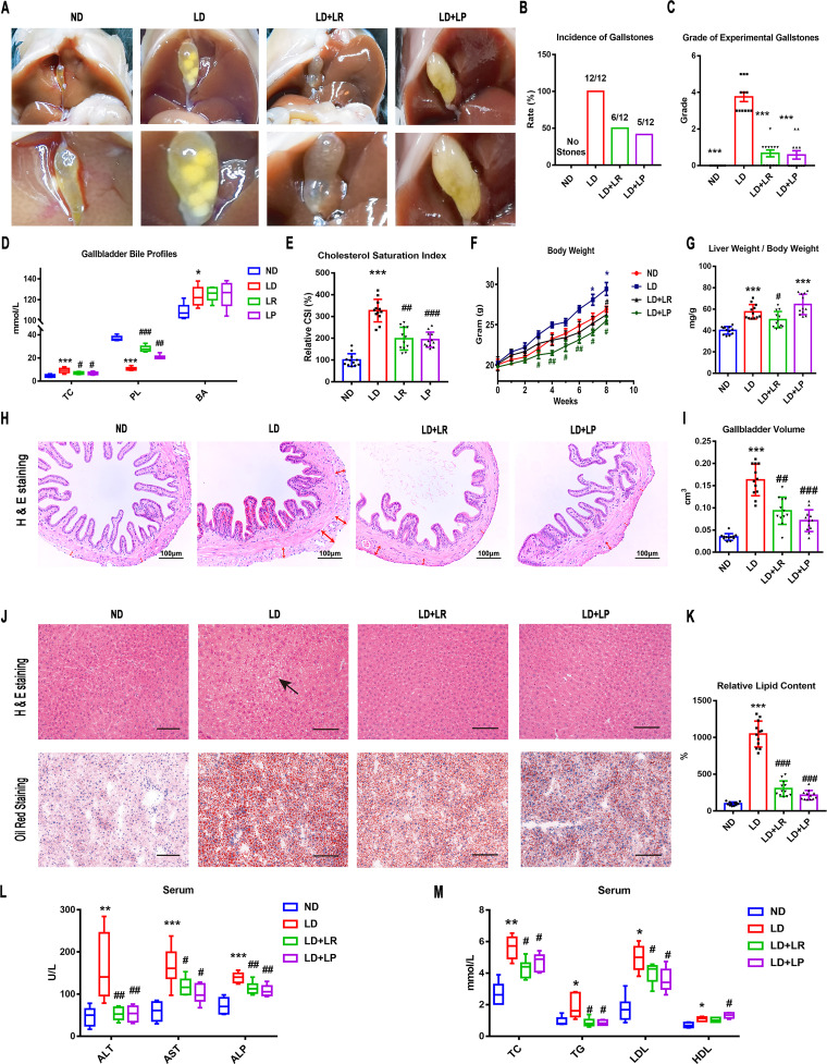
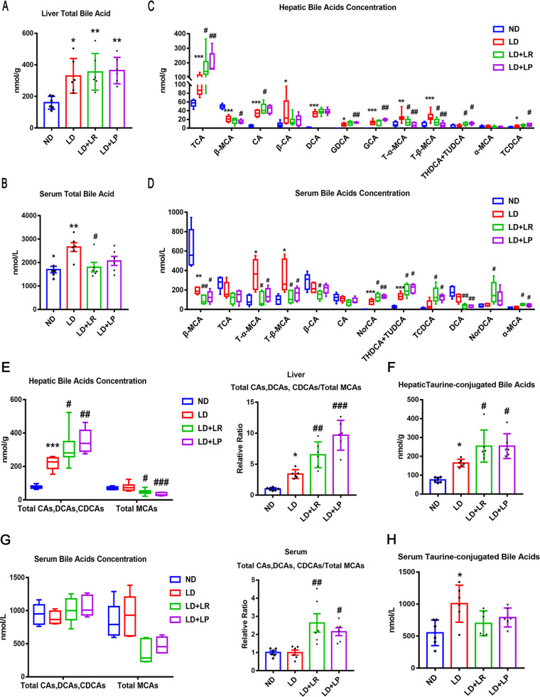
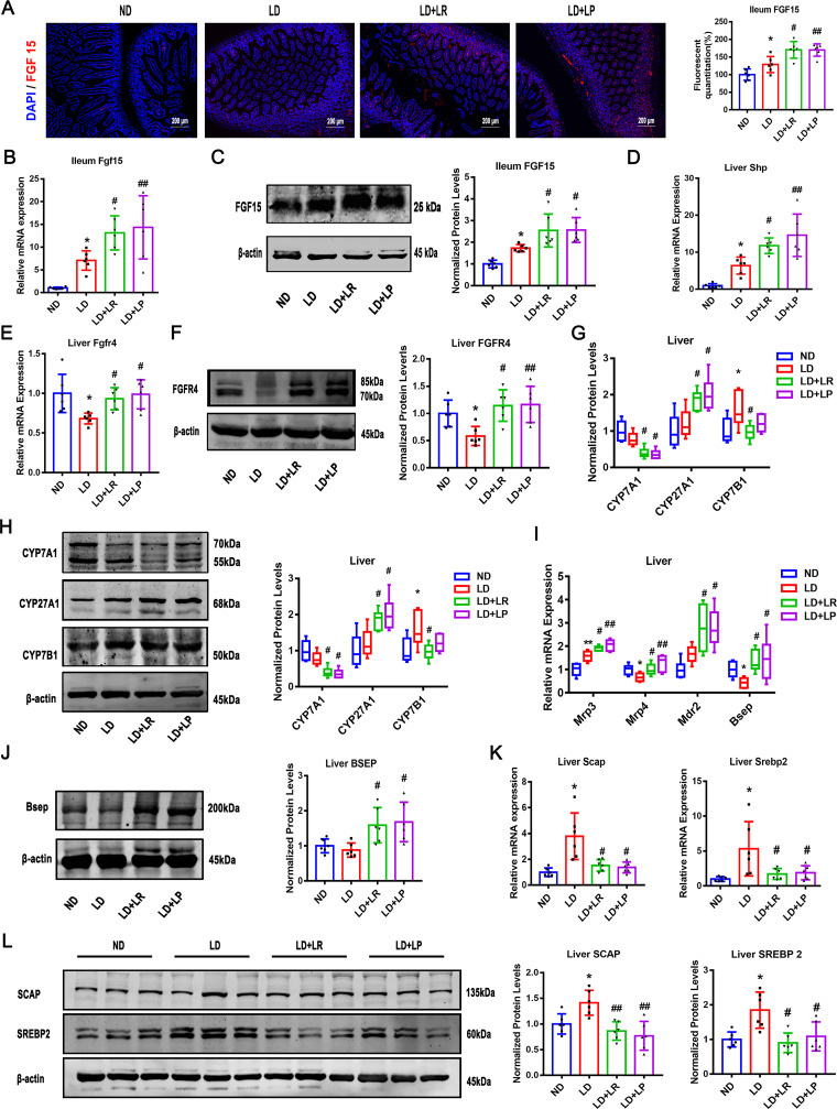
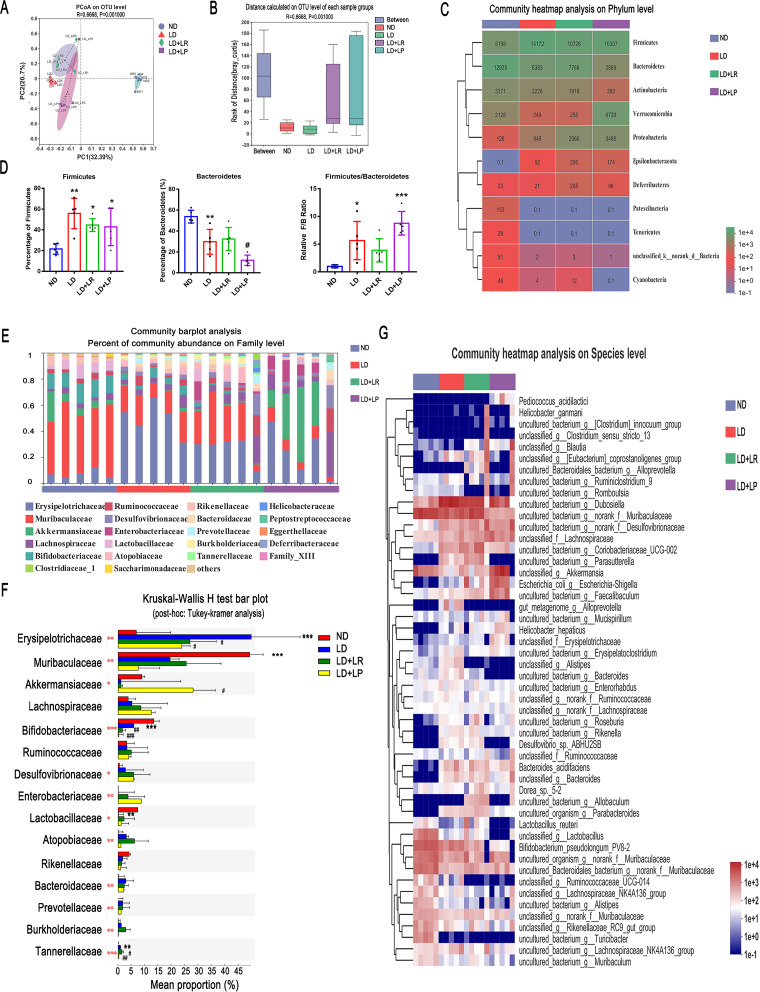
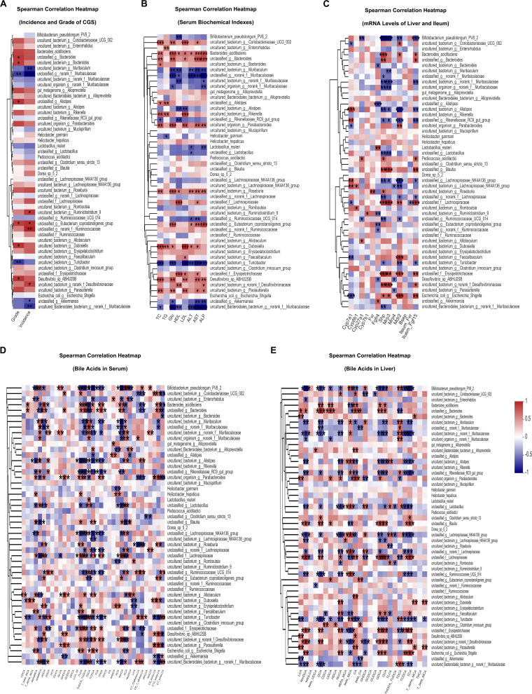
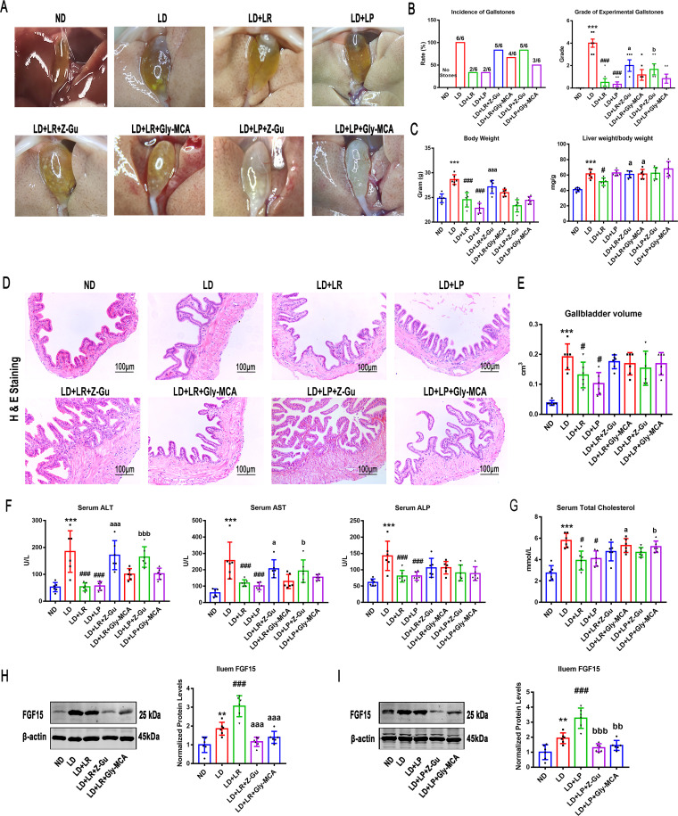
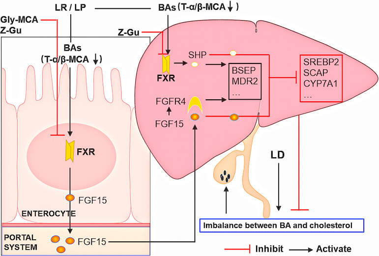
Cholesterol gallstone (CGS) disease is characterized by an imbalance in bile acid (BA) metabolism and is closely associated with gut microbiota disorders. However, the role and mechanism by which probiotics targeting the gut microbiota attenuate cholesterol gallstones are still unknown. In this study, Limosilactobacillus reuteri strain CGMCC 17942 and Lactiplantibacillus plantarum strain CGMCC 14407 were individually administered to lithogenic-diet (LD)-fed mice for 8 weeks. Both Lactobacillus strains significantly reduced LD-induced gallstones, hepatic steatosis, and hyperlipidemia. These strains modulated BA profiles in the serum and liver, which may be responsible for the activation of farnesoid X receptor (FXR). At the molecular level, L. reuteri and L. plantarum increased ileal fibroblast growth factor 15 (FGF15) and hepatic fibroblast growth factor receptor 4 (FGFR4) and small heterodimer partner (SHP). Subsequently, hepatic cholesterol 7α-hydroxylase (CYP7A1) and oxysterol 7α-hydroxylase (CYP7B1) were inhibited. Moreover, the two strains enhanced BA transport by increasing the levels of hepatic multidrug resistance-associated protein homologs 3 and 4 (Mrp3/4), hepatic multidrug resistance protein 2 (Mdr2), and the bile salt export pump (BSEP). In addition, both L. reuteri and L. plantarum reduced LD-associated gut microbiota dysbiosis. L. reuteri increased the relative abundance of Muribaculaceae, while L. plantarum increased that of Akkermansia. The changed gut microbiota was significantly negatively correlated with the incidence of cholesterol gallstones and the FXR-antagonistic BAs in the liver and serum and with the FXR signaling pathways. Furthermore, the protective effects of the two strains were abolished by both global and intestine-specific FXR antagonists. These findings suggest that Lactobacillus might relieve CGS through the FXR signaling pathways. IMPORTANCE Cholesterol gallstone (CGS) disease is prevalent worldwide. None of the medical options for prevention and treatment of CGS disease are recommended, and surgical management has a high rate of recurrence. It has been reported that the factors involved in metabolic syndrome are highly connected with CGS formation. While remodeling of dysbiosis of the gut microbiome during improvement of metabolic syndrome has been well studied, less is known about prevention of CGS formation after regulating the gut microbiome. We used the lithogenic diet (LD) to induce an experimental CGS model in C57BL/6J mice to investigate protection against CGS formation by Limosilactobacillus reuteri strain CGMCC 17942 and Lactiplantibacillus plantarum strain CGMCC 14407. We found that these L. reuteri and L. plantarum strains altered the bile acid composition in mice and improved the dysbiosis of the gut microbiome. These two Lactobacillus strains prevented CGS formation by fully activating the hepatic and ileal FXR signaling pathways. They could be a promising therapeutic strategy for treating CGS or preventing its recurrence.

Keywords: FGF15; FXR; Lactobacillus; bile acid; cholesterol gallstones; gut microbiota.


Conflict of interest statement

The authors declare no conflict of interest.

Figures

FIG 1
FIG 1
L. reuteri (LR) and L. plantarum (LP) treatments reduced LD-induced gallstones and metabolic disorders in mice. Twelve mice were randomly assigned to each group and fed a normal diet (ND) or a lithogenic diet (LD) with or without L. reuteri or L. plantarum treatment (109 CFU/day) for 8 weeks. (A) Gross appearance of gallbladders and gallstones of mice administered different treatments. (B) Percentage of gallstone incidence in each group of mice. (C) The grades of experimental cholesterol gallstones (CGSs) in the mice were based on the observed cholelithiasis. (D) Total cholesterol (TC), bile acids (BA), and phospholipids (PL) in gallbladders. (E) CSI in each group of mice. (F) Body weights of mice were recorded once a week. (G) Ratios of liver weight to body weight. (H) Representative images of H&E-stained gallbladder sections (×200). The tunica adventitia of the gallbladder is indicated by red arrows. (I) Gallbladder volumes were estimated by the length, diameter, and circumference of the gallbladders. (J) Representative images of H&E-stained and oil red O-stained liver sections (×200). (K) Percentages of oil red O-positive areas. (L and M) Serum alanine aminotransferase (ALT), aspartate aminotransferase (AST), alkaline phosphatase (ALP), total cholesterol (TC), triglyceride (TG), high-density lipoprotein (HDL), and low-density lipoprotein (LDL) were determined by using a Siemens fast automatic biochemical analyzer (Advia 2400). Data were analyzed by ANOVA along with the post hoc Tukey test and are presented as the mean values ± SEM (n = 12). *, 0.01 < P ≤ 0.05; **, 0.001 < P ≤ 0.01; ***, P ≤ 0.001 versus ND group; #, 0.01 < P ≤ 0.05; ##, 0.001 < P ≤ 0.01; ###, P ≤ 0.001 versus LD group.
FIG 2
FIG 2
L. reuteri (LR) and L. plantarum (LP) treatments changed the bile acid species in the liver and serum. The mice in each group were sacrificed at the end of the 8th week, and serum and liver were collected. (A) Total bile acids (TBA) in the livers of mice. (B) TBA in the serum of mice. (C) The top 12 most abundant hepatic BA species were analyzed. (D) The top 12 most abundant serum BA species are shown. NorCA, norcholic acid; NorDCA, 23-nordeoxycholic acid. (E) Total CAs, DCAs, and CDCAs, total MCAs, and ratios of total CAs, DCAs, and CDCAs to total MCAs in the livers. (F) Total taurine-conjugated BAs in the livers. (G) Total CAs, DCAs, and CDCAs, total MCAs, and ratios of total CAs, DCAs, and CDCAs to total MCAs in the serum. (H) Total taurine-conjugated BAs in the serum. Data were analyzed by ANOVA along with the post hoc Tukey test and are presented as the mean values ± SEM (n = 6). *, 0.01 < P ≤ 0.05; **, 0.001 < P ≤ 0.01; ***, P ≤ 0.001 versus normal diet (ND) group; #, 0.01 < P ≤ 0.05; ##, 0.001 < P ≤ 0.01; ###, P ≤ 0.001 versus lithogenic diet (LD) group.
FIG 3
FIG 3
L. reuteri (LR) and L. plantarum (LP) treatments activated FXR signaling pathway. The mice in each group were sacrificed at the end of the 8th week, and serum and ileum samples were collected. (A) Immunofluorescence analyses of ileal FGF15 (×400). (B) Ileal mRNA expression of Fgf15. (C) Protein expression and quantification of ileal FGF15. (D and E) Hepatic mRNA levels of Shp and Fgfr4. (F) Protein expression and quantification of hepatic FGFR4. (G and H) Hepatic mRNA and protein expression of CYP7A1, CYP27A1, and CYP7B1 and quantification of the proteins. (I) Hepatic mRNA levels of Mrp3, Mrp4, Mdr2, and Bsep. (J) Protein expression and quantification of hepatic BSEP. (K and L) Hepatic mRNA and protein expression of SCAP and SREBP2 and quantification of the proteins. Data were analyzed by ANOVA along with the post hoc Tukey test and are presented as the mean values ± SEM (n = 6). *, 0.01 < P ≤ 0.05; **, 0.001 < P ≤ 0.01; ***, P ≤ 0.001 versus normal diet (ND) group; #, 0.01 < P ≤ 0.05; ##, 0.001 < P ≤ 0.01; ###, P ≤ 0.001 versus lithogenic diet (LD) group.
FIG 4
FIG 4
L. reuteri (LR) and L. plantarum (LP) treatments changed the CGS-associated gut microbiota composition of LD-fed mice. Fecal samples from ND-fed mice or LD-fed mice with or without Lactobacillus treatment were collected at the 8th week for quantification. (A) Principal coordinate analysis (PCoA) of unweighted UniFrac analysis based on the OTU abundances of different groups (Bray-Curtis ANOSIM, R = 0.6683, P = 0.001). (B) Distance box plots based on the OTU abundances of groups (Bray-Curtis ANOSIM, R = 0.6668, P = 0.001). (C) Relative abundances of phyla in the gut microbiota of ND-fed mice and LD-fed mice with or without L. reuteri or L. plantarum treatment. (D) Percentages of Firmicutes and Bacteroidetes and ratios of Firmicutes/Bacteroidetes (F/B). (E) The relative abundances of fecal bacteria in each group of mice at the family level were analyzed with a community bar plot analysis. (F) Comparison of the top 15 most abundant families in the ND, LD, LD + L. reuteri, and LD + L. plantarum groups by Kruskal-Wallis H test with post hoc (Tukey-Kramer) analysis. (G) The top 50 most abundant species in the gut microbiota in different groups are presented in a heatmap. Data are presented as the mean values ± SEM (n = 5). *, 0.01 < P ≤ 0.05; **, 0.001 < P ≤ 0.01; ***, P ≤ 0.001 versus normal diet (ND) group; #, 0.01 < P ≤ 0.05; ##, 0.001 < P ≤ 0.01; ###, P ≤ 0.001 versus lithogenic diet (LD) group. The red asterisks in panel F show statistically significant differences between the groups by Kruskal-Wallis H test.
FIG 5
FIG 5
Correlation between gut microbiota and host CGS-related parameters. (A) Spearman’s correlation analysis was performed between the top 50 most abundant species levels and the incidence and grades of CGS. (B) Spearman’s correlation analysis was performed between the top 50 most abundant species levels and the serum AST, ALT, ALP, TG, TC, HDL, LDL, and glucose (Glu) levels. (C) Spearman’s correlation analysis was performed between the top 50 most abundant species levels and the mRNA levels of liver Cyp7a1, Cyp8b1, Cyp7b1, Cyp27a1, Mdr2, Mrp3, Mrp4, Bsep, Shp, hepatic Fxr, and Fgfr4 and ileum Fxr and Fgf15. (D) Spearman’s correlation analysis was performed between the top 50 most abundant species levels and 35 kinds of BAs in the serum of mice. (E) Spearman’s correlation analysis was performed between the top 50 most abundant species levels and 26 kinds of BAs in the livers of mice. Red and blue denote positive and negative associations, respectively. n = 5. *, 0.01 < P ≤ 0.05; **, 0.001 < P ≤ 0.01; ***, P ≤ 0.001; |R|0.5.
FIG 6
FIG 6
Inhibition of FXR activation attenuated the protective effects of L. reuteri (LR) and L. plantarum (LP) in LD-fed mice. Forty-eight mice were randomly assigned to 8 groups fed the ND or LD with or without L. reuteri or L. plantarum treatment (109 CFU/day). At the beginning of the L. reuteri or L. plantarum treatment, the mice were gavaged with (Z/E)-guggulsterone (Z-Gu), a global FXR inhibitor, or glycine-β-muricholic acid (Gly-MCA), an intestine-specific FXR inhibitor. (A) Gross appearance of gallbladders and gallstones of mice administered different treatments. (B) Percentage of gallstone incidence in each group of mice and grade of experimental CGS, based on visualized cholelithiasis. (C) Body weights of mice on the last day and ratios of liver weight to body weight. (D) Representative images of H&E-stained gallbladder sections (×200). (E) Gallbladder volumes estimated by the length, diameter, and circumference of the gallbladders. (F and G) Serum ALT, AST, ALP, and TC were determined by using a Siemens fast automatic biochemical analyzer (Advia 2400). (H and I) Protein expression and quantification of ileal FGF15 with or without L. reuteri (H) or L. plantarum (I) treatment. Data were analyzed by ANOVA along with the post hoc Tukey test and are presented as the mean values ± SEM (n = 6). *, 0.01 < P ≤ 0.05; **, 0.001 < P ≤ 0.01; ***, P ≤ 0.001 versus normal diet (ND) group; #, 0.01 < P ≤ 0.05; ##, 0.001 < P ≤ 0.01; ###, P ≤ 0.001 versus lithogenic diet (LD) group; a, 0.01 < P ≤ 0.05; aa, 0.001 < P ≤ 0.01; aaa, P ≤ 0.001 versus LD + L. reuteri; b, 0.01 < P ≤ 0.05; bb, 0.001 < P ≤ 0.01; bbb, P ≤ 0.001 versus LD + L. plantarum.
FIG 7
FIG 7
Schematic diagram summarizing the mechanisms by which L. reuteri and L. plantarum prevent CGS formation.

Similar articles

Cited by

References

    1. Forrest K, Welch C, Williams E, Smart H, Lombard M. 2006. Investigation of cholesterol gallstone disease. Lancet 368:989–990. doi: 10.1016/S0140-6736(06)69412-9. - DOI - PubMed
    1. Lammert F, Gurusamy K, Ko CW, Miquel J-F, Méndez-Sánchez N, Portincasa P, van Erpecum KJ, van Laarhoven CJ, Wang DQ-H. 2016. Gallstones. Nat Rev Dis Primers 2:16024. doi: 10.1038/nrdp.2016.24. - DOI - PubMed
    1. Wang J, Xu C, Cheng Q, Zhao J, Wu S, Li W, Ma W, Liu C, Jiang X. 2020. RNA sequencing revealed signals of evolution from gallbladder stone to gallbladder carcinoma. Front Oncol 10:823. doi: 10.3389/fonc.2020.00823. - DOI - PMC - PubMed
    1. Shabanzadeh DM, Sørensen LT, Jørgensen T. 2017. Association between screen-detected gallstone disease and cancer in a cohort study. Gastroenterology 152:1965–1974.e1. doi: 10.1053/j.gastro.2017.02.013. - DOI - PubMed
    1. Di Ciaula A, Wang DQH, Wang HH, Bonfrate L, Portincasa P. 2010. Targets for current pharmacologic therapy in cholesterol gallstone disease. Gastroenterol Clin North Am 39:245–264. doi: 10.1016/j.gtc.2010.02.005. - DOI - PMC - PubMed

Publication types

MeSH terms

Substances