. 2022 Oct 26;13(5):e0127722.
doi: 10.1128/mbio.01277-22. Epub 2022 Aug 29.

Atomic Structure of IglD Demonstrates Its Role as a Component of the Baseplate Complex of the Francisella Type VI Secretion System

Affiliations

Atomic Structure of IglD Demonstrates Its Role as a Component of the Baseplate Complex of the Francisella Type VI Secretion System

Xiaoyu Liu et al. mBio. .

Abstract

Francisella tularensis, a Tier 1 select agent of bioterrorism, contains a type VI secretion system (T6SS) encoded within the Francisella pathogenicity island (FPI), which is critical for its pathogenesis. Among the 18 proteins encoded by FPI is IglD, which is essential to Francisella's intracellular growth and virulence, but neither its location within T6SS nor its functional role has been established. Here, we present the cryoEM structure of IglD from Francisella novicida and show that the Francisella IglD forms a homotrimer that is structurally homologous to the T6SS baseplate protein TssK in Escherichia coli. Each IglD monomer consists of an N-terminal β-sandwich domain, a 4-helix bundle domain, and a flexible C-terminal domain. While the overall folds of IglD and TssK are similar, the two structures differ in three aspects: the relative orientation between their β-sandwich and the 4-helix bundle domains; two insertion loops present in TssK's β-sandwich domain; and, consequently, a lack of subunit-subunit interaction between insertion loops in the IglD trimer. Phylogenetic analysis indicates that IglD is genetically remote from the TssK orthologs in other T6SSs. While the other components of the Francisella baseplate are unknown, we conducted pulldown assays showing IglJ interacts with IglD and IglH, pointing to a model wherein IglD, IglH, and IglJ form the baseplate of the Francisella T6SS. Alanine substitution mutagenesis further established that IglD's hydrophobic pocket in the N-terminal β-sandwich domain interacts with two loops of IglJ, reminiscent of the TssK-TssG interaction. These results form a framework for understanding the hitherto unexplored Francisella T6SS baseplate. IMPORTANCE Francisella tularensis is a facultatively intracellular Gram-negative bacterium that causes the serious and potentially fatal zoonotic illness, tularemia. Because of its extraordinarily high infectivity and mortality to humans, especially when inhaled, F. tularensis is considered a potential bioterrorism agent and is classified as a Tier 1 select agent. The type VI secretion system (T6SS) encoded within the Francisella pathogenicity island (FPI) is critical to its pathogenesis, but its baseplate components are largely unknown. Here, we report the cryoEM structure of IglD from Francisella novicida and demonstrate its role as a component of the baseplate complex of the Francisella T6SS. We further show that IglD interacts with IglJ and IglH, and propose a model in which these proteins interact to form the Francisella T6SS baseplate. Elucidation of the structure and composition of the Francisella baseplate should facilitate the design of strategies to prevent and treat infections caused by F. tularensis.

Keywords: Francisella; IglD; baseplate complex; contractile injection system; cryo-electron microscopy; intracellular pathogen; type VI secretion system.


Conflict of interest statement

The authors declare no conflict of interest.

Figures
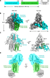
FIG 1
FIG 1
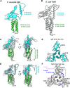
CryoEM structure of IglD. (A) Domain organization of Francisella novicida (Fn) IglD. The full-length protein consists of an N-terminal β sandwich (cyan), middle domain 4-helix bundle (green), and C-terminal domain (light gray). The C-terminal domain was not modeled. (B) Two different views of the cryoEM density map of IglD homotrimer. One protomer is represented with domains shown in the same color scheme as in A, the other two protomers are colored gray. (C) Two different views of IglD homotrimer structure. The structure is colored as in B. The three protomers are labeled as IglD-A, IglD-B, and IglD-C. (D) IglD monomer structure is shown in two different views. Domains are colored as in A. The N termini of the protein are labeled. Cartoon illustrations of full-length IglD monomer with C-terminal domain are shown based on the low-resolution map.
FIG 2
FIG 2
Structure comparison of IglD and TssK. (A) Structure of Fn IglD monomer. Domains are colored as in Fig. 1A. (B) Structure of EAEC TssK monomer (PDB accession no. 5MWN; Chain A). (C) Superposition of Fn IglD (color) and EAEC TssK (gray) monomers by 4-helix bundle domain. (D) Superposition of Fn IglD (color) and EAEC TssK (gray) monomers by N-terminal β-sandwich domain. The two arrows point to two insertion loops in TssK compared with IglD. The magenta one points to loop1 (A108-R124), the blue one points to loop2 (E134-E139). (E) IglD trimer top view, 4-helix bundle domain was omitted for clarity. The three protomers are labeled as IglD-A, IglD-B, and IglD-C. (F) TssK trimer top view, 4-helix bundle domain was omitted for clarity. The three protomers are labeled as TssK-A, TssK-B, and TssK-C. Loop1 in each protomer is colored magenta, and loop2 in each protomer is colored blue. The boxed region shows loop1 from TssK-A and loop2 from TssK-B.
FIG 3
FIG 3
IglJ-His pulls down IglD. Wild-type (WT) Fn or Fn expressing IglJ-His were grown in high KCl, cross-linked with DSP, lysed, and IglJ-associated proteins enriched by nickel-agarose affinity chromatography. (A) SDS-PAGE gel showing proteins eluted in successive fractions with 50 mM and 250 mM imidazole visualized by stain-free UV imaging. (B) Western immunoblot using anti-His epitope antibody identifies IglJ-His in the Fn IglJ-His sample and not in the WT sample. (C and D) Samples eluted with 250 mM imidazole were further analyzed by SDS-PAGE with stain-free UV imaging of proteins (C) and Western immunoblotting using polyclonal antibodies against IglD (D). (E) MS/MS analysis of IglJ-His and WT pulldown samples.
FIG 4
FIG 4
Epitope-tagged IglH pulls down IglD. Wild-type (WT) Fn or Fn expressing IglH with TwinStrep and His-tags were grown in high KCl, cross-linked with DSP, lysed, and IglH-associated proteins enriched by nickel-agarose affinity chromatography. (A) SDS-PAGE gel showing proteins in the lysate, affinity column pass-through (unbound), and eluted in successive fractions with 50 mM and 250 mM imidazole visualized by stain-free UV imaging with glutamate dehydrogenase (GDH) as the most abundant protein eluted by imidazole in both samples. Comparable amounts of GDH demonstrated comparable loading of the affinity resins. (B) Western immunoblot using Streptavidin-peroxidase identifies elution of TwinStrep-tagged IglH in the IglH lanes but not the WT lanes. The endogenous biotinylated protein, AccB (56), was identified at comparable levels in WT and IglH lanes of lysate and unbound samples, again showing comparable loading of the affinity resins. (C) Anti-IglD immunostaining identifies pulldown of IglD in the Fn IglH sample but not in the WT sample. (D) MS/MS analysis of acetone precipitated pellets of the 50 mM and 250 mM eluates of the IglH and WT samples.
FIG 5
FIG 5
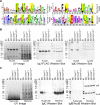
IglD N-terminal domain residues are essential for interaction with IglJ and IglC secretion. (A) Comparison of consensus N-terminal domain sequences of canonical T6SSi TssK, T6SSiii TssK, and Francisella (T6SSii) IglD. Conserved residues contributing to the hydrophobic pocket of T6SSi and T6SSiii TssK and T6SSii IglD are highlighted. (B) Key residues (W8, L14, L19) consistent with TssK-NTD in the IglD structure are shown in stick presentation. Individual protomers of the IglD homotrimer are colored cyan, magenta, and tan. (C) Alanine substitution of IglD NTD residues W8A, L14A, or L19A abolishes the IglJ pulldown of IglD. Stain-free UV image shows comparable lane loading. IglJ-FLAG Western blot shows the capture of IglJ in parental and Ala-substitution strains. IglD Western blot shows pulldown of IglD only in the parental strain. (D) IglD NTD Ala substitution mutants are defective for IglC secretion. Stain-free UV image shows comparable loading of lanes. IglC Western blot shows secretion of IglC into culture supernatant fluid only by the parental strain and comparable levels of IglC in the lysates of all strains. IglB/AccB Western blot shows the absence of leakage of cytosolic proteins IglB or AccB into the culture supernatant fluid.
FIG 6
FIG 6
Alanine substitutions in the C terminus of IglJ disrupt pulldown of IglD and decrease IglC secretion. (A) Comparison of consensus sequences of putative T6SSiii TssG and T6SSii IglJ foot1 and foot2. Putative LG repeat positions of T6SSiii TssG feet (A, top lines) and corresponding sequences conserved in Francisella species (A, bottom lines) are highlighted. (B) Alanine substitution of the three hydrophobic residues of IglJ foot1 (I180, V185, L190) abolishes IglJ pulldown of IglD. Stain-free UV image shows comparable lane loading. IglJ-FLAG Western blot shows the capture of IglJ in parental and Ala-substitution strains. IglD Western blot shows pulldown of IglD in the parental strain and the strain with IglJ foot2 substitutions (V242, E243, L252, L257), but not the strains with alanine substitutions in IgJ foot1 or both foot1 and foot2. (C) Impact of alanine substitutions on IglC secretion. Stain-free UV image shows comparable loading of lanes. IglC Western blot shows that alanine substitution of foot1 markedly decreases IglC secretion, substitution in foot2 even more dramatically decreases IglC secretion, and substitution in both feet abolishes IglC secretion. Western blot of the bacterial lysates shows comparable levels of IglC in all of the strains. IglB/AccB Western blot shows the absence of leakage of cytosolic proteins IglB or AccB into the culture supernatant fluid.
FIG 7
FIG 7
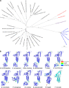
Phylogenetic relationships of IglD and TssK as inferred by using the Maximum Likelihood method and JTT matrix-based model (37). (A) T6SSi are shown in black; T6SSii (Francisella) are shown in blue, and T6SSiii are shown in red. The tree with the highest log likelihood (-29351.96) is shown. The percentage of trees in which the associated taxa clustered together is shown next to the branches. Initial tree(s) for the heuristic search were obtained automatically by applying Neighbor-Join and BioNJ algorithms to a matrix of pairwise distances estimated using the JTT model, and then selecting the topology with a superior log likelihood value. The tree is drawn to scale, with branch lengths measured in the number of substitutions per site. This analysis involved 38 amino acid sequences. Evolutionary analyses were conducted in MEGA11 (39). (B) AlphaFold2 prediction results for TssK/IglD from representative species in (A). The source organism is labeled below each structure. The AlphaFold2 prediction models are colored according to pLDDT confidence. The last one is the Fn IglD structure solved in this study.
FIG 8
FIG 8
Schematic illustration of Fn T6SS baseplate assembly. (A) IglD trimer (colored green), IglJ (magenta), and IglH (light blue) are components of the Fn T6SS baseplate. IglJ interacts with two IglD homotrimers via foot1 and foot2. (B) The baseplate docks to the DotU-PdpB-IglE membrane complex (orange) and further recruits the tail complex (dark blue and dark green). The question mark within the baseplate indicates the unknown component(s). In particular, the Francisella sheath initiator protein, i.e., the TssE ortholog, has not been identified. OM, outer membrane; IM, inner membrane.

References

    1. Oyston PC, Sjostedt A, Titball RW. 2004. Tularaemia: bioterrorism defence renews interest in Francisella tularensis. Nat Rev Microbiol 2:967–978. doi:10.1038/nrmicro1045. - DOI - PubMed
    1. Ellis J, Oyston PC, Green M, Titball RW. 2002. Tularemia. Clin Microbiol Rev 15:631–646. doi:10.1128/CMR.15.4.631-646.2002. - DOI - PMC - PubMed
    1. Telford SR, 3rd, Goethert HK. 2020. Ecology of Francisella tularensis. Annu Rev Entomol 65:351–372. doi:10.1146/annurev-ento-011019-025134. - DOI - PMC - PubMed
    1. Saslaw S, Eigelsbach HT, Prior JA, Wilson HE, Carhart S. 1961. Tularemia vaccine study. II. Respiratory challenge. Arch Intern Med 107:702–714. doi:10.1001/archinte.1961.03620050068007. - DOI - PubMed
    1. Saslaw S, Eigelsbach HT, Wilson HE, Prior JA, Carhart S. 1961. Tularemia vaccine study. I. Intracutaneous challenge. Arch Intern Med 107:689–701. doi:10.1001/archinte.1961.03620050055006. - DOI - PubMed

Publication types

MeSH terms

LinkOut - more resources