. 2022 Aug 2;63(9):32.
doi: 10.1167/iovs.63.9.32.

Topical Glaucoma Therapy Is Associated With Alterations of the Ocular Surface Microbiome

Affiliations

Topical Glaucoma Therapy Is Associated With Alterations of the Ocular Surface Microbiome

Chih-Chiun J Chang et al. Invest Ophthalmol Vis Sci. .

Abstract

Purpose: To investigate the ocular surface microbiome of patients with unilateral or asymmetric glaucoma being treated with topical ophthalmic medications in one eye and to determine whether microbial community changes were related to measures of ocular surface disease.

Methods: V3-V4 16S rRNA sequencing was conducted on ocular surface swabs collected from both eyes of 17 subjects: 10 patients with asymmetric/unilateral glaucoma using topical glaucoma therapy on only one eye and seven age-matched, healthy controls with no history of ocular disease or eyedrop use. Samples were categorized into three groups: patients' glaucomatous eye treated with eyedrops, patients' contralateral eye without eyedrops, and healthy control eyes. Comparisons were made for microbial diversity and composition, with differences in composition tested for association with ocular surface disease measures including tear meniscus height, tear break-up time, and Dry Eye Questionnaire.

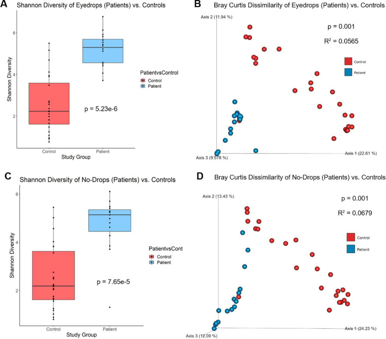
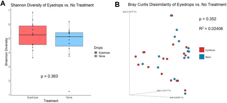
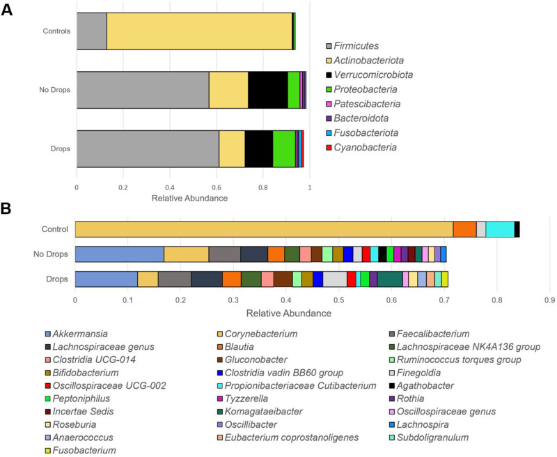
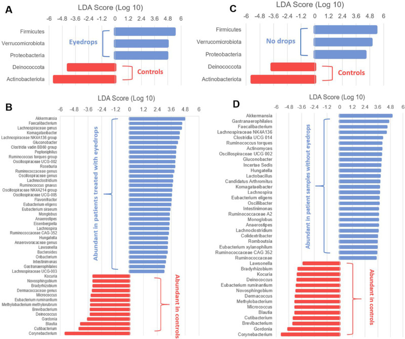
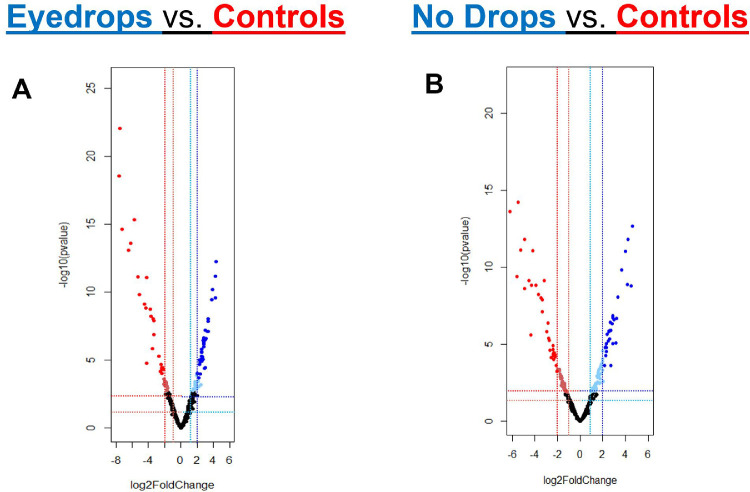
Results: Samples obtained from the patients' treated and untreated eyes both had significantly greater alpha-diversity and relative abundance of gram-negative organisms compared to healthy controls. The microbial composition of patient eyes was associated with decreased tear meniscus height and tear break-up time, whereas metagenomic predictions, based on 16S rRNA data, suggested increased synthesis of lipopolysaccharide.

Conclusions: The ocular surface microbiome of patients taking unilateral preserved glaucoma drops is characterized by a highly diverse array of gram-negative bacteria that is significantly different from the predominantly gram-positive microbes detected on healthy control eyes. These compositional differences were associated with decreased tear film measures and distinct inferred protein synthesis pathways, suggesting a potential link between microbial alterations and ocular surface inflammation.


Conflict of interest statement

Disclosure: C.-C.J. Chang, None; K. Somohano, None; C. Zemsky, None; A.-C. Uhlemann, None; J. Liebmann, None; G.A. Cioffi, None; L.A. Al-Aswad, None; S.V. Lynch, Siolta Therapeutics, Inc. (E, I, C); B.J. Winn, None

Figures

Figure 1.
Figure 1.
Alpha and Beta Diversity for patient eyes treated with eyedrops and no-drops compared to healthy control eyes. (A) Patient eyes treated with eyedrops (n = 16) exhibited significantly greater alpha-diversity measures compared to healthy control eyes (n = 24) in Shannon diversity (P = 5.23e-6, Kruskal-Wallis). There were also significant differences in microbial composition, demonstrated by principal coordinate analysis of beta diversity, between patient eye samples with eyedrops compared to controls. (B) Differences were seen in beta-diversity based on Bray Curtis distances (P = 0.001, R2 = 0.0565, PERMANOVA). (C) Patient eyes without eyedrops (n = 15) also exhibited significantly greater alpha-diversity measures compared to healthy controls (n = 24) in Shannon diversity (P = 7.65e-5, Kruskal-Wallis). There were significant differences in microbial composition between patient eye samples without eyedrops compared to controls. (D) Differences were seen in beta-diversity based on Bray Curtis distances (P = 0.001, R2 = 0.0679, PERMANOVA). The axis values in the beta-diversity plot are the percentage of variance of phylogenetic beta diversity.
Figure 2.
Figure 2.
Alpha and Beta Diversity for patient eyes treated with eyedrops (“Eyedrops”) and contralateral eyes without eyedrops (“No Treatment”). Patients were defined as study participants with a diagnosis of unilateral glaucoma, receiving eyedrop treatment in the glaucomatous eye (“eyedrops,” treated) but not the contralateral eye (“no treatment,” untreated). (A) There were no differences in alpha-diversity measures between the treated (n = 16) and untreated eye samples (n = 15) in Shannon diversity (P = 0.363, Kruskal-Wallis). There were no significant differences in microbiome composition between patient eyes treated with drops compared with untreated eyes, as no differences were seen in beta-diversity based on Bray Curtis distances (P = 0.352, R2 = 0.02406, PERMANOVA). The axis values in beta-diversity plots are the percentage of variance of phylogenetic beta diversity.
Figure 3.
Figure 3.
Taxonomy bar plot of microbial composition in control and patient (“no drops” and “drops”) eye samples. Patient samples are divided into “Drops” for samples obtained from eyes receiving topical glaucoma medications and “No Drops” for samples obtained from contralateral eyes not receiving topical eyedrops. Only species with relative abundance >1.0% were represented in the figure and considered in pairwise comparisons. Relative abundance values for each individual sample were first calculated by dividing the number of read counts assigned to the amplicon sequence variants corresponding to individual microbes by the total read count of the sample. The relative abundance values represented in the figure are averages across all samples of the respective category (“Controls,” “No Drops,” “Drops”). (A) Composition of organisms based on relative abundance at the phyla level. Patient samples contained higher abundance of Firmicutes, Verrucomicrobiota, Proteobacteria, Patescibacteria, Bacteroidota, Fusobacteriota, and Cyanobacteria (P < 0.05, LDA > 2) compared to control samples. Control samples contained higher abundance of Actinobacteriota (P < 0.05, LDA > 2). (B) Composition of organisms based on relative abundance at the genus level. Patient samples had higher abundance of Akkermansia, Faecalibacterium, Lachnospiraceae genus, Lachnospiraceae NK4A136 group, Clostridia UCG-014, Gluconobacter, Ruminococcus torques group, Bifidobacterium, Clostridia vadin B860 group, Oscillospiraceae UCG-002, Peptoniphilus, and Rothia compared to control samples (P < 0.05, LDA > 2). Control samples had increased abundance in Corynebacterium and Propionibacteriaceae Cutibacterium (P < 0.05, LDA > 2). No differences in relative abundance were found between eyes treated with drops and untreated patient eye samples at both the phyla (A) and genus (B) level. LDA - linear discriminant analysis, statistical test used after performing centered log-ratio (CLR) transformation of raw read counts.
Figure 4.
Figure 4.
Linear discriminant analysis (LDA) effect size (LEfSe) plot of taxonomic biomarkers identified in the ocular surface microbiome of patients and controls. LDA scores are effect size estimates for a particular taxonomic marker in a specific group, with values interpreted as the relative magnitude of abundance compared to the other group. Specific bacterial sequence variants at the phyla (A, C) and genus (B, D) level were sorted based on patient eyes treated with eyedrops (A, B) and patient eyes without treatment (C, D) with healthy control samples used as the comparison group. Patient eyes treated with eyedrops (A) and untreated patient eyes (C) had greater abundance of Firmicutes, Verrucomicrobiota, and Proteobacteria and lower abundance of Deinococcota and Actinobacteriota compared to controls. The relatively abundant bacterial sequence variants at the genus level are labelled for eye samples with drops (B) and without drops (D) in blue and controls in red (B, D). All LDA scores and bacterial sequence variants presented are statistically significant, with P < 0.05 (Kruskal-Wallis and pairwise Wilcoxon test) and LDA score ≥ 2.0 the criteria for meeting significance.
Figure 5.
Figure 5.
Volcano Plots for ASVs. Sequence variants (each individual point) and their log2foldchange are plotted comparing (A) patient eye samples receiving eyedrop treatment compared to controls and (B) patient eye samples without eyedrop treatment compared to controls. The log2fold change values are outputs based on DeSEQ2 workflow. Sequence variants found to be relatively abundant with P < 0.05 and log2foldchange > 1.0 are denoted in light blue (relatively abundant in patients) and light red (relatively abundant in controls). Sequence variants found to be relatively abundant with P < 0.01 and log2foldchange > 2.0 are denoted in blue (relatively abundant in patients) and red (relatively abundant in controls). The sequence variants and taxonomic assignments associated with individual data points are noted in Supplemental Tables S1, S2.

References

    1. Liu SA, Zhao ZN, Sun NN, Han Y, Chen J, Fan ZG.. Transitions of the understanding and definition of primary glaucoma. Chin Med J. 2018; 131: 2852–2859. - PMC - PubMed
    1. Halpern DL, Grosskreutz CL.. Glaucomatous optic neuropathy: mechanisms of disease. Ophthalmol Clin North Am. 2002; 15(1): 61–68. - PubMed
    1. Dunn N, Mullee M, Perry H, Holmes C.. Association between dementia and infectious disease: evidence from a case-control study. Alzheimer Dis Assoc Dis. 2005; 19: 91–94. - PubMed
    1. Tanito M, Kaidzu S, Takai Y, Ohira A.. Association between systemic oxidative stress and visual field damage in open-angle glaucoma. Sci Rep. 2016; 6: 25792. - PMC - PubMed
    1. Jaenen N, Baudouin C, Pouliquen P, Manni G, Figueiredo A, Zeyen T.. Ocular symptoms and signs with preserved and preservative-free glaucoma medications. Eur J Ophthalmol. 2007; 17: 341–349. - PubMed

Publication types

MeSH terms