. 2022 Aug 29;16(8):e0010659.
doi: 10.1371/journal.pntd.0010659. eCollection 2022 Aug.

Highly specific and sensitive detection of Burkholderia pseudomallei genomic DNA by CRISPR-Cas12a

Affiliations

Highly specific and sensitive detection of Burkholderia pseudomallei genomic DNA by CRISPR-Cas12a

Somsakul Pop Wongpalee et al. PLoS Negl Trop Dis. .

Abstract

Detection of Burkholderia pseudomallei, a causative bacterium for melioidosis, remains a challenging undertaking due to long assay time, laboratory requirements, and the lack of specificity and sensitivity of many current assays. In this study, we are presenting a novel method that circumvents those issues by utilizing CRISPR-Cas12a coupled with isothermal amplification to identify B. pseudomallei DNA from clinical isolates. Through in silico search for conserved CRISPR-Cas12a target sites, we engineered the CRISPR-Cas12a to contain a highly specific spacer to B. pseudomallei, named crBP34. The crBP34-based detection assay can detect as few as 40 copies of B. pseudomallei genomic DNA while discriminating against other tested common pathogens. When coupled with a lateral flow dipstick, the assay readout can be simply performed without the loss of sensitivity and does not require expensive equipment. This crBP34-based detection assay provides high sensitivity, specificity and simple detection method for B. pseudomallei DNA. Direct use of this assay on clinical samples may require further optimization as these samples are complexed with high level of human DNA.


Conflict of interest statement

The authors have declared that no competing interests exist.

Figures
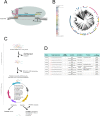
Fig 1
Fig 1. Designing B. pseudomallei-specific crRNA.
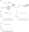
(A) A schematic representation of CRISPR-Cas12a recognizing its target DNA. In forming a CRISPR-Cas12a RNP complex, Cas12a protein (grey lobes) needs to interact with crRNA. The crRNA consists of two essential elements—a repeat (pink dots) and a spacer (blue dots). The repeat is a structural scaffold of the crRNA, required for crRNA-Cas12a interaction, while the spacer is used for target recognition. An extra 3’ extension (green dots) of the RNA is added in this study (see main text). Recognition of CRISPR-Cas12a RNP complex to its target DNA is specified by base-pairing interaction between a 20-nt spacer of crRNA and a template strand of the target DNA (dark grey dots). PAM (TTTV; orange dots), however, is a prerequisite for this interaction. The target recognition leads to cleavage of the DNA at the PAM-distal sites (black arrow heads) and trans cleavage. (B) A phylogeny of global B. pseudomallei population (n = 3,341) collected between years of 1935 and 2018 (see Materials and Methods). The outer ring is colored coded by major lineages denoted as the Burkholderia Bayesian cluster (bb) 1 to 22. Blue points located in the inner ring highlight 30 representative genomes used for CRISPR-Cas12a target scanning in Fig 1C. (C) A schematic representation of the bioinformatic analysis used for identifying B. pseudomallei-specific CRISPR-Cas12a target sites. The analysis consists of 2 steps—CRISPR selection and CRISPR filtering. In the CRISPR selection, common candidate target sites are generated from 30 representative B. pseudomallei genomes found in Fig 1B. These candidates are then filtered through 1,071 genomes of possible cross-reactive pathogens during the CRISPR filtration step. Candidate target sites that are unique or contain significant mismatches are selected as final candidates. (D) A table showing details of eight CRISPR-Cas12a target sites (patent pending) identified from Fig 1C. Target sequences are shown, consisting of a 20-nucleotide spacer, preceded by PAM (red text). Percentage coverage denotes occurrence of each final candidate in the B. pseudomallei global genome collection (n = 3,341). Annotation is based on that of the K96243 strain (assembly GCF_000011545.1).
Fig 2
Fig 2. Specific detection of B. pseudomallei genomic DNA by the designed CRISPR-Cas12a.
(A) A schematic representation of DETECTR assay. First, a region containing the CRISPR-Cas12a target site in the pathogen’s genomic DNA is pre-amplified by flanking primers (magenta arrows) in an isothermal amplification reaction called RPA. This amplified DNA is then combined with the pre-assembled CRISPR-Cas12a RNP complex in a CRISPR reaction that contains FAM-Quencher ssDNA probes (F-Q). Recognition of CRISPR RNP to its target self-activates its trans cleavage (collateral cleavage) activity. This results in cleavage of the FAM fluorophore from its quencher, hence the generation of a fluorescent signal, which can be detected by fluorometer. (B, C, D) Collateral cleavage activity of crBP34, crBP36 and crBP38, respectively. Each CRISPR-Cas12a RNP complex was tested against genomic DNA from a human cell line and 11 other pathogens, including B. thailandensis and B. psedomallei. ~10,000 copies of genomic DNA were used in 40-μl RPA reactions, of which 3 μl were used in 100-μl CRISPR reactions. Fluorescent signals were collected every three minutes. Signals from CRISPR reactions that contained water-input RPA were used for background subtraction. Each data point represents an average from six CRISPR reactions derived from two independent sets of RPA reactions. Note that some data points sporadically become slightly negative after background subtraction, causing them to be invisible on the scale. Standard deviation is also plotted. a.u.: arbitrary units.
Fig 3
Fig 3. crBP34-DETECTR shows comparable sensitivity to real-time PCR.
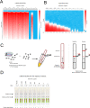
(A) A heatmap showing fluorescent signals generated by crBP34-DETECTR assay. Known copy numbers of B. pseudomallei genomic DNA ranging from 40,000–0 copies were used in 50-μl RPA reactions, of which 15 μl were used in 100-μl CRISPR reactions. Fluorescent signals were collected at three-minute intervals. The heatmap denotes fluorescence at indicated times compared to fluorescence at 0 minutes (F/F0). For each copy number, two independent RPA reactions were performed; each was subjected to three replicates of CRISPR reactions (total of six CRISPR reactions). A scale bar is shown on the right. (B) A heatmap showing fluorescent signals generated by real-time PCR assay. Known copy numbers of B. pseudomallei genomic DNA ranging from 40,000–0 copies were used in 20-μl PCR reactions. The heatmap denotes fluorescent signals (ΔRn) as a function of the PCR cycle. Datasets from three replicates are presented. A scale bar is shown on the right. A black arrowhead indicates a threshold value that is used for calculating threshold cycles (Ct), presented under the heatmap. (C) A schematic representation of a lateral flow dipstick readout. RPA and CRISPR reactions were performed similar to Fig 2A, except that FAM-Quencher probes are replaced with FAM-Biotin probes (F-B). Note that RNP assembly is done simultaneously in the CRISPR reaction. Following this, a sample pad of a dipstick is directly immerged into the CRISPR reaction to allow labeling of the probes by anti-FAM antibodies and diffusion into the dipstick. Positive detection is indicated by the presence of an intensified band at the anti-IgG line. The streptavidin line serves as an internal control (see main text). (D) Lateral flow dipstick readout retains sensitivity of the crBP34-DETECTR assay. Known copy numbers of B. pseudomallei genomic DNA ranging from 40,000–0 copies were used in 30-μl RPA reactions, of which 5 μl were used in 50-μl CRISPR reactions that contained FAM-Biotin probes. Dipsticks were used as a readout of the assay, as explained in c). Two sets of RPA reactions were performed. Visual interpretation was done by comparing the intensity of a positive band to its background at 0 copy genomic DNA.
Fig 4
Fig 4. crBP34 specifically detects clinically isolated B. pseudomallei.
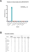
(A) A graph displaying fluorescent activation (F20/F0) values from crBP34-DETECTR assay. 1 ng of genomic DNA from each bacterial isolate was used in 30-μl RPA reactions, of which 5 μl were used in 100-μl CRISPR reactions. Fluorescent signals were collected at time points 0 and 20 minutes and used for calculating F20/F0 values. Each dot represents a single replicate from each isolate. (B) A table detailing statistical data of Fig 4A.

Similar articles

Cited by

References

    1. Gassiep I, Armstrong M, Norton R. Human Melioidosis. Clin Microbiol Rev. 2020;33: e00006–19. doi: 10.1128/CMR.00006-19 - DOI - PMC - PubMed
    1. Limmathurotsakul D, Golding N, Dance DAB, Messina JP, Pigott DM, Moyes CL, et al.. Predicted global distribution of Burkholderia pseudomallei and burden of melioidosis. Nat Microbiol. 2016;1: 15008. doi: 10.1038/nmicrobiol.2015.8 - DOI - PubMed
    1. Wiersinga WJ, Currie BJ, Peacock SJ. Melioidosis. New England Journal of Medicine. 2012;367: 1035–1044. doi: 10.1056/NEJMra1204699 - DOI - PubMed
    1. Chaowagul W, White NJ, Dance DA, Wattanagoon Y, Naigowit P, Davis TM, et al.. Melioidosis: a major cause of community-acquired septicemia in northeastern Thailand. J Infect Dis. 1989;159: 890–899. doi: 10.1093/infdis/159.5.890 - DOI - PubMed
    1. Simpson AJ, Suputtamongkol Y, Smith MD, Angus BJ, Rajanuwong A, Wuthiekanun V, et al.. Comparison of imipenem and ceftazidime as therapy for severe melioidosis. Clin Infect Dis. 1999;29: 381–387. doi: 10.1086/520219 - DOI - PubMed

Publication types