. 2022 Aug 29;17(8):e0273509.
doi: 10.1371/journal.pone.0273509. eCollection 2022.

Chemical and enzymatic modifications of 5-methylcytosine at the intersection of DNA damage, repair, and epigenetic reprogramming

Affiliations

Chemical and enzymatic modifications of 5-methylcytosine at the intersection of DNA damage, repair, and epigenetic reprogramming

Tuvshintugs Baljinnyam et al. PLoS One. .

Abstract

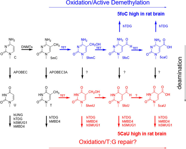
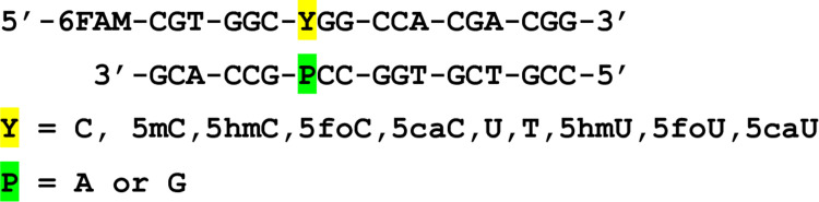
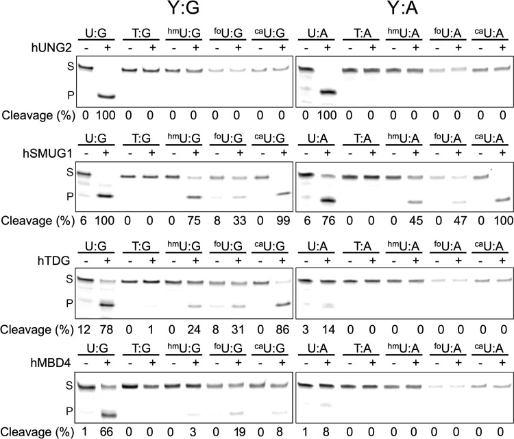
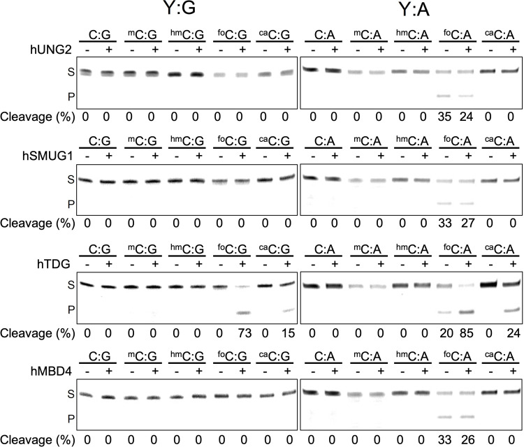
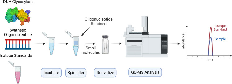
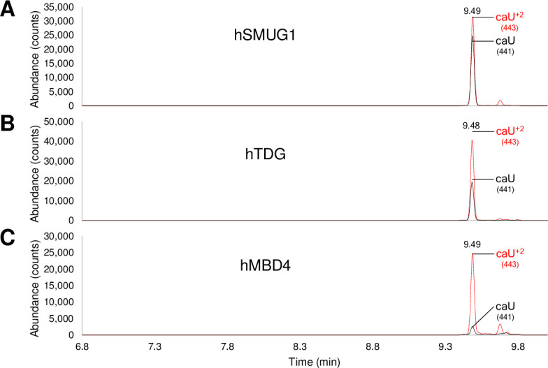
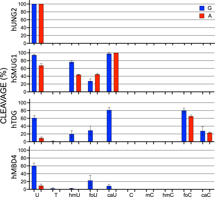
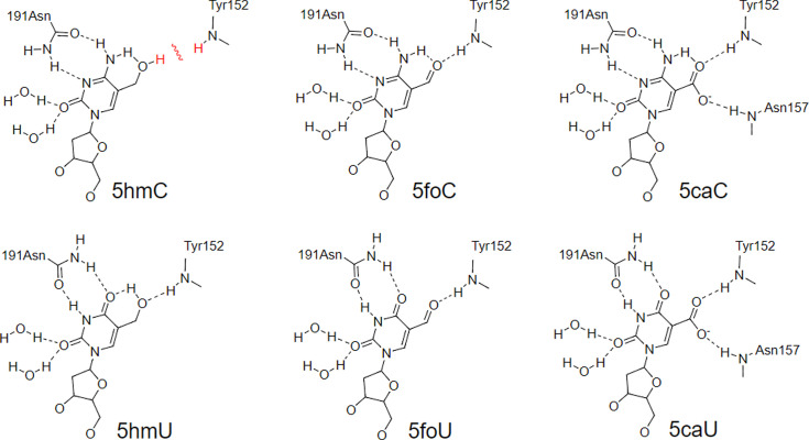
The DNA of all living organisms is persistently damaged by endogenous reactions including deamination and oxidation. Such damage, if not repaired correctly, can result in mutations that drive tumor development. In addition to chemical damage, recent studies have established that DNA bases can be enzymatically modified, generating many of the same modified bases. Irrespective of the mechanism of formation, modified bases can alter DNA-protein interactions and therefore modulate epigenetic control of gene transcription. The simultaneous presence of both chemically and enzymatically modified bases in DNA suggests a potential intersection, or collision, between DNA repair and epigenetic reprogramming. In this paper, we have prepared defined sequence oligonucleotides containing the complete set of oxidized and deaminated bases that could arise from 5-methylcytosine. We have probed these substrates with human glycosylases implicated in DNA repair and epigenetic reprogramming. New observations reported here include: SMUG1 excises 5-carboxyuracil (5caU) when paired with A or G. Both TDG and MBD4 cleave 5-formyluracil and 5caU when mispaired with G. Further, TDG not only removes 5-formylcytosine and 5-carboxycytosine when paired with G, but also when mispaired with A. Surprisingly, 5caU is one of the best substrates for human TDG, SMUG1 and MBD4, and a much better substrate than T. The data presented here introduces some unexpected findings that pose new questions on the interactions between endogenous DNA damage, repair, and epigenetic reprogramming pathways.


Conflict of interest statement

The authors have declared that no competing interests exist.

Figures

Fig 1
Fig 1. Possible active demethylation pathways resulting from the modification of 5mC.
The oxidation of 5mC to 5foC and 5caC, and the removal of 5foC and 5caC by hTDG (blue) have been established. The glycosylase removal of 5hmU, 5foU and 5caU is established here (red). However, the enzymatic conversion of T to 5hmU, 5foU and 5caU (red) remains controversial.
Fig 2
Fig 2. Sequence of the oligonucleotide duplex used in this study.
See Fig 1 for structures of the pyrimidine analogs, ‘Y’.
Fig 3
Fig 3
The excision specificity of human uracil family glycosylases for a series of 5-substituted uracil analogs (Y) paired with G (left) or A (right). FAM-labelled oligonucleotide duplexes (2.5 pmol) in buffer appropriate for each enzyme were incubated with a glycosylase at 37°C for 1 h. Duplexes paired with G were then incubated with hyTDG-lyase (Y163K) (65°C, 1 h) and those paired with A incubated with NaOH solution (96°C, 10 min) to cleave the abasic site. Oligonucleotides were resolved on a 20% polyacrylamide/urea gel, visualized on a STORM imager, and quantified with “Image quant” software. The amount of cleavage is shown below the lanes in each gel (S = substrate, P = product). Some artifact cleavage of 5foU-containing oligonucleotides is observed due to its thermal lability. Replicate gel images are shown in S1 and S2 Figs in S1 File.
Fig 4
Fig 4
The excision specificity of human uracil family glycosylases for a series of 5-substituted cytosine analogs (Y) paired with G (left) or A (right). FAM-labelled oligonucleotide duplexes (2.5 pmol) in buffer appropriate for each enzyme were incubated with a glycosylase at 37°C for 1 h. Duplexes paired with G were then incubated with hyTDG-lyase (Y163K) (65°C, 1 h) and those paired with A incubated with NaOH solution (96°C, 10 min) to cleave the abasic site. Oligonucleotides were resolved on a 20% polyacrylamide/urea gel, visualized on a STORM imager, and quantified with “Image quant” software. The amount of cleavage is shown below the lanes in each gel (S = substrate, P = product). Some artifact cleavage of 5foC-containing oligonucleotides is observed due to its alkaline lability. Replicate gel images are shown in S3 and S4 Fig in S1 File.
Fig 5
Fig 5. GC-MS scheme.
This workflow illustrates the GC-MS based glycosylase assay to determine excision and base release from a synthetic oligonucleotide using stable-isotope standards.
Fig 6
Fig 6. Confirmation of 5caU removal measured by GC-MS.
A) 5caU:G treated with hSMUG1, B) 5caU:G treated with hTDG, and C) 5caU:G treated with hMBD4. A mixture of duplex oligo, containing a 5caU:G mispair (110 pmol) with a 2-fold excess of the complementary strand, and a 5caU+2 isotope labeled standard (55 pmol) was prepared in buffer: 10 mM potassium phosphate pH 7.7, 30 mM sodium chloride and 40 mM potassium chloride. The oligonucleotides and free base standard were incubated with approximately 100 pmol of either hSMUG1, hTDG or hMBD4 at 37°C for 2 h in a 110 μL reaction volume. Free bases were separated from enzymes and oligonucleotides by spin filtration, 90% of the sample was dried and analyzed by GC-MS. Derivatized free bases were separated by GC and identified by MS with selected ion monitoring. The ions for 5caU released by glycosylases (441 m/z, black) and 5caU+2 isotope enriched standard (443 m/z, red) were monitored, and the relative sizes of the integrated peak areas were used to calculate the amount of 5caU released. There was approximately 39, 24 and, 5 pmoles 5caU released by hSMUG1, hTDG and hMBD4, respectively. The overall removal of 5caU when paired opposite G by human glycosylases was hSMUG1 > hTDG > hMBD4, consistent with the trend seen by gel-based analysis of excision.
Fig 7
Fig 7. Summary for human glycosylase specificity for substrate.
U, T, 5hmU, 5foU, 5caU, C, 5mC, 5hmC, 5foC and 5caC (mispaired with G (blue) or A (red)) oligonucleotides were treated with indicated enzymes for 1 hour at 37°C, and subsequently treated with hyTDG-lyase (Y163K) or NaOH to break the DNA backbone at the abasic site, mixed with an equal volume of formamide, and separated in 20% polyacrylamide denaturing gel. Gels were visualized using a STORM gel imager. Data presented as average value ± SD (n = 3).
Fig 8
Fig 8. Potential hydrogen bonding interactions in the catalytic pocket of hTDG that allows recognition of 5foC, 5caC, 5hmU, 5foU, and 5caU, but not 5hmC.

Similar articles

Cited by

References

    1. Lindahl T, Nyberg B. Rate of Depurination of Native Deoxyribonucleic Acid. Biochemistry. 1972;11: 3610–3618. doi: 10.1021/bi00769a018 - DOI - PubMed
    1. Lindahl T, Andersson A. Rate of Chain Breakage at Apurinic Sites Double-Stranded Deoxyribonucleic Acid. Biochemistry. 1972;11: 3618–3623. doi: 10.1021/bi00769a019 - DOI - PubMed
    1. Lindahl T, Nyberg B. Heat-induced deamination of cytosine residues in DNA. Biochemistry. 1974;13: 3405–3410. - PubMed
    1. Lindahl T. My Journey to DNA Repair. Genomics, Proteomics Bioinforma. 2013;11: 2–7. doi: 10.1016/j.gpb.2012.12.001 - DOI - PMC - PubMed
    1. ImLay J, Linn S. DNA Damage and Oxygen Radical Toxicity. Science (80-). 1988;240: 1302–1309. doi: 10.1126/science.3287616 - DOI - PubMed

Publication types