

Cell cycle control of kinetochore assembly
- PMID: 36037227
- PMCID: PMC9427032
- DOI: 10.1080/19491034.2022.2115246
Cell cycle control of kinetochore assembly
Abstract
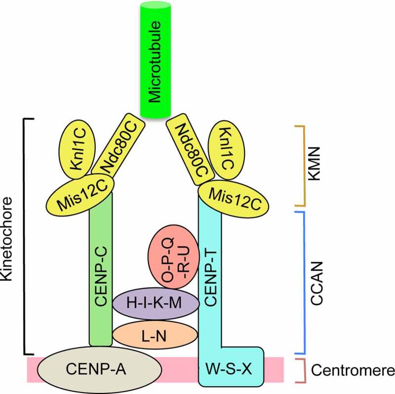
The kinetochore is a large proteinaceous structure assembled on the centromeres of chromosomes. The complex machinery links chromosomes to the mitotic spindle and is essential for accurate chromosome segregation during cell division. The kinetochore is composed of two submodules: the inner and outer kinetochore. The inner kinetochore is assembled on centromeric chromatin and persists with centromeres throughout the cell cycle. The outer kinetochore attaches microtubules to the inner kinetochore, and assembles only during mitosis. The review focuses on recent advances in our understanding of the mechanisms governing the proper assembly of the outer kinetochore during mitosis and highlights open questions for future investigation.
Keywords: CCAN; CENP-A; CENP-C; CENP-T; KMN; Kinetochore assembly; cell cycle; centromere; phosphorylation.
Conflict of interest statement
No potential conflict of interest was reported by the author(s).
Figures
Similar articles
-
Structure of the inner kinetochore CCAN complex assembled onto a centromeric nucleosome.Nature. 2019 Oct;574(7777):278-282. doi: 10.1038/s41586-019-1609-1. Epub 2019 Oct 2. Nature. 2019. PMID: 31578520 Free PMC article.
-
Where is the right path heading from the centromere to spindle microtubules?Cell Cycle. 2019 Jun;18(11):1199-1211. doi: 10.1080/15384101.2019.1617008. Epub 2019 May 20. Cell Cycle. 2019. PMID: 31075048 Free PMC article. Review.
-
Phosphorylation of CENP-R by Aurora B regulates kinetochore-microtubule attachment for accurate chromosome segregation.J Mol Cell Biol. 2022 Sep 27;14(7):mjac051. doi: 10.1093/jmcb/mjac051. J Mol Cell Biol. 2022. PMID: 36069839 Free PMC article.
-
MEL-28/ELYS and CENP-C coordinately control outer kinetochore assembly and meiotic chromosome-microtubule interactions.Curr Biol. 2022 Jun 6;32(11):2563-2571.e4. doi: 10.1016/j.cub.2022.04.046. Epub 2022 May 23. Curr Biol. 2022. PMID: 35609608 Free PMC article.
-
An updated view of the kinetochore architecture.Trends Genet. 2023 Dec;39(12):941-953. doi: 10.1016/j.tig.2023.09.003. Epub 2023 Sep 27. Trends Genet. 2023. PMID: 37775394 Review.
Cited by
-
Emerging Role of Deuterium/Protium Disbalance in Cell Cycle and Apoptosis.Int J Mol Sci. 2023 Feb 4;24(4):3107. doi: 10.3390/ijms24043107. Int J Mol Sci. 2023. PMID: 36834518 Free PMC article. Review.
-
High-resolution analysis of human centromeric chromatin.Life Sci Alliance. 2025 Jan 23;8(4):e202402819. doi: 10.26508/lsa.202402819. Print 2025 Apr. Life Sci Alliance. 2025. PMID: 39848706 Free PMC article.
-
A role for the kinetochore protein, NUF2, in ribosome biogenesis.Mol Biol Cell. 2025 Feb 1;36(2):ar16. doi: 10.1091/mbc.E24-08-0337. Epub 2024 Dec 20. Mol Biol Cell. 2025. PMID: 39705402 Free PMC article.
-
Nonessential kinetochore proteins contribute to meiotic chromosome condensation through polo-like kinase.Mol Biol Cell. 2025 Feb 1;36(2):ar14. doi: 10.1091/mbc.E24-08-0348. Epub 2024 Dec 20. Mol Biol Cell. 2025. PMID: 39705398 Free PMC article.
-
Tetraploidy in normal tissues and diseases: mechanisms and consequences.Chromosoma. 2025 Mar 21;134(1):3. doi: 10.1007/s00412-025-00829-1. Chromosoma. 2025. PMID: 40117022 Free PMC article. Review.
References
-
- Sharp LW. Introduction to cytology. New York: McGraw-Hill Book Company, inc; 1934.
-
- Cheeseman IM, Desai A. Molecular architecture of the kinetochore-microtubule interface. Nat Rev Mol Cell Biol. 2008;9(1):33–46. - PubMed
-
- Cleveland DW, Mao Y, Sullivan KF. Centromeres and kinetochores: from epigenetics to mitotic checkpoint signaling. Cell. 2003;112(4):407–421. - PubMed
Publication types
MeSH terms
Grants and funding
LinkOut - more resources
Full Text Sources