. 2022 Sep 6;119(36):e2122170119.
doi: 10.1073/pnas.2122170119. Epub 2022 Aug 29.

False-positive IRESes from Hoxa9 and other genes resulting from errors in mammalian 5' UTR annotations

Affiliations

False-positive IRESes from Hoxa9 and other genes resulting from errors in mammalian 5' UTR annotations

Christina Akirtava et al. Proc Natl Acad Sci U S A. .

Abstract

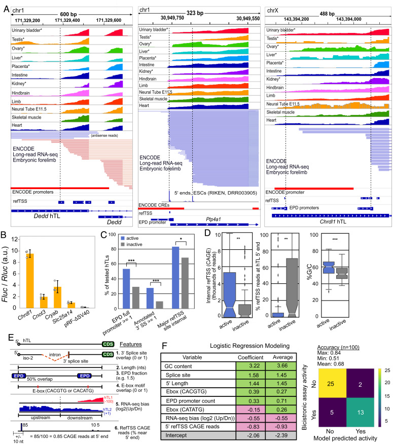
Hyperconserved genomic sequences have great promise for understanding core biological processes. It has been recently proposed that scores of hyperconserved 5' untranslated regions (UTRs), also known as transcript leaders (hTLs), encode internal ribosome entry sites (IRESes) that drive cap-independent translation, in part, via interactions with ribosome expansion segments. However, the direct functional significance of such interactions has not yet been definitively demonstrated. We provide evidence that the putative IRESes previously reported in Hox gene hTLs are rarely included in transcript leaders. Instead, these regions function independently as transcriptional promoters. In addition, we find the proposed RNA structure of the putative Hoxa9 IRES is not conserved. Instead, sequences previously shown to be essential for putative IRES activity encode a hyperconserved transcription factor binding site (E-box) that contributes to its promoter activity and is bound by several transcription factors, including USF1 and USF2. Similar E-box sequences enhance the promoter activities of other putative Hoxa gene IRESes. Moreover, we provide evidence that the vast majority of hTLs with putative IRES activity overlap transcriptional promoters, enhancers, and 3' splice sites that are most likely responsible for their reported IRES activities. These results argue strongly against recently reported widespread IRES-like activities from hTLs and contradict proposed interactions between ribosomal expansion segment ES9S and putative IRESes. Furthermore, our work underscores the importance of accurate transcript annotations, controls in bicistronic reporter assays, and the power of synthesizing publicly available data from multiple sources.

Keywords: Hox; IRES; UTR; bicistronic; translation.


Conflict of interest statement

The authors declare no competing interest.

Figures

Fig. 1.
Fig. 1.
The proposed structure of the putative Hoxa9 IRES element is not conserved. (A) Rainbow graphs depict the probability of base pairing for the Hoxa9 IRES-like region from mouse (Top), human (Middle), and zebrafish (Bottom). Pairing regions (stems) are numbered as in ref. . Base pairing probabilities were determined using RNAstructure. The mouse model is highly consistent with the published model (3). Red brackets indicate the frequency of P3a and P4 helix formation in 10,000 predicted suboptimal structures. Human and mouse Hoxa9 share P1 and P4, but lack P3, which was reported to be essential for IRES activity (3). Zebrafish Hoxa9 does not share any structural similarity with mammalian homologs, despite driving bicistronic reporter activity (3). (B) Secondary structure model of mouse Hoxa9 putative IRES region (3). Corresponding zebrafish sequences are shown in red. Most proposed base pairs are not conserved. Zebrafish has insertions (asterisks) and deletions (Ns) in the critical P3 and P4 elements. (C) Results of R-scape analysis of mutual information for the mouse Hoxa9 putative IRES region using alignments from 208 mammals and 23 other vertebrates. The number of covarying sites (y axis) is given for different e-value cutoffs (x axis). Although the alignment has the power to detect ∼10 compensatory pairs (red point; Dataset S1), covarying base pairs are less common than expected by chance in the IRES-like element (blue line).
Fig. 2.
Fig. 2.
The putative extended 5′ UTR and IRES regions of mouse Hoxa9 are not expressed at biologically meaningful levels. A genome browser view showing Refseq annotations of the mouse Hoxa10/Mir196b/Hoxa9 in red, green, and blue, respectively, is shown. The putative TL(5′ UTR) and IRES regions are shown in pink. Promoters from the EPD and the ENCODE project are shown above the Refseq gene models. Illumina short-read (Upper) and PacBio full-length (Lower) RNA-seq data from the ENCODE consortium show negligible levels of RNA over the putative 5′ UTR and IRES regions. Similarly, two 5′ CAGE-seq studies [Ivanov et al. (56) and Abugessaisa et al. (53)] show no TSSs at the putative 5′ UTR, and a strong TSS peak downstream of the putative IRES. ChIP-seq peaks show RNAPII is found immediately downstream of EPD Hoxa9 and Hoxa10 promoters in mouse embryonic forelimbs [Eng et al. (29)]. PacBio RNA-seq detects Hoxa9/a10 and Hoxa9/Mir196b fusion transcripts. Regions corresponding to the PCR amplicons used previously to detect (RT-PCR) the putative Hoxa9 IRES and quantify (RT-qPCR) Hoxa9 mRNA are shown in red. The Hoxa9 RT-qPCR amplicon used for expression and polysome analysis is not specific to spliced Hoxa9 mRNA, and can amplify fusion transcripts, unspliced transcripts, and truncated transcripts initiating at refTSS annotated start sites within Hoxa9 introns.
Fig. 3.
Fig. 3.
The putative IRES-like domains of Hoxa9 and other Hox genes encode functional promoters. (A) The putative Hoxa9 IRES is a promoter. The SV40 promoter was deleted from the pRF bicistronic vector. Putative IRES regions were cloned between Renilla luciferase (Rluc) and Fluc and tested for activity in C3H10T1/2 cells. Bar graphs show the Fluc to Rluc ratio indicating promoter activity from mouse and human Hoxa9 regions. The extended UTR and IRES-like regions function as independent promoters in the forward orientation. (B) Putative IRES-like regions from other mouse Hoxa genes function as promoters. Annotated transcript leaders from each Hoxa gene were tested as in A. TLs containing putative IRES-like elements drove expression, while non-IRES TLs had background expression levels. Error bars show 95% CIs with n = 3.
Fig. 4.
Fig. 4.
Putative IRES-like elements in Hoxa genes contain functional E-boxes recognized by USF2. (A) Genome browser view of CUT&RUN sequencing data shows the binding location of human USF2 (37). The dashed box shows the location of a hyperconserved E-box motif (also see SI Appendix, Fig. S7). (B) Sequence alignment from diverse representative vertebrate genomes upstream of mouse Hoxa9, including the EPD promoter region. The diagram includes 5′ CAGE-seq data (53). The location of P4 domain sequences and the nonconserved G-rich ES9S interaction site are shown above, while the hyperconserved E-box and CAAT-box are noted below the alignment. The CAAT box and a TATA-like element are noted on the annotated Hoxa9 transcript leader in light blue and magenta, respectively (SI Appendix, Fig. S8). (C) The Fluc and Rluc reporter genes were moved to two independent plasmids to test the functions of E-box elements. (D) Mutation of the E-box motif from mouse and human Hoxa9 decreased expression in C3H10T1/2 cells. The siRNA codepletion of USF1 and USF2 decreased expression of wild-type (Welch’s one-tail t test, *P < 0.05; **P < 0.006), but not E-box mutant, reporters (n.s. signifies “not significant”). (E) Deletion of E-boxes from putative IRESes of Hoxa3, Hoxa5, Hoxa7, and Hoxa11 decrease promoter activity. Single E-box mutations show slight increases in Hoxa7 promoter activity, while the double mutation eliminated promoter function. Error bars show 95% CIs with n = 3.
Fig. 5.
Fig. 5.
Putative IRES-like hTLs can be explained by promoters and 3′ splice sites due to 5′ UTR annotation errors. (A) Examples of promoter overlap commonly seen in putative IRES-like hTLs. Short-read (Upper) and long-read (Lower) RNA-seq data show transcription often initiates internally, coinciding with annotated promoters (ENCODE and EPD) and TSSs (refTSS). (B) The hTLs from four putative mouse IRESes have promoter activity in pRF-ΔSV40 transfected C3H10T1/2 cells. Error bars show 95% CIs with n = 3. (C) The hTLs with putative IRES-like activity are enriched in EPD promoters, 3′ splice sites, and major internal TSS sites (Χ2 tests). (D) IRES-active hTLs have significantly more internal CAGE 5′ reads, a lower fraction of TSS reads at annotated 5′ ends, and higher G/C content than IRES-inactive hTLs (Wilcoxon rank-sum tests). (E) Features for logistic regression modeling. RNA-seq bias is the ratio of reads in upstream and downstream hTL halves across GWIPs-viz RNA-seq datasets. refTSS CAGE reads are the percentage of 5′ end reads mapped near the annotated TSS (data from ref. 28). (F) Logistic regression modeling of IRES-like and non-IRES hTLs. Features associated with internal promoters guanine-cytosine (GC content, EPD promoter count fraction, E-boxes) and splice sites are positively correlated with bicistronic reporter expression, while features associated with full-length TLs (CAGE reads at annotated 5′ ends and RNA-seq 5′ end bias) are negatively correlated with bicistronic reporter activity. One hundred models were generated, with an average accuracy of 68%; *, **, and *** denote P < 0.05, 0.01, and 0.001, respectively.

References

    1. Hinnebusch A. G., Ivanov I. P., Sonenberg N., Translational control by 5′-untranslated regions of eukaryotic mRNAs. Science 352, 1413–1416 (2016). - PMC - PubMed
    1. Pelletier J., Sonenberg N., The organizing principles of eukaryotic ribosome recruitment. Annu. Rev. Biochem. 88, 307–335 (2019). - PubMed
    1. Xue S., et al. , RNA regulons in Hox 5′ UTRs confer ribosome specificity to gene regulation. Nature 517, 33–38 (2015). - PMC - PubMed
    1. Leppek K., et al. , Gene- and species-specific Hox mRNA translation by ribosome expansion segments. Mol. Cell 80, 980–995.e13 (2020). - PMC - PubMed
    1. Leppek K., Byeon G. W., Fujii K., Barna M., VELCRO-IP RNA-seq reveals ribosome expansion segment function in translation genome-wide. Cell Rep. 34, 108629 (2021). - PMC - PubMed

Publication types

LinkOut - more resources