. 2022;14(6):1295-1310.
doi: 10.1016/j.jcmgh.2022.08.008. Epub 2022 Aug 28.

Culture-Associated DNA Methylation Changes Impact on Cellular Function of Human Intestinal Organoids

Affiliations

Culture-Associated DNA Methylation Changes Impact on Cellular Function of Human Intestinal Organoids

Rachel D Edgar et al. Cell Mol Gastroenterol Hepatol. 2022.

Abstract

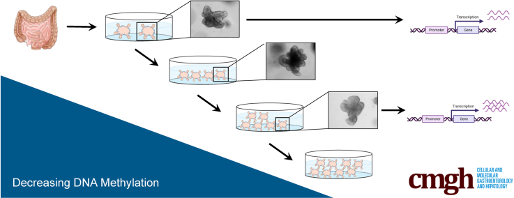
Background & aims: Human intestinal epithelial organoids (IEOs) are a powerful tool to model major aspects of intestinal development, health, and diseases because patient-derived cultures retain many features found in vivo. A necessary aspect of the organoid model is the requirement to expand cultures in vitro through several rounds of passaging. This is of concern because the passaging of cells has been shown to affect cell morphology, ploidy, and function.

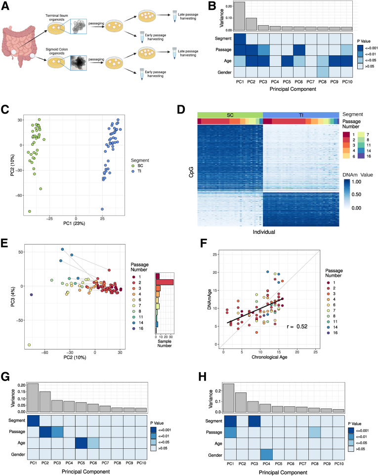
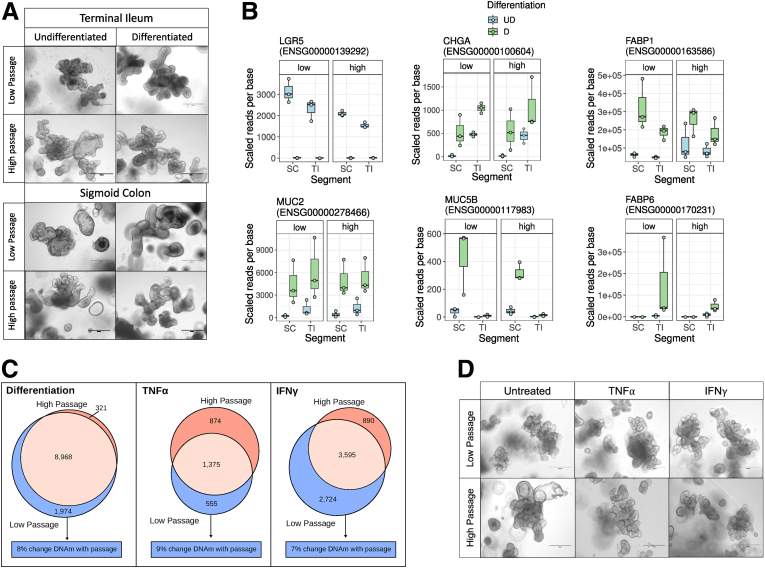
Methods: Here, we analyzed 173 human IEO lines derived from the small and large bowel and examined the effect of culture duration on DNA methylation (DNAm). Furthermore, we tested the potential impact of DNAm changes on gene expression and cellular function.

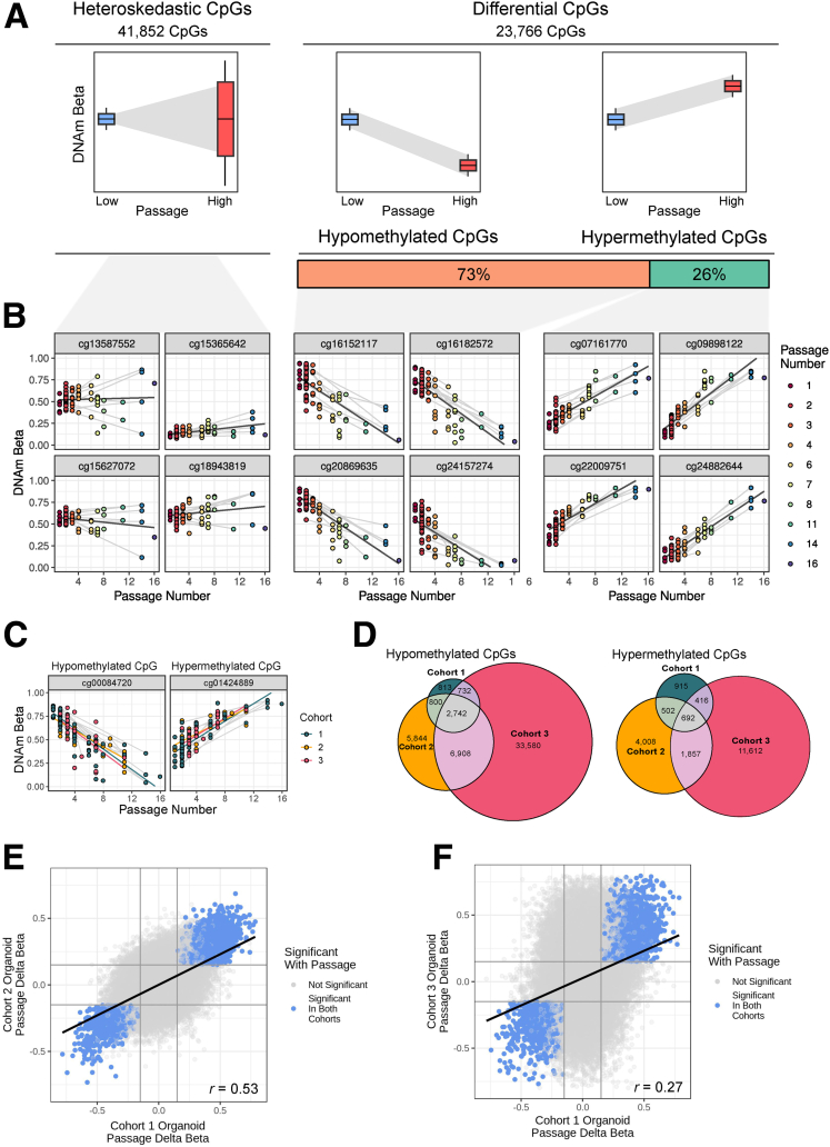
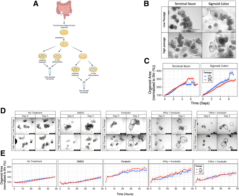
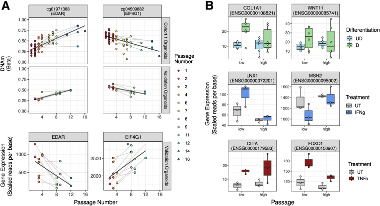
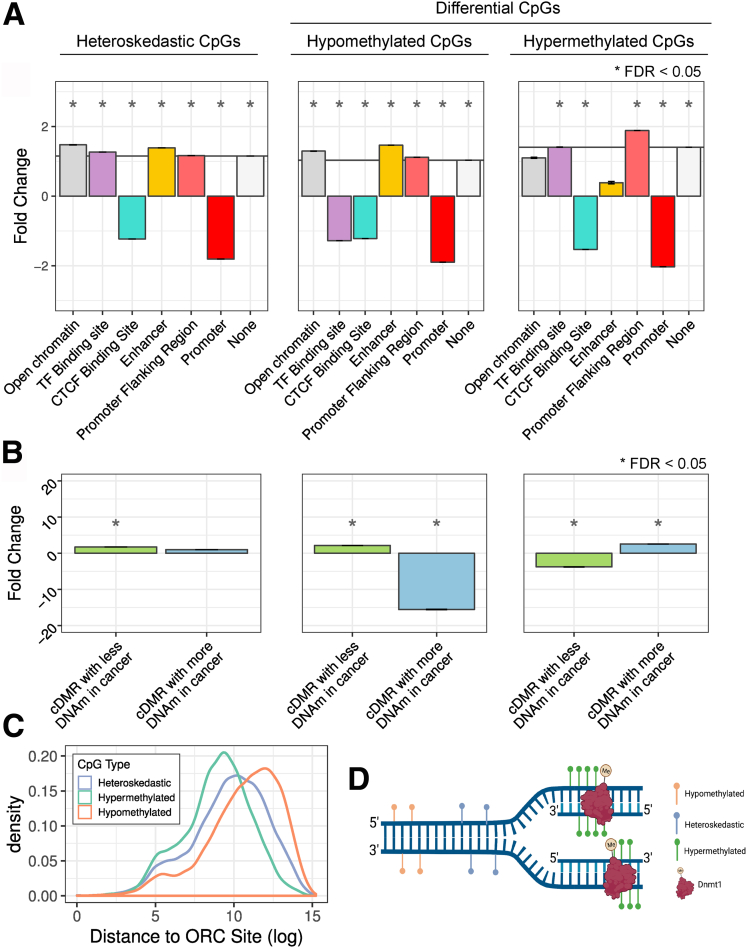
Results: Our analyses show a reproducible effect of culture duration on DNAm in a large discovery cohort as well as 2 publicly available validation cohorts generated in different laboratories. Although methylation changes were seen in only approximately 8% of tested cytosine-phosphate-guanine dinucleotides (CpGs) and global cellular function remained stable, a subset of methylation changes correlated with altered gene expression at baseline as well as in response to inflammatory cytokine exposure and withdrawal of Wnt agonists. Importantly, epigenetic changes were found to be enriched in genomic regions associated with colonic cancer and distant to the site of replication, indicating similarities to malignant transformation.

Conclusions: Our study shows distinct culture-associated epigenetic changes in mucosa-derived human IEOs, some of which appear to impact gene transcriptomic and cellular function. These findings highlight the need for future studies in this area and the importance of considering passage number as a potentially confounding factor.

Keywords: Culture Conditions; Epigenetics; Intestinal Epithelium; Organoid.


Figures

None
Graphical abstract
Figure 1
Figure 1
Sampling site of origin and IEO passage number are associated with the main components of DNAm variation. (A) Outline of study design. (B) Scree plot showing DNAm variance accounted for by each PC and association with sample variables. P values were generated with a Spearman correlation for continuous variables or an analysis of variance for categoric variables. (C) PC1 and PC2 of IEO genome-wide DNAm profiles (cohort 1). (D) Heatmap showing DNAm of the top 500 CpGs differentially DNAm between small- and large-bowel IEOs. (E) PC2 and PC3 of IEO DNAm profiles. Samples are colored by passage number. Lines connect samples derived from the same patient profiled at different passages. (F) The association between chronological and epigenetic age is shown, with points colored by the passage of the IEO. As in panel B, variance was accounted for by PCs in validation cohorts. (G) Cohort 2. (H) Cohort 3. SC, sigmoid colon.
Figure 2
Figure 2
Long-term culture effects on IEO DNAm are validated in independent cohorts. (A) Schematic of the 3 types of passage-associated DNAm changes at individual CpGs and the proportion of passage CpG split by type of change in DNAm. (B) Representative CpGs with DNAm significantly associated with passage. Samples are colored by passage number and grey lines connect samples derived from the same patient. Regression lines between passage and DNAm are shown in black. (C) Representative CpGs with significant differential DNAm associated with passage in all 3 cohorts. Samples are colored by cohort and grey lines connect samples derived from the same patient. Regression lines between passage and DNAm are shown separately for each cohort. (D) Overlap of CpGs in cohorts 1–3 with significant differential DNAm associated with passage. (E and F) Direction of effect is consistent between cohorts. The delta betas from 2 cohorts are shown as points with CpGs significantly associated with passage highlighted.
Figure 3
Figure 3
High- and low-passage IEOs are functionally similar. (A) Outline of experimental design. (B) Bright-field images of TI- and sigmoid colon–derived IEOs at low and high passage taken by the EVOS FL system (Life Technologies). Scale bars: 300 μm. (C) Comparison of growth curves between low- and high-passage TI- and SC-derived IEOs (n = 2, 6 technical replicates per each biological replicate). (D) Representative images of TI organoids in standard medium Non-treated (NT), vehicle control medium (DMSO), cultured with forskolin IFNγ+forskolin or TNFα+forskolin taken with by Incucyte. Scale bars: 800 μm. (E) Comparison of organoid size between early and late-passage organoids after proinflammatory cytokines and forskolin treatments (n = 2, 4 technical replicates per each biological replicate).
Figure 4
Figure 4
High- and low-passage IEOs are similar upon differentiation and proinflammatory cytokine stimulation. (A) High- and low-passage IEOs look similar before and after differentiation. Bright-field images of TI- and sigmoid colon (SC)-derived IEOs after in vitro differentiation, respectively, taken by the EVOS FL system (Life Technologies). Scale bars: 300 μm. Outline of experimental design. (B) Gene expression of selected differentiation markers in IEOs by gut segment and passage (high or low). (C) Venn diagram illustrating overlap of differentially expressed genes in high- and low-passage IEOs upon differentiation and co-culture with IFNγ or TNFα. (D) Bright-field images of TI-derived IEOs at low and high passage after proinflammatory cytokine treatment (TNFα or IFNγ), taken by the EVOS FL system (Life Technologies). Scale bars: 300 μm. D, Differentiated; UD, Undifferentiated.
Figure 5
Figure 5
Genes are differentially expressed with IEO passage. (A) Representative CpGs and genes showing DNAm and mRNA expression associated significantly with passage in cohort 1 and a validation cohort. Samples are colored by passage number and grey lines connect samples derived from the same patient. Regression lines between passage and DNAm/expression are in black. (B) mRNA expression of representative genes showing passage-dependent differences in response to in vitro differentiation, IFNγ, or TNFα. D, Differentiated; UD, Undifferentiated; UT, Untreated.
Figure 6
Figure 6
Passage affects DNAm in specific regions of the genome. (A and B) Enrichment of passage-associated CpGs across genomic regions expressed as fold change relative to the total number of CpGs present on the EPIC array. Standard error bars indicate mean fold change for the error across 1000 random samplings. (C) Distance of passage-associated CpGs from origin replication complex (ORC) sites. (D) Schematic showing location of passage-associated DNAm changes relative to the origins of replication.

References

    1. Yui S., Nakamura T., Sato T., Nemoto Y., Mizutani T., Zheng X., Ichinose S., Nagaishi T., Okamoto R., Tsuchiya K., Clevers H., Watanabe M. Functional engraftment of colon epithelium expanded in vitro from a single adult Lgr5 + stem cell. Nat Med. 2012;18:618–623. - PubMed
    1. Matano M., Date S., Shimokawa M., Takano A., Fujii M., Ohta Y., Watanabe T., Kanai T., Sato T. Modeling colorectal cancer using CRISPR-Cas9–mediated engineering of human intestinal organoids. Nat Med. 2015;21:256–262. - PubMed
    1. Broutier L., Mastrogiovanni G., Verstegen M.M., Francies H.E., Gavarró L.M., Bradshaw C.R., Allen G.E., Arnes-Benito R., Sidorova O., Gaspersz M.P., Georgakopoulos N., Koo B.-K., Dietmann S., Davies S.E., Praseedom R.K., Lieshout R., IJzermans J.N.M., Wigmore S.J., Saeb-Parsy K., Garnett M.J., van der Laan L.J., Huch M. Human primary liver cancer–derived organoid cultures for disease modeling and drug screening. Nat Med. 2017;23:1424–1435. - PMC - PubMed
    1. Berkers G., van Mourik P., Vonk A.M., Kruisselbrink E., Dekkers J.F., de Winter-de Groot K.M., Arets H.G.M., Marck-van der Wilt R.E.P., Dijkema J.S., Vanderschuren M.M., Houwen R.H.J., Heijerman H.G.M., van de Graaf E.A., Elias S.G., Majoor C.J., Koppelman G.H., Roukema J., Bakker M., Janssens H.M., van der Meer R., Vries R.G.J., Clevers H.C., de Jonge H.R., Beekman J.M., van der Ent C.K. Rectal organoids enable personalized treatment of cystic fibrosis. Cell Rep. 2019;26:1701–1708.e3. - PubMed
    1. Driehuis E., Hoeck A van, Moore K., Kolders S., Francies H.E., Gulersonmez M.C., Stigter E.C.A., Burgering B., Geurts V., Gracanin A., Bounova G., Morsink F.H., Vries R., Boj S., Es J van, Offerhaus G.J.A., Kranenburg O., Garnett M.J., Wessels L., Cuppen E., Brosens L.A.A., Clevers H. Pancreatic cancer organoids recapitulate disease and allow personalized drug screening. Proc Natl Acad Sci U S A. 2019;116:26580–26590. - PMC - PubMed

Publication types