. 2022 Sep 27;16(9):14792-14806.
doi: 10.1021/acsnano.2c05647. Epub 2022 Aug 29.

Engineering Lipid Nanoparticles for Enhanced Intracellular Delivery of mRNA through Inhalation

Affiliations

Engineering Lipid Nanoparticles for Enhanced Intracellular Delivery of mRNA through Inhalation

Jeonghwan Kim et al. ACS Nano. .

Abstract

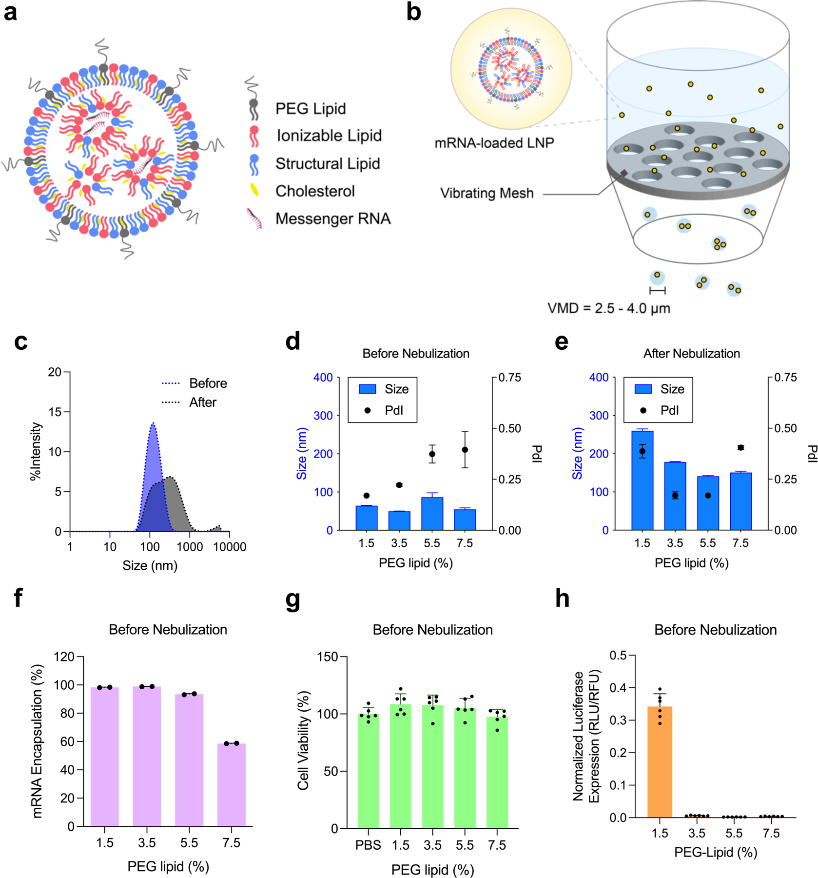
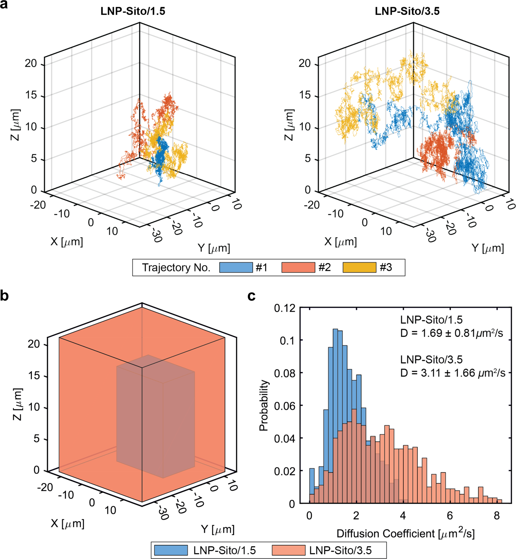
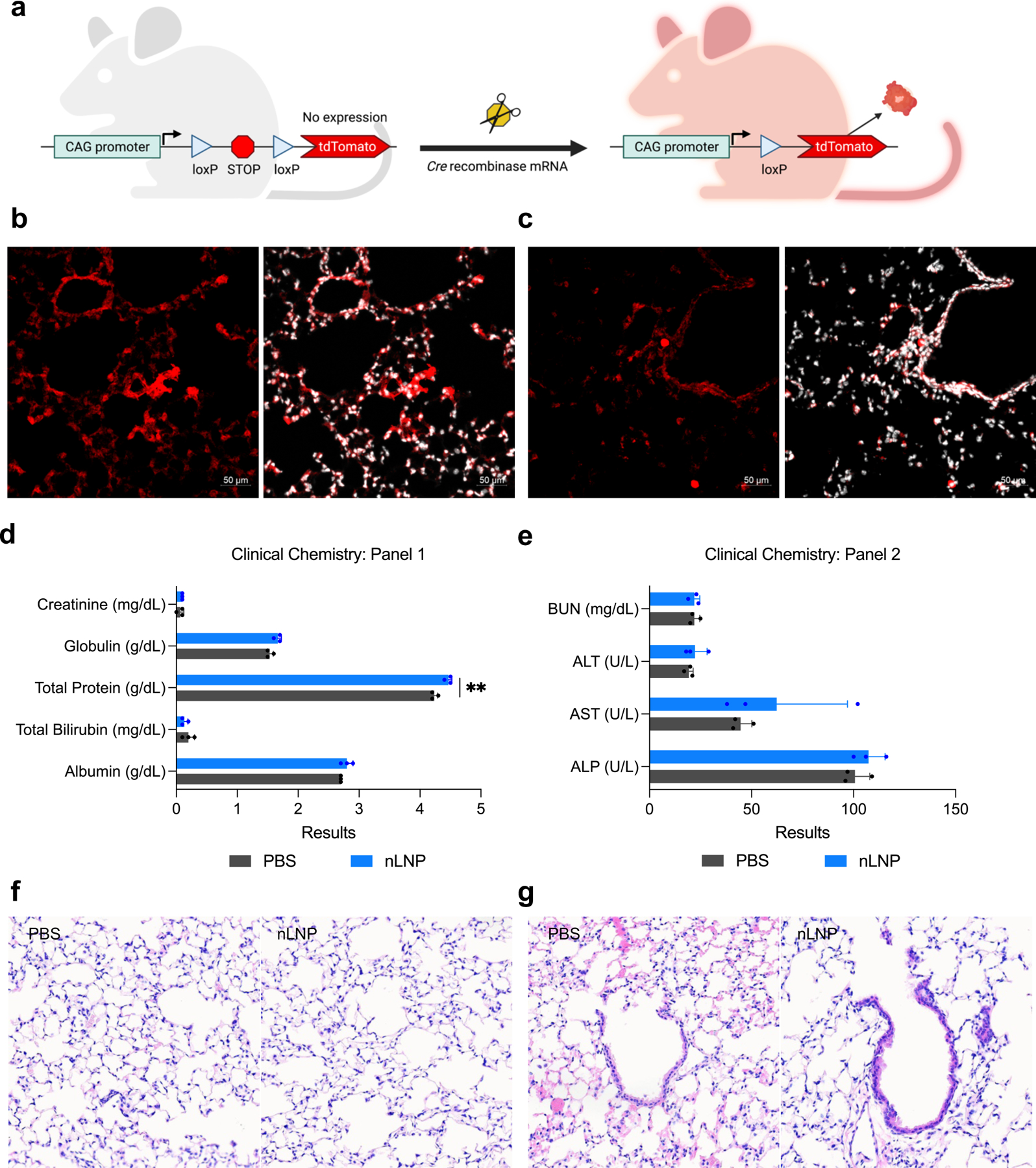
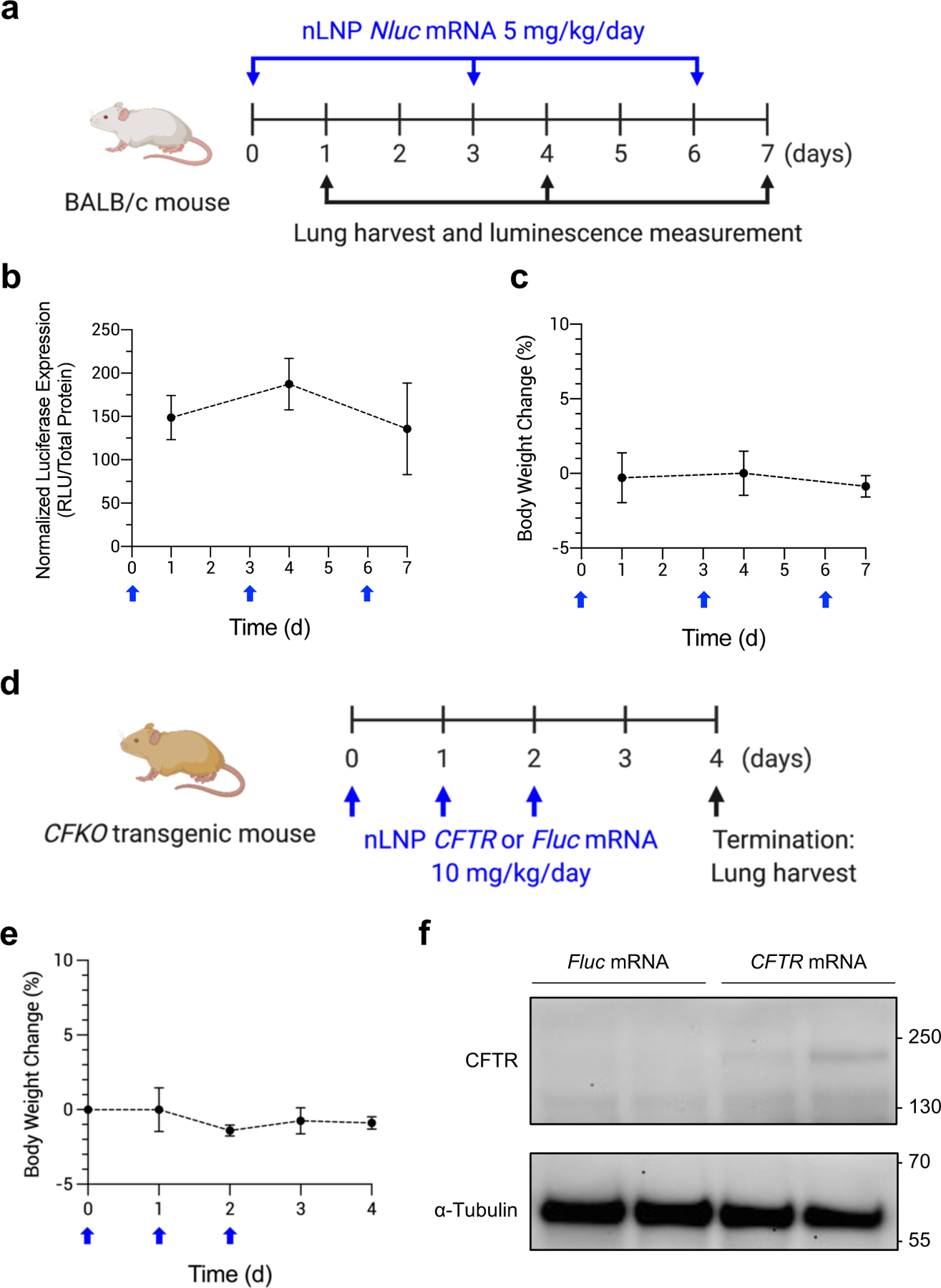
Despite lipid nanoparticles' (LNPs) success in the effective and safe delivery of mRNA vaccines, an inhalation-based mRNA therapy for lung diseases remains challenging. LNPs tend to disintegrate due to shear stress during aerosolization, leading to ineffective delivery. Therefore, LNPs need to remain stable through the process of nebulization and mucus penetration, yet labile enough for endosomal escape. To meet these opposing needs, we utilized PEG lipid to enhance the surficial stability of LNPs with the inclusion of a cholesterol analog, β-sitosterol, to improve endosomal escape. Increased PEG concentrations in LNPs enhanced the shear resistance and mucus penetration, while β-sitosterol provided LNPs with a polyhedral shape, facilitating endosomal escape. The optimized LNPs exhibited a uniform particle distribution, a polyhedral morphology, and a rapid mucosal diffusion with enhanced gene transfection. Inhaled LNPs led to localized protein production in the mouse lung without pulmonary or systemic toxicity. Repeated administration of these LNPs led to sustained protein production in the lungs. Lastly, mRNA encoding the cystic fibrosis transmembrane conductance regulator (CFTR) was delivered after nebulization to a CFTR-deficient animal model, resulting in the pulmonary expression of this therapeutic protein. This study demonstrated the rational design approach for clinical translation of inhalable LNP-based mRNA therapies.

Keywords: cystic fibrosis; inhalation; lung delivery; mRNA therapy; nebulization; pulmonary delivery; β-sitosterol.


Conflict of interest statement

The authors declare the following competing financial interest(s): G.S. is an inventor in patent application US20200129445A1 that details LNP-Sito. G.S and J.K have submitted a US provisional patent application (No. 63/225,766) related to this work. G.S. is a co-founder of EnterX Bio and RNAvax Bio, and has an advisory role to Saliogen Therapeutics Inc., Rare Air Inc., Circ Bio Inc., and Sanofi.

Figures

Figure 1.
Figure 1.
Nebulization process influence physicochemical properties of LNP. (a) A schematic representation of mRNA-loaded LNP. Ionizable lipid (red), cholesterol (yellow), structural lipid (blue), and PEG lipid (gray) form a spherical nanoparticle, encapsulating mRNA inside. (b) A schematic of nebulization process of mRNA-loaded LNP. VMD; volume mean diameter. (c) Size change of Onpattro® LNP formulation during nebulization. Blue; before nebulization, Gray; after nebulization. (d-h) Characterization of LNP-Chol with various PEG lipid contents. (d,e) Size and PdI of LNP-Chol (d) before and (e) after nebulization. (f) mRNA encapsulation of LNP-Chol with various PEG lipid contents. (g) Cell viability and (h) normalized luciferase expression of HeLa cells treated with various LNP-Chol at 50 ng mRNA per well for 24 h. Data were presented in mean ± standard deviation.
Figure 2.
Figure 2.
Enhanced mRNA transfection by substitution of cholesterol to β-sitosterol in LNP. (a,b) Chemical structures of (a) cholesterol and (b) β-sitosterol. β-sitosterol has an additional alkyl group (red) in comparison to cholesterol. (c-f) Physicochemical characterization of LNP-Chol and LNP-Sito containing various amounts of PEG lipid. (c,e) DLS analysis and (d,f) mRNA encapsulation of LNP (c,d) before and (e,f) after nebulization. (g,h) Normalized luciferase expression in HeLa cells transfected with (g) LNP solution and (h) LNP aerosol at 50 ng and 1 μg mRNA per well, respectively. Data were presented in mean ± standard deviation. ***p<0.001, ****p<0.0001; significant analysis by two-way ANOVA with Sidak’s multiple comparison test.
Figure 3.
Figure 3.
Cryogenic transmission electron microscopy (cryoTEM) imaging of LNP-Chol (left) and LNP-Sito (right) containing varying amounts of PEG lipids. Scale bars indicate 50 nm.
Figure 4.
Figure 4.
LNP inhalation produces selective lung transfection. (a) A representative bioluminescent image of isolated organs showing a localized luciferase expression in the lungs. (b) A diagram of mouse lung anatomy showing five lung lobes (left), and ex vivo bioluminescent images of mouse lung lobes 24 h after LNP inhalation (right, n=4). (c) Quantification of total flux in individual lung lobes harvested from BALB/c mouse at 24 h after LNP-Sito/1.5 inhalation at a dose of 100 μg mRNA (n=4). (d) Normalized Nluc expression in mouse lung homogenates harvested from BALB/c mice at 24 h after inhalation of LNP-Chol and LNP-Sito containing Nluc mRNA and various amount of PEG lipid. Data were presented in mean ± standard deviation. ****p<0.001, **p<0.01; significant analysis by two-way ANOVA with Sidak’s multiple comparison test (n=8).
Figure 5.
Figure 5.
3D-SMART was performed for high-speed 3D particle tracking to capture nanoparticle diffusion. (a) Three 40 second-long representative trajectories of LNP-Sito/1.5 and LNP-Sito/3.5. The measured 3D position is plotted at 1 ms temporal resolution. (b) The boundary of the representative trajectories drawn as boxes, demonstrating the dramatically increased travel range of the LNP-Sito/3.5 (orange) compared to LNP-Sito/1.5 (blue). (c) Analysis of trajectories for LNP-Sito/1.5 (blue, n=323) and LNP-Sito/3.5 (orange, n=353), mean ± standard deviation. The two formulations show significant differences in diffusion coefficient (p<0.0001).
Figure 6.
Figure 6.
Inhaled nLNP transfects airway epithelium without causing acute toxicity. (a) A diagram of Cre-lox recombination in Ai9 transgenic Cre reporter mouse that expresses tdTomato when loxP recombination has occurred. (b-c) Representative immunohistochemistry of Ai9 mouse lung sections displaying tdTomato expression after inhalation of nLNP encapsulating Cre mRNA. tdTomato (red) and nuclei (white) were detected in (b) alveolar spaces and (e) airway bronchioles. 20x magnification. Scale bars refer to 50 μm. (d,e) Clinical chemistry test of mouse sera harvested at 24 h after PBS or nLNP inhalation. Data were presented in mean ± standard deviation. **p<0.01, significant analysis by multiple unpaired t-test (n=3). (f,g) Histopathological analysis of (f) alveolar spaces and (g) bronchial epithelium of murine lungs harvested at 24 h after PBS (left) or nLNP (right) inhalation. Hematoxylin and eosin; 20x magnification. Arrows indicate the bronchial epithelium.
Figure 7.
Figure 7.
Repeated inhalation of nLNP to sustain production of therapeutic protein. (a) A dosing regimen for persistent inhalation of nLNP encapsulating Nluc mRNA. nLNP was administered via inhalation every 3 days at a dose of 5 mg/kg/day mRNA (blue arrows). At 24 h after each dose, mouse lungs were collected to measure luciferase expression (black arrows). (b) Normalized luciferase expression in mouse lungs and (c) body weight change of BALB/c after repeat dosing (blue arrow). Data were presented in mean ± standard deviation (n=4–12). (d) A dosing regimen for CFTR mRNA delivery via inhalation. nLNP encapsulating CFTR or Fluc mRNA was administered to CFKO mice daily for 3 d (blue arrows), followed by harvesting lungs to detect CFTR proteins (black arrow). (e) Body weight change of CFKO transgenic mice after repeat dosing (blue arrow). (f) Western blot images after immunoprecipitation using an anti-CFTR antibody. mRNA delivered by nLNP is noted above the images. Upper and lower blots were probed using anti-CFTR and anti-α-Tubulin antibodies, respectively. Approximate molecular weights are marked on the left sides of the images.

References

    1. O’Sullivan BP; Freedman SD Cystic Fibrosis. The Lancet 2009, 373, 1891–1904. 10.1016/S0140-6736(09)60327-5. - DOI - PubMed
    1. De Boeck K; Amaral MD Progress in Therapies for Cystic Fibrosis. Lancet Respir. Med 2016, 4, 662–674. 10.1016/S2213-2600(16)00023-0. - DOI - PubMed
    1. Mukherjee A; MacDonald KD; Kim J; Henderson MI; Eygeris Y; Sahay G Engineered Mutant α-ENaC Subunit MRNA Delivered by Lipid Nanoparticles Reduces Amiloride Currents in Cystic Fibrosis–Based Cell and Mice Models. Sci. Adv 2020, 6, eabc5911. 10.1126/sciadv.abc5911. - DOI - PMC - PubMed
    1. Haque AKMA; Dewerth A; Antony JS; Riethmüller J; Schweizer GR; Weinmann P; Latifi N; Yasar H; Pedemonte N; Sondo E; Weidensee B; Ralhan A; Laval J; Schlegel P; Seitz C; Loretz B; Lehr C-M; Handgretinger R; Kormann MSD Chemically Modified HCFTR MRNAs Recuperate Lung Function in a Mouse Model of Cystic Fibrosis. Sci. Rep 2018, 8, 16776. 10.1038/s41598-018-34960-0. - DOI - PMC - PubMed
    1. Suzuki S; Crane AM; Anirudhan V; Barillà C; Matthias N; Randell SH; Rab A; Sorscher EJ; Kerschner JL; Yin S; Harris A; Mendel M; Kim K; Zhang L; Conway A; Davis BR Highly Efficient Gene Editing of Cystic Fibrosis Patient-Derived Airway Basal Cells Results in Functional CFTR Correction. Mol. Ther 2020, 28, 1684–1695. 10.1016/j.ymthe.2020.04.021. - DOI - PMC - PubMed

Publication types

LinkOut - more resources