Case Reports
. 2022 Aug 29;15(8):e245179.
doi: 10.1136/bcr-2021-245179.

Microgeodic disease in an infant

Affiliations
Case Reports

Microgeodic disease in an infant

Katrina Frances Stranks et al. BMJ Case Rep. .

Abstract

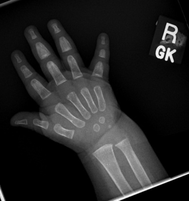
We describe an infant presenting with intermittent discolouration and swelling of her fingers and toes occurring with changes in ambient temperature. Extensive investigations revealed raised inflammatory markers as well as sclerotic lesions within the phalanges and diffuse marrow oedema. Infectious and inflammatory causes were considered and excluded based on the clinical presentation and investigation findings. The persistence of symptoms prompted further investigation with MRI. Correlation of the MRI findings with previous case reports resulted in a diagnosis of microgeodic disease-an uncommon, self-limiting condition thought to be due to cold-induced vasospasm leading to avascular necrosis of the bone.

Keywords: Dermatology; Paediatrics; Primary Care; Rheumatology.


Conflict of interest statement

Competing interests: None declared.

Figures

Figure 1
Figure 1
Right hand at presentation.
Figure 2
Figure 2
Generalised oedema and erythema of the right four fingers extending to the palm at presentation.
Figure 3
Figure 3
X-ray of right hand at presentation demonstrating rarefaction and periosteal reaction in the third, fourth and fifth middle phalanges, distal fourth phalanx, proximal third and fifth phalanges. Associated cortical punched out metaphyseal lesions and swelling of the overlying soft tissues are evident.
Figure 4
Figure 4
Ultrasound image showing hypoechoic thickened periosteum over the fifth proximal phalanx (PP).
Figure 5
Figure 5
Colour Doppler ultrasound image showing hyperaemia of the periosteum of the fifth proximal phalynx (PP).
Figure 6
Figure 6
Coronal T2-weighted fat suppressed MRI of the right hand showing diffuse marrow oedema of the involved phalanges and intramedullary lytic areas with extensive surrounding soft-tissue inflammation.
Figure 7
Figure 7
X-ray of right hand at resolution.

References

    1. Davis A. Microgeodic disease. Christchurch: DermNet NZ; 2013. https://dermnetnz.org/topics/microgeodic-disease/
    1. Maroteaux P. [5 cases of microgeodic disease of phalanges of unknown etiology in infants]. Ann Radiol 1970;13:229–36. - PubMed
    1. Lee RKL, Griffith JF, Read JW, et al. . Phalangeal microgeodic disease: report of two cases and review of imaging. Skeletal Radiol 2013;42:451–5. 10.1007/s00256-012-1561-8 - DOI - PubMed
    1. Prindaville B, Antaya RJ. Chilblains and microgeodic disease diagnosed concurrently in a child’s toe. Pediatr Dermatol 2013;30:269–70. 10.1111/j.1525-1470.2011.01665.x - DOI - PubMed
    1. Yang Y-Z, Ma X-H, Yang X-H, et al. . Phalangeal microgeodic syndrome in childhood. Pediatr Radiol 2018;48:1108–12. 10.1007/s00247-018-4137-5 - DOI - PubMed

Publication types

LinkOut - more resources