Review
. 2022:154:1-69.
doi: 10.1016/bs.ai.2022.07.001. Epub 2022 Aug 22.

Antibody-mediated immunity to SARS-CoV-2 spike

Affiliations
Review

Antibody-mediated immunity to SARS-CoV-2 spike

John M Errico et al. Adv Immunol. 2022.

Abstract

Despite effective spike-based vaccines and monoclonal antibodies, the SARS-CoV-2 pandemic continues more than two and a half years post-onset. Relentless investigation has outlined a causative dynamic between host-derived antibodies and reciprocal viral subversion. Integration of this paradigm into the architecture of next generation antiviral strategies, predicated on a foundational understanding of the virology and immunology of SARS-CoV-2, will be critical for success. This review aims to serve as a primer on the immunity endowed by antibodies targeting SARS-CoV-2 spike protein through a structural perspective. We begin by introducing the structure and function of spike, polyclonal immunity to SARS-CoV-2 spike, and the emergence of major SARS-CoV-2 variants that evade immunity. The remainder of the article comprises an in-depth dissection of all major epitopes on SARS-CoV-2 spike in molecular detail, with emphasis on the origins, neutralizing potency, mechanisms of action, cross-reactivity, and variant resistance of representative monoclonal antibodies to each epitope.

Keywords: Coronavirus cross-reactivity; N-terminal domain antibody; Neutralizing variants of concern; Receptor-binding domain antibody; S2 antibody; SARS-CoV-2 neutralizing antibody; SARS-CoV-2 spike antibody; Spike epitopes.


Conflict of interest statement

Disclosures The authors declare no competing interests.

Figures

Fig. 1
Fig. 1
Overview of SARS-CoV-2 spike structure. (A) Linear diagram showing the domains and subdomains of SARS-CoV-2 spike with residue numbers listed below each segment. Black hexagons indicate the location of N-linked glycans. S1/S2 furin cleavage motif and S2′ dibasic cleavage motif are listed above their respective sites. NTD, N-terminal domain; RBD, receptor binding domain; RBM, receptor binding motif; SD1, subdomain 1; SD2- subdomain 2; FP, fusion peptide; FPPR, fusion peptide proximal region; HR1, heptad repeat 1; CH, central helix; CD, connector domain; SH, stem helix; HR2, heptad repeat 2; TM, transmembrane anchor. (B) Structures of the S1 portion of SARS-CoV-2 spike in both the all “down” configuration (PDB: 6XR8) and the “one up” configuration (PDB: 6VSB). One monomer of the trimer is displayed as a cartoon ribbon, while the other 2 are shown as surface representations. Subdomains of S1 are colored as in (A). N-linked glycans are shown as blue spheres. (C) Close up of the RBD (PDB: 6W41). RBM motif is colored as in (A); N343 glycan shown as ball and stick representation with carbon atoms colored light blue, oxygen atoms red and nitrogen atoms dark blue. Secondary structural elements are labeled as previously described (Lan et al., 2020). (D) Close up of the NTD (PDB: 7B62). The galectin-like fold is shown in gold, with glycans shown in ball and stick representation. Secondary structural elements identified via DSSP are labeled. (E) Pre-fusion structure of S2 (PDBnn6XR8). One monomer of S2 is displayed as a cartoon ribbon and colored according to (A). The remaining two S2 protomers are rendered as gray surfaces. S1 cap is shown as a clipped surface representation and colored according to (A). N-linked glycans are shown as blue spheres. All structural depictions were generated using UCSF ChimeraX (Goddard et al., 2018).
Fig. 2
Fig. 2
RBD substitutions in SARS-CoV-2 variants. SARS-CoV-2 receptor binding domain (RBD) with substitutions from SARS-CoV-2 variants shaded red. Black outline indicates human ACE2 residues contact that define the RBM (Lan et al., 2020). Labels indicate amino acid changes and variants encoding each change using WHO nomenclature (α, B.1.1.7; β, B.1.351; ɣ, P.1; δ, B.1.617.2; ο.1, B.1.1.529.1/BA.1; ο.2, B.1.1.529.2/BA.2; ο, Both BA.1 and BA.2).
Fig. 3
Fig. 3
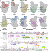
Epitopes on the RBD. (A) RBD with epitopes from prototypical antibodies representing each class or antigenic site depicted as shaded region on RBD surface. Labels indicate specific strands and secondary structure elements involved in each epitope, depicted as cartoon ribbons underneath transparent RBD surface. (B) Multiple sequence alignment of SARS-CoV-2 RBD residues 333–527 for wild-type Wuhan-Hu-1 and variant strains as well as SARS-CoV; dots indicate fully conserved residues relative to Wuhan-Hu-1 reference strain. SARS-COV-2 RBD Secondary structure is diagrammed above Wuhan-Hu-1 sequence as described (Lan et al., 2020). Representative antibodies with epitope class (Barnes et al., 2020) and antigenic site (Piccoli et al., 2020) indicated in parentheses have their RBD contacts highlighted over the variant RBD sequences and are unrelated to the variant sequence they are overlaid on. Half-shaded residues indicate binding by antibodies from both colors. Yellow triangles for S2M11 indicate quaternary contacts on an adjacent RBD. RBM motif is underlined; hACE2 contacts are identified with stars (Lan et al., 2020). Branch icon above N343 indicates N-linked glycan. RBD depictions use PDB: 6M0J. Epitope contacts were identified by buried surface area measurement of atomic models (CB6, PDB: 7C01; S2E12, PDB: 7R6X; 2B04, PDB: 7K9I; S2M11, PDB: 7K43; S309, PDB: 7R6W; REGN10987, PDB: 6XDG; S2X35, PDB: 7R6W; CR3022, PDB: 6W41; COVA1–16, PDB: 7JMW; S2H97, PDB: 7M7W) using UCSF ChimeraX with a probe radius of 1.4 Å and the default cutoff of 1.0 Å2 (Goddard et al., 2018).
Fig. 4
Fig. 4
Structure of the NTD. SARS-CoV-2 N-terminal domain (NTD) with loops N1 through N5 shaded green and glycans shown as light blue blobs. The NTD is otherwise colored yellow. On the right, the RBD is shown in pink, with the remainder of the spike colored gray.
Fig. 5
Fig. 5
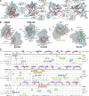
Epitopes of the NTD. (A) NTD with mutations from SARS-CoV-2 variants shaded red. Labels indicate amino acid changes and variants encoding each change using WHO nomenclature (α, B.1.1.7; β, B.1.351; ɣ, P.1; δ, B.1.617.2; ο.1, B.1.1.529.1/BA.1; ο.2, B.1.1.529.2/BA.2; ο, Both BA.1 and BA.2). A green tripod designates the tetrapyrrole-binding site. (B) NTD with epitopes from antibodies representing each antigenic site depicted as shaded region on NTD surface. Labels indicate secondary structures or specific loops, designated by name (N1 through N5) or flanking beta-strands, depicted as cartoon ribbons underneath transparent NTD surface. (C) Multiple sequence alignment of SARS-CoV-2 NTD residues 14–307 for wild-type Wuhan-Hu-1 and variant strains as well as SARS-CoV. Dots indicate fully conserved residues relative to Wuhan-Hu-1 reference strain; dashes indicate deletions, and gaps represent insertions. SARS-COV-2 NTD Secondary structure is diagrammed above Wuhan-Hu-1 sequence determined via DSSP annotation of PDB: 7B62 in ChimeraX (Goddard et al., 2018), and loops N1 through N5 are delineated in green. NTD contacts for representative antibodies targeting distinct antigenic sites are highlighted over NTD sequences and are unrelated to the underlying variant sequence. NTD depictions use PDB: 7B62. Epitope contacts were identified by buried surface area measurement of atomic models (S2M28, PDB: 7LY3; S2X303, PDB: 7SOF; S2L20, PDB: 7N8I; P008_056, PDB: 7NTC; DH1052, PDB: 7LAB; 5–7, PDB: 7RW2) using ChimeraX with a probe radius of 1.4 Å and the default cutoff of 1.0 Å2 (Goddard et al., 2018).
Fig. 6
Fig. 6
Epitopes on S2. (A) Pre- (left) and post-fusion (right) structures of SARS-CoV-2 spike S2 colored by domain. Approximate orientations of viral and host membranes at each stage of fusion depicted by curved dashed lines. (B) Ribbon diagram showing the fusion peptide epitope (residues 809–833). Purple side chains indicate important contact residues for COV44–62 and COV44–79. (C) Ribbon diagram showing the stem-helix epitope. Contact residues for CV3–25, S2P6, CC40.8, and B6 are colored blue and labeled. Exposed indicates side of stem-helix that is solvent accessible on the prefusion conformation; cryptic label indicates solvent inaccessible side of stem-helix in the prefusion conformation. Red star indicates H1159 clash predicted to prevent B6 neutralization of sarbecoviruses. (D) Multiple sequence alignment of the fusion peptide (left) and stem-helix (right) epitope for β-coronaviruses SARS-CoV-2, SARS-CoV, MERS-CoV, hCOV-OC43, hCOV-HKU1, and bCoV-HKU4; α-coronaviruses NL63 and 229E; and δ-coronavirus porcine deltacoronavirus (PDCoV). Dots indicate conserved residues relative to SARS-CoV-2 Wuhan-Hu-1 reference strain (top). Horizontal highlights mark contact residues for antibodies listed below alignment, either previously reported (COV44–62 & COV44–79 (Dacon et al., 2022)) or identified by buried surface area analysis in UCSF ChimeraX (S2P6, PDB: 7RNJ; B6, PDB: 7M55; CC40.8, PDB: 7SJS; CV3–25, PDB: 7NAB) with a probe radius of 1.4 Å and default cutoff of 1.0 Å2 (Goddard et al., 2018). Horizontal highlights are not related to the originating viral sequence they cover. Vertical green outlines indicate totally conserved residues between all listed coronaviruses (FP) or only β-coronaviruses (SH). Stars above alignment indicate contact residues important for binding by each antibody if data is available. Branched icon above N1158 indicates N-linked glycan site on SARS-CoV-2.

References

    1. Abdool Karim S.S., de Oliveira T. New SARS-CoV-2 variants—Clinical, public health, and vaccine implications. New England Journal of Medicine. 2021;384(19):1866–1868. doi: 10.1056/NEJMc2100362. - DOI - PMC - PubMed
    1. Abu-Raddad L.J., Chemaitelly H., Butt A.A. Effectiveness of the BNT162b2 Covid-19 Vaccine against the B.1.1.7 and B.1.351 Variants. New England Journal of Medicine. 2021;385(2):187–189. doi: 10.1056/NEJMc2104974. - DOI - PMC - PubMed
    1. Akgun E., Tuzuner M.B., Sahin B., Kilercik M., Kulah C., Cakiroglu H.N., et al. Proteins associated with neutrophil degranulation are upregulated in nasopharyngeal swabs from SARS-CoV-2 patients. PLoS One. 2020;15(10) doi: 10.1371/journal.pone.0240012. - DOI - PMC - PubMed
    1. Alsoussi W.B., Turner J.S., Case J.B., Zhao H., Schmitz A.J., Zhou J.Q., et al. A potently neutralizing antibody protects mice against SARS-CoV-2 infection. Journal of Immunology (Baltimore, Md.: 1950) 2020;205(4):915–922. doi: 10.4049/jimmunol.2000583. - DOI - PMC - PubMed
    1. Altomare C.G., Adelsberg D.C., Carreno J.M., Sapse I.A., Amanat F., Ellebedy A.H., et al. Structure of a vaccine-induced, germline-encoded human antibody defines a neutralizing epitope on the SARS-CoV-2 Spike N-terminal domain. mBio. 2022;13(3) doi: 10.1128/mbio.03580-21. - DOI - PMC - PubMed

Publication types

Supplementary concepts