. 2022 Aug 29;13(8):745.
doi: 10.1038/s41419-022-05196-8.

Targeting proliferative retinopathy: Arginase 1 limits vitreoretinal neovascularization and promotes angiogenic repair

Affiliations

Targeting proliferative retinopathy: Arginase 1 limits vitreoretinal neovascularization and promotes angiogenic repair

Abdelrahman Y Fouda et al. Cell Death Dis. .

Abstract

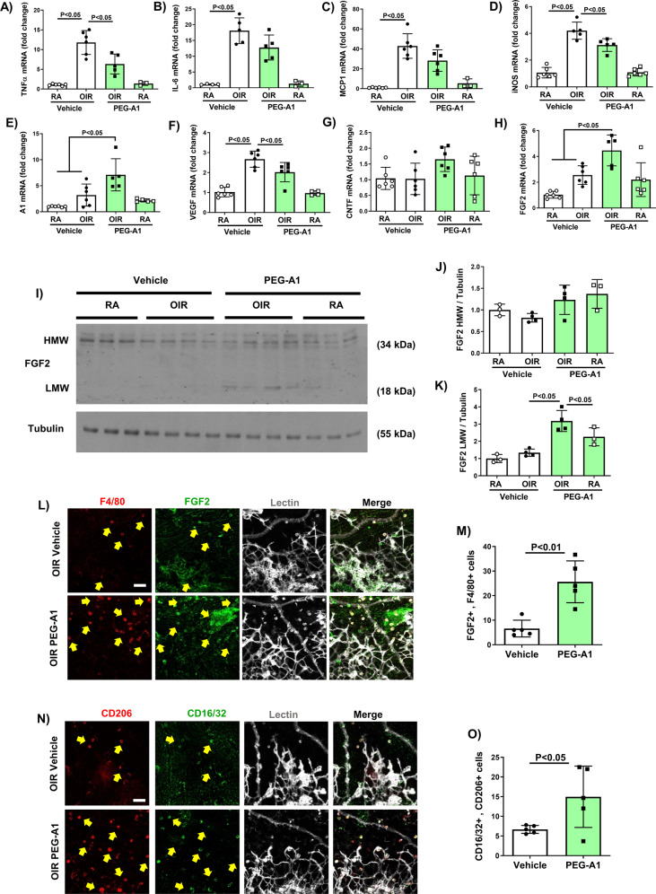
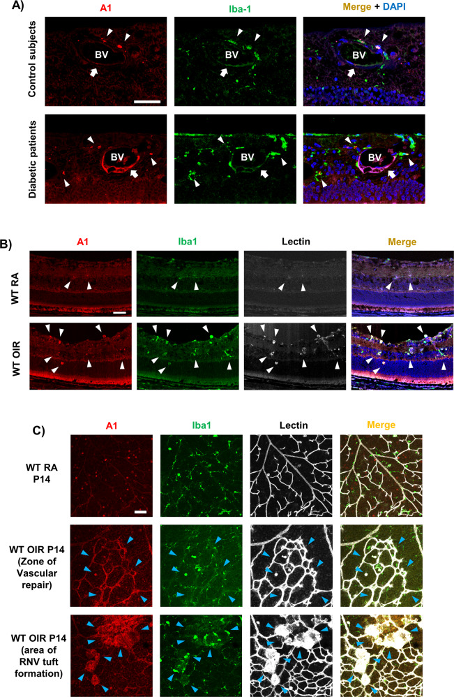
Current therapies for treatment of proliferative retinopathy focus on retinal neovascularization (RNV) during advanced disease and can trigger adverse side-effects. Here, we have tested a new strategy for limiting neurovascular injury and promoting repair during early-stage disease. We have recently shown that treatment with a stable, pegylated drug form of the ureohydrolase enzyme arginase 1 (A1) provides neuroprotection in acute models of ischemia/reperfusion injury, optic nerve crush, and ischemic stroke. Now, we have determined the effects of this treatment on RNV, vascular repair, and retinal function in the mouse oxygen-induced retinopathy (OIR) model of retinopathy of prematurity (ROP). Our studies in the OIR model show that treatment with pegylated A1 (PEG-A1), inhibits pathological RNV, promotes angiogenic repair, and improves retinal function by a mechanism involving decreased expression of TNF, iNOS, and VEGF and increased expression of FGF2 and A1. We further show that A1 is expressed in myeloid cells and areas of RNV in retinal sections from mice with OIR and human diabetic retinopathy (DR) patients and in blood samples from ROP patients. Moreover, studies using knockout mice with hemizygous deletion of A1 show worsened RNV and retinal injury, supporting the protective role of A1 in limiting the OIR-induced pathology. Collectively, A1 is critically involved in reparative angiogenesis and neuroprotection in OIR. Pegylated A1 may offer a novel therapy for limiting retinal injury and promoting repair during proliferative retinopathy.


Conflict of interest statement

AF, RBC, and RWC have a pending patent on the use of arginase 1 as a treatment for ischemic retinopathies. PN-MC is the chief executive officer of Bio-Cancer Treatment International Limited and holds stocks or shares in Bio-Cancer Treatment International Limited.

Figures

Fig. 1
Fig. 1. PEG-A1 promotes vessel sprouting and reduces the AVA in OIR retinas at P17.
AC OIR mice were injected intravitreally with either PEG-A1 or vehicle on P12 and sacrificed at p17. Quantitation of lectin-labeled retinal flatmounts showed significant decreases in AVA and RNV tuft formation with PEG-A1 treatment. Scale bar = 100 µm. D, E Higher magnification images and quantification showed a significant increase in vessel sprouts (white-arrow heads) entering the AVA with PEG-A1 treatment. Scale bar = 20 µm. F, G Fluorescein angiography at P50 showed normalization of OIR-induced vessel tortuosity with PEG-A1 treatment.
Fig. 2
Fig. 2. PEG-A1 treatment reduces OIR-induced retinal dysfunction, limits loss of horizontal cells, and reduces PARP cleavage.
A OIR reduced the retinal photopic B-wave ERG response at P50, which was partially rescued by PEG-A1 treatment. B OIR reduced visual acuity at P50 as measured by OptoMotry and this was rescued by PEG-A1 treatment. C, D Immunofluorescence labeling of P17 retinal sections with the horizontal cell marker Calbindin at P17 showed a decrease in numbers of horizontal cells in Vehicle-OIR retinas as compared to RA. PEG-A1 treatment reduced this loss. Scale bar = 50 µm. E, F Western blotting and quantification of P17 retinal lysates showed increased PARP cleavage in the OIR retinas, which was significantly reduced with PEG-A1 treatment.
Fig. 3
Fig. 3. PEG-A1 treatment ameliorates the OIR-induced inflammatory response and reduces VEGF while increasing FGF2.
AH Analysis of mRNA levels showed upregulation of TNF, IL-6, MCP1, and VEGF in OIR retinas at P13. PEG-A1 treatment significantly reduced TNF and VEGF and showed a trend towards a reduction in IL-6 and MCP1. PEG-A1 treatment also showed a trend towards increasing the neurotrophic factor CNTF at P13. Analysis at P17 showed upregulation of iNOS mRNA after OIR, which was significantly reduced with PEG-A1 treatment. Furthermore, PEG-A1 treatment increased the mRNA levels of A1 and FGF2 at P17. IK Western blotting of retina lysates at P17 showed upregulation of low molecular weight (LMW) FGF2 (18 kDa band) with PEG-A1 treatment as compared to vehicle with a similar increase in the high molecular weight (HMW) FGF2 but the later did not reach statistical significance. L, M Immunolabeling of retina flatmounts at P17 showed co-localization of FGF2 with F4/80 (microglia/macrophage marker) and lectin (blood vessel marker) (arrows). Image quantification showed upregulation of FGF2/F4/80 double-positive cells with PEG-A1 treatment. Scale bar = 20 µm. N, O Immunolabeling of retina flatmounts at P17 showed co-localization of the M2 macrophage marker CD206 with the M1 marker CD16/32. Image quantification showed upregulation of CD16/32 / CD206 double-positive cells with PEG-A1 treatment. Scale bar = 20 µm.
Fig. 4
Fig. 4. A1 deletion increases pathological neovascularization and decreases vascular repair at P17.
AC WT and A1 KO littermates were subjected to OIR at P7 and prepared for analysis on P17. Avascular area (AVA, yellow outline) and pathological RNV (tufts, highlighted in white) were significantly increased in A1 KO retinas. Scale bar = 100 µm. D, E Higher magnification images show decreased vessel sprouts at the AVA border zone of the P17 lectin-labeled A1 KO retina flatmounts. Scale bar = 20 µm. F, G Fluorescein angiography images show persistent vessel tortuosity in the WT OIR retinas through P50, which was increased in the A1 KO retinas. Data are presented as mean ± SD in all the figures.
Fig. 5
Fig. 5. A1 deletion worsens retinal thinning, apoptosis, horizontal cells loss, and glial activation after OIR.
AD SD-OCT analysis of 12-week-old mice retinas showed a significant reduction in thickness of the total retina, ganglion cell complex (GCC), and outer nuclear layer plus inner segments (ONL + IS) in the A1 KO OIR retinas as compared to WT OIR retinas. E, F TUNEL labeling and quantification at P9 showed a significant increase in TUNEL-positive cells in A1 KO OIR retina sections as compared to WT OIR retinas. Scale bar = 50 µm. G, H Calbindin labeling and quantitation of retina sections at P17 showed horizontal cell loss after OIR that was further aggravated in A1 KO OIR retinas. Scale bar = 50 µm. IK GFAP immunolabeling of retina cross-sections at P17 showed increased expression of GFAP in Müller cells beyond their end-feet after OIR, which was further increased in A1 KO retinas. Western blotting on P17 retina homogenates and quantification confirmed the immunolabeling results. Scale bar = 50 µm.
Fig. 6
Fig. 6. A1 is upregulated in human DR and mouse OIR retinal sections.
A Immunolabeling of retina sections from human donor with PDR showed increased co-localization of A1 with Iba-1 and blood vessel (BV)-like structures as compared with non-diabetic control. Scale bar = 50 µm. B Immunolabeling of P17 mouse retina sections showed co-localization of A1 with Iba-1-positive myeloid cells (microglia/macrophages) and lectin-positive blood vessels after OIR. Scale bar = 20 µm. C Immunolabeling of mouse retina flatmount at P14 showed A1 upregulation and co-localization with Iba-1-positive microglia/macrophages and lectin-positive blood vessels in both the areas of vascular repair (VR) and RNV tufts (RNV). Scale bar = 20 µm.
Fig. 7
Fig. 7. Endothelial but not myeloid A1 deletion decreases pathological angiogenesis in OIR.
AC Analysis of P17 OIR retinas or using lectin staining showed a significant decrease in pathological neovascularization (NV, highlighted in white) in endothelial A1 knockout mice (E-A1−/−) as compared to littermate floxed controls (A1f/f), while the AVA (yellow outline) showed a decreasing trend that was not statistically significant. Scale bar = 100 µm. DF Analysis of myeloid A1 knockout mice (M-A1−/−) OIR retinas showed no change in AVA or NV tuft formation at P17 as compared to littermate floxed controls (A1f/f). Scale bar = 100 µm. G, H Choroidal angiogenesis assay and quantification showed marked suppression of the angiogenic response with endothelial A1 deletion or PEG-A1 treatment. IL Treatment of bovine retinal endothelial cells with PEG-A1 for 6 h led to increased ERK phosphorylation as measured by western blotting while 24 h treatment increased FGF2 protein levels.

References

    1. Nicholson BP, Schachat AP. A review of clinical trials of anti-VEGF agents for diabetic retinopathy. Graefe’s Arch Clin Exp Ophthalmol. 2010;248:915–30. doi: 10.1007/s00417-010-1315-z. - DOI - PubMed
    1. Liegl R, Hellstrom A, Smith LE. Retinopathy of prematurity: the need for prevention. Eye Brain. 2016;8:91–102. - PMC - PubMed
    1. Hartnett ME. Advances in understanding and management of retinopathy of prematurity. Surv Ophthalmol. 2017;62:257–76. doi: 10.1016/j.survophthal.2016.12.004. - DOI - PMC - PubMed
    1. Nuzzi R, Tridico F. Local and systemic complications after intravitreal administration of anti-vascular endothelial growth factor agents in the treatment of different ocular diseases: a five-year retrospective study. Semin Ophthalmol. 2015;30:129–35. doi: 10.3109/08820538.2013.835833. - DOI - PubMed
    1. Falavarjani KG, Nguyen QD. Adverse events and complications associated with intravitreal injection of anti-VEGF agents: a review of literature. Eye (Lond, Engl) 2013;27:787–94. doi: 10.1038/eye.2013.107. - DOI - PMC - PubMed

Publication types