. 2022 Aug 29;13(8):742.
doi: 10.1038/s41419-022-05192-y.

Long noncoding RNA LINC00239 inhibits ferroptosis in colorectal cancer by binding to Keap1 to stabilize Nrf2

Affiliations

Long noncoding RNA LINC00239 inhibits ferroptosis in colorectal cancer by binding to Keap1 to stabilize Nrf2

Yuying Han et al. Cell Death Dis. .

Abstract

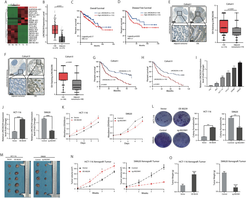
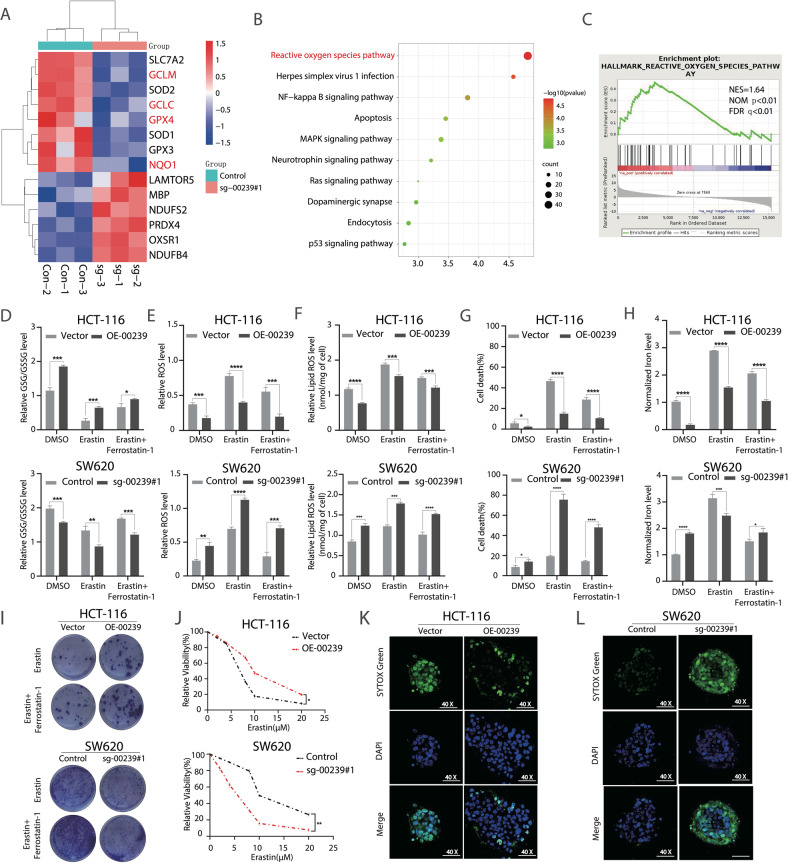
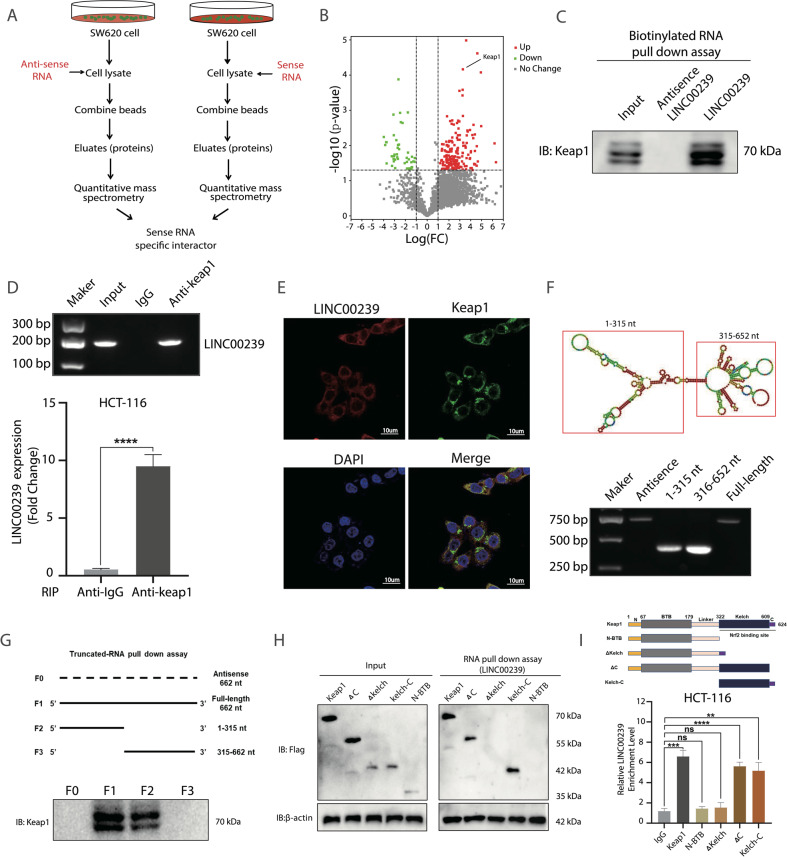
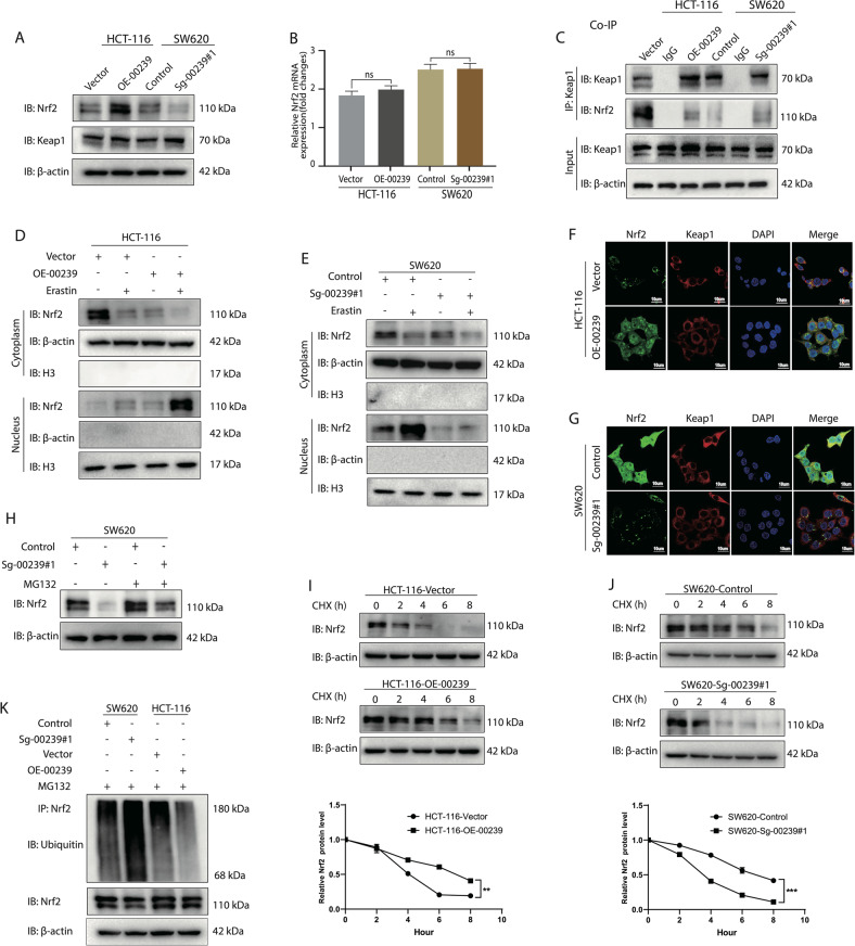
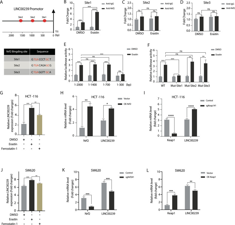
Ferroptosis, a novel regulated cell death induced by iron-dependent lipid peroxidation, plays an important role in tumor development and drug resistance. Long noncoding RNAs (lncRNAs) are associated with various types of cancer. However, the precise roles of many lncRNAs in tumorigenesis remain elusive. Here we explored the transcriptomic profiles of lncRNAs in primary CRC tissues and corresponding paired adjacent non-tumor tissues by RNA-seq and found that LINC00239 was significantly overexpressed in colorectal cancer tissues. Abnormally high expression of LINC00239 predicts poorer survival and prognosis in colorectal cancer patients. Concurrently, we elucidated the role of LINC00239 as a tumor-promoting factor in CRC through in vitro functional studies and in vivo tumor xenograft models. Importantly, overexpression of LINC00239 decreased the anti-tumor activity of erastin and RSL3 by inhibiting ferroptosis. Collectively, these data suggest that LINC00239 plays a novel and indispensable role in ferroptosis by nucleotides 1-315 of LINC00239 to interact with the Kelch domain (Nrf2-binding site) of Keap1, inhibiting Nrf2 ubiquitination and increasing Nrf2 protein stability. Considering the recurrence and chemoresistance constitute the leading cause of death in colorectal cancer (CRC), ferroptosis induction may be a promising therapeutic strategy for CRC patients with low LINC00239 expression.


Conflict of interest statement

The authors declare no competing interests.

Figures

Fig. 1
Fig. 1. Overexpression of LINC00239 promotes CRC metastasis and indicates a worse prognosis.
A Heatmap of K-means clustering of differentially expressed lncRNAs (log2FC > 5.0, Padj. < 0.05) during three paired CRC (T1–T3) and adjacent normal (N1–N3) tissues. LINC00239 is marked in red. B Statistical analysis of LINC00239 expression in TCGA of colorectal cancer and normal tissues, paired t test. Red indicates colorectal cancer tissue (n = 275), and gray indicates adjacent tissue (n = 275). C Kaplan–Meier overall survival (OS) analysis of LINC00239 status in CRC patients (TCGA, n = 269). D Kaplan–Meier disease-free survival (DFS) analysis of LINC00239 status in CRC patients (TCGA, n = 269). E, F The representative image of ISH staining of LINC00239 in CRC and adjacent nontumor tissues microarray. The scale bars represent 250 μm (low magnification) and 50 μm (high magnification). Right: The ISH score of LINC00239 in CRC and adjacent nontumor samples in Cohort I (n = 174) and Cohort II (n = 180). G, H Kaplan–Meier analysis of the relationships of LINC00239 expression and overall survival times or the recurrence rates in two independent CRC cohorts. I Real-time PCR analyses of the expression of LINC00239 in CRC cell lines and normal intestinal epithelial cells (FHC). J Real-time PCR analysis of LINC00239 expression in HCT-116 and SW620 cells after lentivirus transfection. K Cell viability in the indicated CRC cell lines by CCK-8 assay. L Cell viability in the indicated CRC cell lines by colony-formation assay. MO Nude mice are shown after injection of HCT-116 and SW620 cells stably expressing the control vector, LINC00239 or Ko-LINC00239 expression plasmids. Tumor formation was monitored at the images (M), indicated times (N), and weights (O) were recorded (n = 5). Data shown represent mean ± SD from three independent experiments. ns P > 0.05, *P < 0.05, **P < 0.01, ***P < 0.001, ****P < 0.0001, Student’s t test.
Fig. 2
Fig. 2. LINC00239 upregulates colorectal cancer cell proliferation by inhibiting ferroptosis.
A RNA-seq-based heatmap indicating the changes of ferroptosis-related genes in SW620 cells knockout LINC00239. B KEGG analysis of LINC00239-related gene co-expression networks from the sequencing data. C GSEA analysis of LINC00239-related gene co-expression networks from the sequencing data. DH GSH/GSSG ratios (D), ROS levels (E), Lipid ROS (F), cell viability (G), and Fe2+ concentration (H) was measured in the four indicated cell lines after treated with 10 μM erastin for 48 h and the addition of 2 µM ferrostatin-1 (Fer-1). I, J Colony-formation and CCK-8 assay to evaluate the cell viability of LINC00239 on colorectal cancer cells after treated with 2 μM erastin and the addition of 2 µM ferrostatin-1 (Fer-1). K, L Spheroids generated from the indicated cell lines were cultured for 96 h and treated with 15 μM erastin for 48 h. Dead cells were stained by SYTOX Green (original magnification, ×40). Data shown represent mean ± SD from three independent experiments. ns P > 0.05, *P < 0.05, **P < 0.01, ***P < 0.001, ****P < 0.0001, Student’s t test.
Fig. 3
Fig. 3. LINC00239 interacts with Keap1.
A Proteomic approach to identify the LINC00239-specific interactors. B Proteome-wide accurate quantification and significance. C Western blot of the proteins from anti-sense LINC00239 and LINC00239 pull-down assays. D RNA immunoprecipitation experiments were performed using anti-Keap1 antibody, and specific primers were used to detect LINC00239. E ImmunoFISH images of LINC00239 and Keap1 in SW620 cells. F Top, the predicted secondary structure of LINC00239. Bottom, the in vitro-transcribed full-length LINC00239 and deletion fragments with the correct sizes indicated. G Deletion mapping of the Keap1-binding domain in LINC00239. Top, diagrams of full-length LINC00239 and the deletion fragments. Bottom, immunoblot analysis for Keap1 in the protein samples pulled down by different LINC00239 constructs. H The RNA Pull-down analysis of FLAG-tagged Keap1 versus domain truncation mutants) retrieved by in vitro-transcribed biotinylated LINC00239. I RIP assays show the association of the Kelch domain with LINC00239. Data shown represent mean ± SD from three independent experiments. ns P > 0.05, **P < 0.01, ***P < 0.001, ****P < 0.0001, Student’s t test.
Fig. 4
Fig. 4. LINC00239 interacts with Keap1 and regulate the stability of Nrf2.
A Western blot analysis of the expression levels of Keap1 and Nrf2. B The mRNA level of Nrf2 encoding gene (NFE2L2) was not significantly different in CRC cells lacking or overexpressing LINC00239. C LINC00239 reduced the interaction between Nrf2 and Keap1. CRC cells were treated with 10 μM erastin for 12 h. Cell lysates were immunoprecipitated with an anti-Keap1 antibody and blotted with an anti-Nrf2 antibody. D, E LINC00239 regulates the subcellular localization of Nrf2. Subcellular fractionation was used to isolate cytoplasmic and nuclear proteins, and immunoblotting was performed to examine the localization of Nrf2 following the downregulation or overexpression of LINC00239. F, G Immunofluorescence was used to localize Nrf2 in cells lacking or overexpressing LINC00239. All cells were treated with 10 μM erastin for 12 h. CRC cells were labeled with anti-Nrf2 (green), anti-Keap1 (red), and DAPI (blue). Scale bar: 10 μm. H LINC00239 reduced the protein degradation of Nrf2. SW620 cells transfected with LINC00239-knockdown and control plasmids were left untreated or treated with 10 μM of MG132 for 6 h to block the degradation of ubiquitinated proteins. I, J LINC00239 stabilized Nrf2 under basal conditions. All cells were left untreated or treated with 50 μg/mL CHX and incubated for the indicated time periods. Lysates were analyzed by western blotting. K LINC00239 reduced the ubiquitination of Nrf2. All cells treated with 10 μM of MG132 for 6 h and subjected to an in vivo ubiquitination assay to detect ubiquitin-conjugated endogenous Nrf2 proteins. Lysates were denatured, immunoprecipitated with anti-Nrf2 and blotted with an anti-ubiquitin antibody. Data shown represent mean ± SD from three independent experiments. ns P > 0.05, **P < 0.01, ***P < 0.001, ****P < 0.0001, Student’s t test.
Fig. 5
Fig. 5. LINC00239 regulates ferroptosis by modulating the expression of the Keap1/Nrf2 signaling pathway.
A, B Dose-dependent toxicity of erastin-induced cell death. CF GSH/GSSG ratios (C), ROS levels (D), lipid ROS (E), and cell viability (F) was measured in the four indicated cell lines after treated with 10 μM erastin for 48 h and the addition of 2 µM ferrostatin-1 (Fer-1). G, H Colony-formation assay to evaluate the cell viability of LINC00239 on colorectal cancer cells after treated with 2 μM erastin and the addition of 2 µM ferrostatin-1 (Fer-1). IN Nude mice are shown after injection of HCT-116-OE-00239 and SW620-sg-00239#1 cells stably expressing the control vector, sgNrf2#1, OE-Nrf2, sgKeap1#1, or OE-Keap1 expression plasmids. Tumor formation was monitored at the images (I, J), indicated times (K, L), and weights (M, N) were recorded (n = 5). All experiments were thereafter treated with erastin three times a week. Data shown represent mean ± SD from three independent experiments. ns P > 0.05, **P < 0.01, ***P < 0.001, ****P < 0.0001, Student’s t test.
Fig. 6
Fig. 6. Nrf2 mediates positive feedback transcript activation of LINC00239 in CRC.
A Schematic representation (upper panel) of the LINC00239 promoters from humans. Different symbols represent the Nrf2-binding sites. TSS transcriptional start site. BD Chromatin immunoprecipitation (ChIP-qRT-PCR) analysis erastin increased the binding of Nrf2 to the LINC00239 promoter. SW620 cells were treated with DMSO or 10 μM erastin, and chromatin immunoprecipitation was performed using Nrf2-specific antibodies. E, F Erastin increased the binding of Nrf2 to the LINC00239 promoter. SW620 cells were transiently transfected with a luciferase reporter driven by the LINC00239 promoter (1–2000 bp, 1–1400 bp, 1–700 bp, or 1–300 bp), LINC00239 promoter (WT, mut-Site1, mut-Site2, or mut-Site3), and then were left untreated or treated with 10 μM erastin for 24 h. G HCT-116 cells were treated with DMSO, 10 µM erastin or 2 µM ferrostatin-1 (Fer-1) for 24 h. qPCR analysis the levels of LINC00239 in HCT-116 cells. H HCT-116 cells were transfected with control or Nrf2-overexpression plasmids. qPCR analysis the levels of LINC00239 and Nrf2 in HCT-116 cells. I HCT-116 cells were transfected with control or Keap1-knockdown (sgKeap1#1) plasmids. qPCR analysis the levels of LINC00239 and Keap1 in HCT-116 cells. J SW620 cells were treated with DMSO, 10 µM erastin, or 2 µM ferrostatin-1 (Fer-1) for 24 h. qPCR analysis the levels of LINC00239 in SW620 cells. K SW620 cells were transfected with control or Nrf2-knockdown (sgNrf2#1) plasmids. qPCR analysis the levels of LINC00239 and Nrf2 in SW620 cells. L SW620 cells were transfected with control or Nrf2-overexpression plasmids. qPCR analysis the levels of LINC00239 and Nrf2 in SW620 cells. Data shown represent mean ± SD from three independent experiments. ns P > 0.05, **P < 0.01, ***P < 0.001, ****P < 0.0001, Student’s t test.
Fig. 7
Fig. 7. LINC00239 expression has a positive correlation with Nrf2 and GPX4 expression in CRC tissues.
A Representative images of IHC or ISH staining of LINC00239, Nrf2, and GPX4 expression in human CRC samples in Cohort I (n = 174). The scale bars represent 250 μm (low magnification) and 50 μm (high magnification). B Correlation analysis of LINC00239 and Nrf2 expression in the CRC tissues in cohort I (n = 174). C Correlation analysis of LINC00239 and GPX4 expression in the CRC tissues in cohort I (n = 174). D, E Kaplan–Meier’s curves generated with the data from the CRC patients with negative versus positive LINC00239, Nrf2, or GPX4 expression. The correlation between LINC00239 and Nrf2 (D) or GPX4 (E) expression and overall survival or recurrence in patients with CRC in cohort I (n = 174). F Representative images of IHC or ISH staining of LINC00239, Nrf2, and GPX4 expression in human CRC samples in cohort II (n = 180). The scale bars represent 250 μm (low magnification) and 50 μm (high magnification). G Correlation analysis of LINC00239 and Nrf2 expression in the CRC tissues in cohort II (n = 180). H Correlation analysis of LINC00239 and GPX4 expression in the CRC tissues in cohort II (n = 180). I, J Kaplan–Meier’s curves generated with the data from the CRC patients with negative versus positive LINC00239, Nrf2 or GPX4 expression. The correlation between LINC00239 and Nrf2 (I) or GPX4 (J) expression and overall survival or recurrence in patients with CRC in cohort II (n = 180). K, L Real-time PCR of LINC00239 and GPX4 expression in adjacent nontumor samples and CRC samples (n = 22). M LINC00239 expression positively correlated with the expression levels of GPX4 in CRC samples (n = 22). N Proposed model of the relationship between LINC00239 and Nrf2 in CRC cells. Data shown represent mean ± SD from three independent experiments. ns P > 0.05, **P < 0.01, ***P < 0.001, ****P < 0.0001, Student’s t test.

References

    1. Siegel RL, Miller KD, Jemal A. Cancer statistics, 2019. CA Cancer J Clin. 2019;69:7–34. doi: 10.3322/caac.21551. - DOI - PubMed
    1. Bray F, Ferlay J, Soerjomataram I, Siegel RL, Torre LA, Jemal A. Global cancer statistics 2018: GLOBOCAN estimates of incidence and mortality worldwide for 36 cancers in 185 countries. CA Cancer J Clin. 2018;68:394–424. doi: 10.3322/caac.21492. - DOI - PubMed
    1. Keum N, Giovannucci E. Global burden of colorectal cancer: emerging trends, risk factors and prevention strategies. Nat Rev Gastroenterol Hepatol. 2019;16:713–32. doi: 10.1038/s41575-019-0189-8. - DOI - PubMed
    1. Dekker E, Rex DK. Advances in CRC prevention: screening and surveillance. Gastroenterology. 2018;154:1970–84. doi: 10.1053/j.gastro.2018.01.069. - DOI - PubMed
    1. Wu Y, Zhang S, Gong X, Tam S, Xiao D, Liu S, et al. The epigenetic regulators and metabolic changes in ferroptosis-associated cancer progression. Mol Cancer. 2020;19:39. doi: 10.1186/s12943-020-01157-x. - DOI - PMC - PubMed

Publication types

MeSH terms

Substances