. 2022 Aug 29;15(1):122.
doi: 10.1186/s13045-022-01345-w.

CircRNA-CREIT inhibits stress granule assembly and overcomes doxorubicin resistance in TNBC by destabilizing PKR

Affiliations

CircRNA-CREIT inhibits stress granule assembly and overcomes doxorubicin resistance in TNBC by destabilizing PKR

Xiaolong Wang et al. J Hematol Oncol. .

Abstract

Background: Circular RNAs (circRNAs) represent a novel type of regulatory RNA characterized by high evolutionary conservation and stability. CircRNAs are expected to be potential diagnostic biomarkers and therapeutic targets for a variety of malignancies. However, the regulatory functions and underlying mechanisms of circRNAs in triple-negative breast cancer (TNBC) are largely unknown.

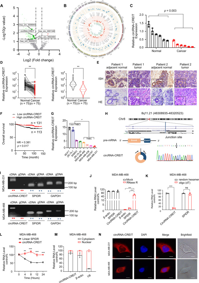
Methods: By using RNA high-throughput sequencing technology, qRT-PCR and in situ hybridization assays, we screened dysregulated circRNAs in breast cancer and TNBC tissues. Then in vitro assays, animal models and patient-derived organoids (PDOs) were utilized to explore the roles of the candidate circRNA in TNBC. To investigate the underlying mechanisms, RNA pull-down, RNA immunoprecipitation (RIP), co immunoprecipitation (co-IP) and Western blotting assays were carried out.

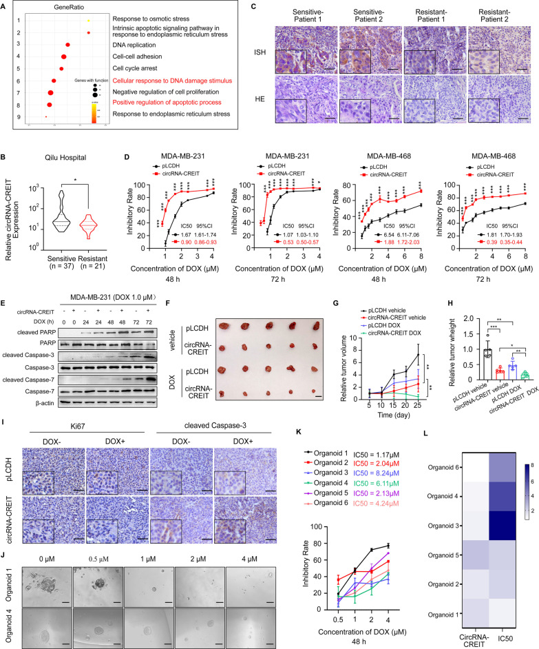
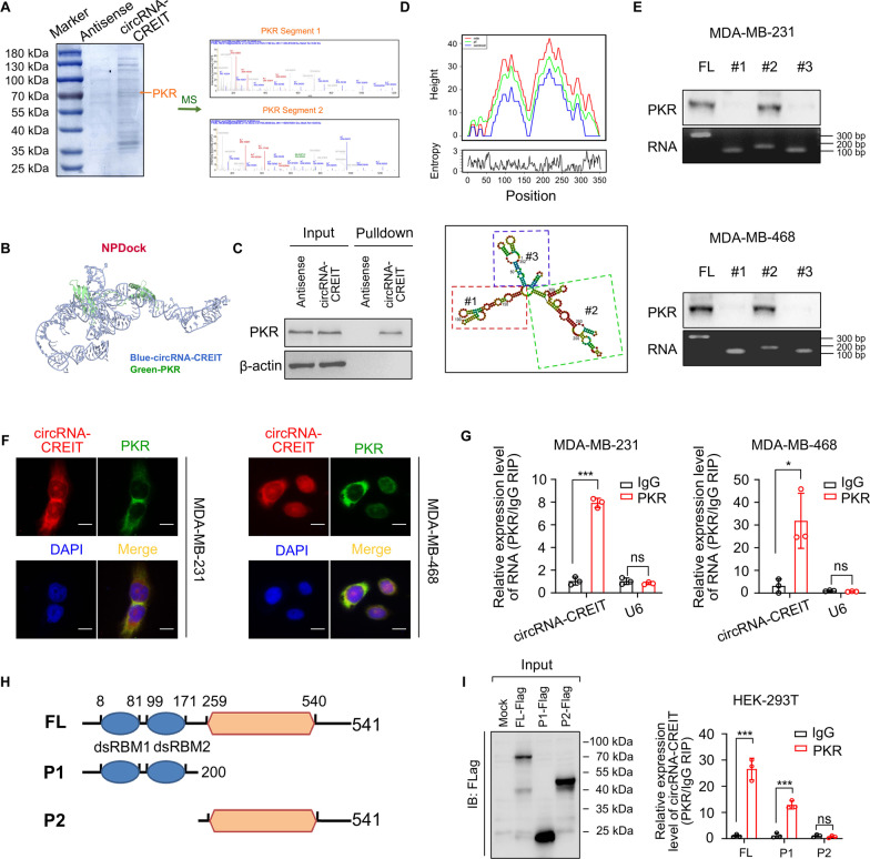
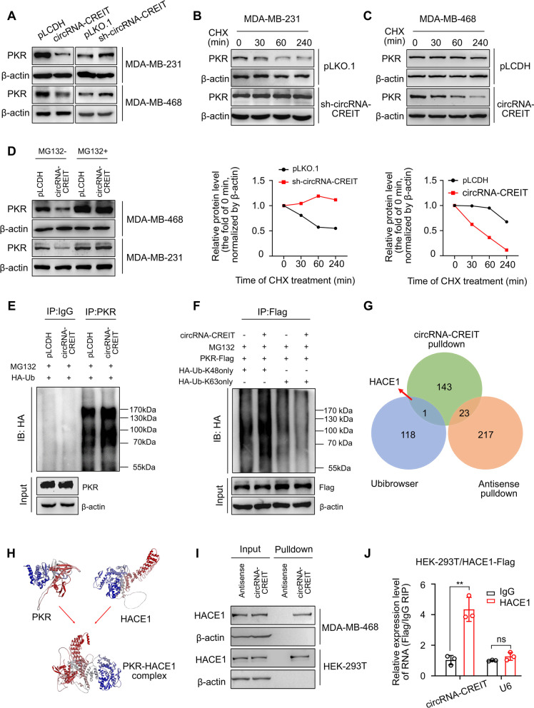
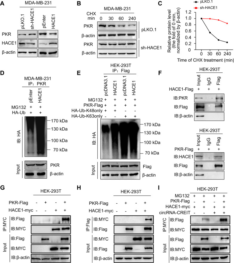
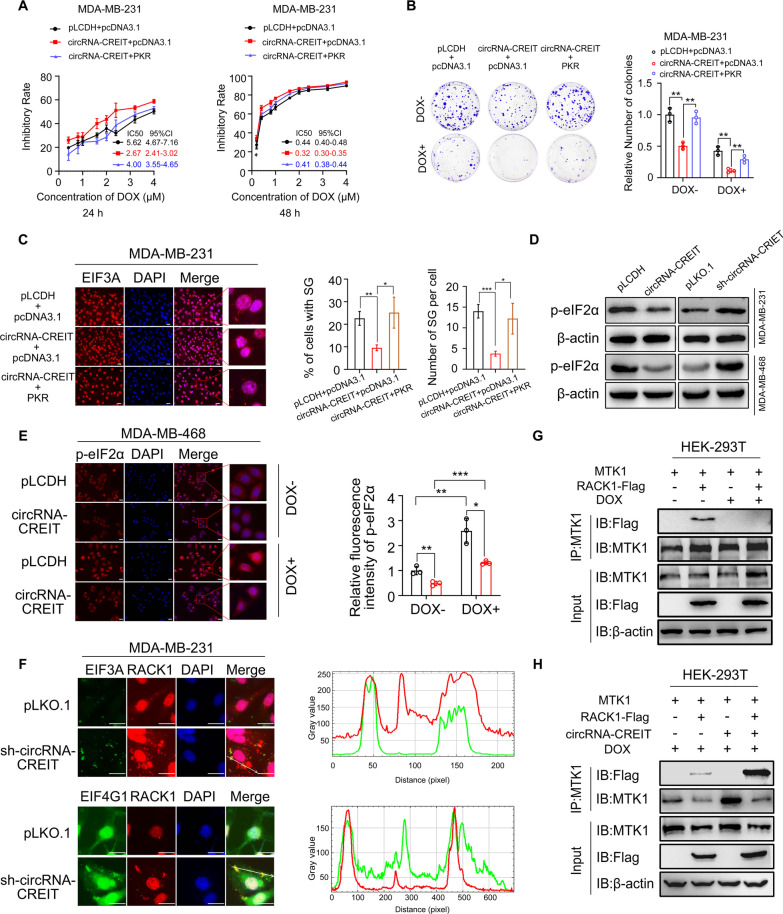
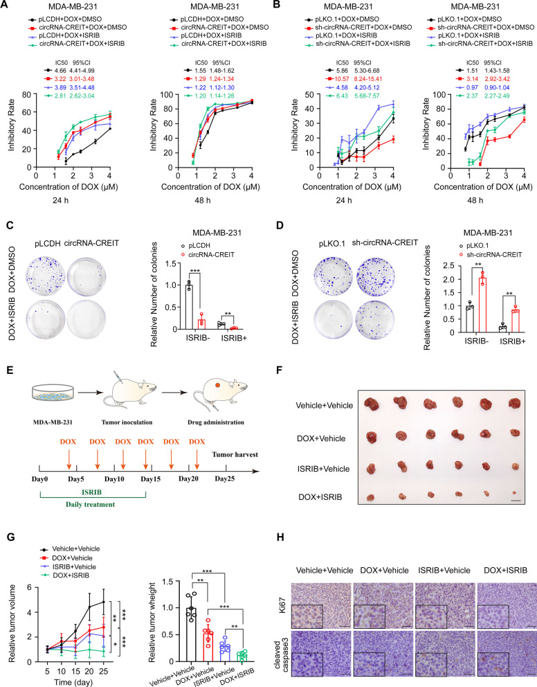
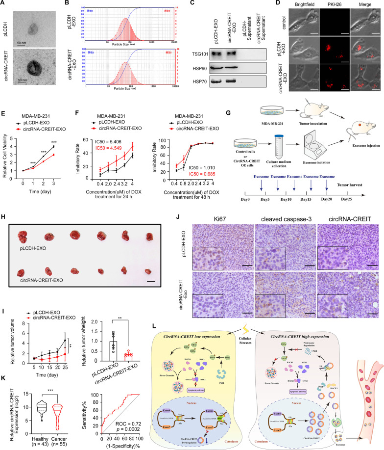
Results: In this study, we demonstrated that circRNA-CREIT was aberrantly downregulated in doxorubicin resistant triple-negative breast cancer (TNBC) cells and associated with a poor prognosis. The RNA binding protein DHX9 was responsible for the reduction in circRNA-CREIT by interacting with the flanking inverted repeat Alu (IRAlu) sequences and inhibiting back-splicing. By utilizing in vitro assays, animal models and patient-derived organoids, we revealed that circRNA-CREIT overexpression significantly enhanced the doxorubicin sensitivity of TNBC cells. Mechanistically, circRNA-CREIT acted as a scaffold to facilitate the interaction between PKR and the E3 ligase HACE1 and promoted proteasomal degradation of PKR protein via K48-linked polyubiquitylation. A reduced PKR/eIF2α signaling axis was identified as a critical downstream effector of circRNA-CREIT, which attenuated the assembly of stress granules (SGs) to activate the RACK1/MTK1 apoptosis signaling pathway. Further investigations revealed that a combination of the SG inhibitor ISRIB and doxorubicin synergistically inhibited TNBC tumor growth. Besides, circRNA-CREIT could be packaged into exosomes and disseminate doxorubicin sensitivity among TNBC cells.

Conclusions: Our study demonstrated that targeting circRNA-CREIT and SGs could serve as promising therapeutic strategies against TNBC chemoresistance.

Keywords: Chemoresistance; CircRNA-CREIT; Stress granules; TNBC.


Conflict of interest statement

All the authors declare they have no competing interests.

Figures

Fig. 1
Fig. 1
CircRNA-CREIT was downregulated in TNBC. A A volcano plot showing the upregulated and downregulated circRNAs in breast cancer tissues. B Circos plot indicating the differentially expressed circRNAs. The outermost circle shows the chromosomal distribution of the circRNAs. The second circle shows the expression levels of the indicated circRNAs. The third circle indicates the circBase ID of the circRNAs. The fourth circle shows the logFC of the indicated circRNAs between the two groups. The innermost circle shows the logp values. C qRT-PCR assay was performed to detect the expression of circRNA-CREIT in the 6 paired breast cancer tissues and their normal counterparts. D Expression of circRNA-CREIT in 75 breast cancer tissues and normal mammary tissues detected by qRT-PCR assay. E ISH of circRNA-CREIT in breast cancer tissues and paired normal mammary tissues. Scale bars = 200 μm. F Kaplan–Meier survival analysis of circRNA-CREIThigh and circRNA-CREITlow patients. G Expression of circRNA-CREIT in breast cancer cell lines with different hormone receptor statuses. H (upper) Diagram showing the locus of circRNA-CREIT in the genome. (lower) Schematic illustration showing that exons 6–7 of human SPIDR circularize to form circRNA-CREIT. The black arrow represents the back-splicing site of circRNA-CREIT confirmed by Sanger sequencing. I Convergent and divergent primers were used to validate the loop structure of circRNA-CREIT. J The expression of circRNA-CREIT, linear SPIDR and β-actin in TNBC cells with or without RNase R treatment. K qRT-PCR analysis of circRNA-CREIT expression in cDNA reverse transcribed with random hexamer or oligo (dT) primers. L Relative RNA levels of circRNA-CREIT and linear SPIDR after actinomycin D treatment detected by qRT-PCR. M Detection of circRNA-CREIT expression in cytoplasmic and nuclear fractions of RNAs extracted from TNBC cells. N RNA FISH assay for circRNA-CREIT with the nucleus distinguished by DAPI. Scale bars = 10 μm. ns no significance; *p < 0.05; **p < 0.01; ***p < 0.001 compared with the controls
Fig. 2
Fig. 2
CircRNA-CREIT significantly enhanced the chemosensitivity of TNBC cells in vitro and in vivo. A GO analysis of differentially expressed genes in circRNA-CREIT-overexpressing MDA-MB-231 cells based on RNA-seq data. B Violin plot showing circRNA-CREIT expression in chemosensitive and chemoresistant breast cancer tissues, detected by qRT-PCR assays. C ISH assay of circRNA-CREIT in chemosensitive and chemoresistant breast cancer tissues. Scale bars = 200 μm. D The impact of circRNA-CREIT overexpression on the cytotoxic effects of DOX is shown. Cell viability was detected by MTT assays. The IC50 values with the 95% CIs are presented. E Western blotting was performed to detect the expression of apoptosis pathway markers. F Images of the xenograft tumors. Scale bars = 10 mm. G Growth curves of xenograft tumors in different groups after treatment. H The tumor weights of the xenograft tumors. I Representative images of IHC staining for Ki67 and cleaved caspase-3 in different groups. Scale bars = 100 μm. J Representative morphologies of breast cancer patient-derived organoids (PDOs) treated with increasing DOX concentrations. Scale bars = 200 μm. K The IC50 values and 95% CIs of the PDOs are shown. Cell viability was measured by CCK8 assays. L Heatmap showing the IC50 value and relative circRNA-CREIT expression level of each PDO. Three independent experiments were conducted for each result. *p < 0.05; **p < 0.01; ***p < 0.001 compared with the controls
Fig. 3
Fig. 3
CircRNA-CREIT physically interacted with PKR. A CircRNA-CREIT probes and control probes were biotinylated and incubated with MDA-MB-231 cell lysates for RNA pull-down assays. (left) Photograph presenting Coomassie brilliant blue staining for the proteins precipitated in the RNA pull-down assays. The orange arrow indicates the size of the PKR protein. (right) Two segments of PKR proteins identified by mass spectrometry (MS). B Graphical representation of the molecular docking between circRNA-CREIT and the PKR protein using NPDock. C Western blotting of independent RNA pull-down assays verified the specific association of PKR protein with circRNA-CREIT using MDA-MB-231 cells. D The secondary structure of circRNA-CREIT was predicted by the online tool RNAfold web server (lower). (upper) Mountain plot representing the minimum free energy (MFE, red), the thermodynamic ensemble (green) and the centroid structures (blue) of circRNA-CREIT. CircRNA-CREIT was divided into three truncates representing three stem loop structures. E Western blotting analysis of PKR pulled down by different circRNA-CREIT truncates. F FISH and IF assays showing the colocalization of PKR and circRNA-CREIT. Scale bars = 10 μm. G RIP assay verifying the binding between PKR and circRNA-CREIT. H Diagrams of full-length (FL) PKR proteins and truncates with domain depletion. I (left) Western blotting analysis of PKR with full-length and truncated PKR proteins in the lysates of the HEK-293 T cells. (right) RIP assay for circRNA-CREIT enrichment in cells transfected with flag-tagged PKR (FL) overexpression vectors and truncated PKR expression vectors. Three independent experiments were conducted for each result. ns, no significance; *p < 0.05; ***p < 0.001 compared with the controls
Fig. 4
Fig. 4
CircRNA-CREIT promoted PKR degradation via the ubiquitin–proteasome system. A Western blotting for PKR protein levels after circRNA-CREIT overexpression or knockdown. B, C TNBC cells with circRNA-CREIT overexpression or knockdown were treated with cycloheximide (CHX) for the indicated times. Western blotting analysis (upper) and statistical analysis (lower) of PKR levels upon CHX treatment are presented, with the level at 0 h as a control. D Western blotting analysis of PKR protein levels regulated by circRNA-CREIT with or without MG132 treatment. E Effects of circRNA-CREIT overexpression on the ubiquitination of PKR proteins. MDA-MB-231 cells were cotransfected with circRNA-CREIT overexpression plasmids and HA-tagged ubiquitin expression plasmids or the corresponding empty vectors. The cell lysates were incubated with anti-PKR or anti-IgG antibodies and protein A/G magnetic beads. The proteins precipitated in the co-IP assay were analyzed by Western blotting. IB immunoblot. F Western blotting assay showing the upregulation of K48-linked ubiquitination in circRNA-CREIT-overexpressing MDA-MB-231 cells. IB: immunoblot. HA-Ub-K48only: the cells were transfected with plasmids expressing HA-tagged ubiquitin with all lysines mutated except K48. HA-Ub-K63only: the cells were transfected with plasmids expressing HA-tagged ubiquitin with all lysines mutated except K63. G Identification of the candidate E3 ligases of PKR by bioinformatics prediction and MS analysis for RNA pull-down products, illustrated by a Venn diagram. H The interaction between PKR and HACE1 proteins was predicted by the ZDOCK server. The predicted structure was visualized by Discovery Studio software. I, J The interaction between circRNA-CREIT and HACE1 was verified by RNA pull-down assays (I) and RIP assay (J). Three independent experiments were conducted for each result. ns no significance; **p < 0.01 compared with the controls
Fig. 5
Fig. 5
CircRNA-CREIT enhanced the binding of HACE1 and PKR proteins. A Western blotting for PKR protein levels after HACE1 overexpression or knockdown in TNBC cells. B, C TNBC cells stably transfected with sh-HACE1 plasmids or empty vectors were treated with CHX for the indicated times. Western blotting (B) and statistical analysis (C) of PKR protein levels are shown, with the level at 0 h as a control. D The impact of HACE1 on the PKR ubiquitination level was verified by co-IP assays and subsequent Western blotting. IB: immunoblot. E K48-linked ubiquitination of PKR was enhanced by HACE1 overexpression. IB: immunoblot. HA-Ub-K48only: the cells were transfected with plasmids expressing HA-tagged ubiquitin with all lysines mutated except K48. HA-Ub-K63only: the cells were transfected with plasmids expressing HA-tagged ubiquitin with all lysines mutated except K63. F HEK-293 T cells transfected with HACE1-Flag or PKR-Flag were lysed, immunoprecipitated with anti-Flag and then subjected to Western blotting assays using anti-PKR or anti-HACE1, respectively. G, H HEK-293 T cells co-transfected with PKR-Flag and HACE1-Myc or the corresponding empty vectors were lysed, immunoprecipitated with anti-MYC (G) or anti-Flag (H), and subjected to Western blotting analysis. I HEK-293 T cells were cotransfected with PKR-Flag, HACE1-Myc and circRNA-CREIT overexpression vectors or the corresponding empty plasmids. Co-IP assays validated that circRNA-CREIT increased the PKR-Flag level precipitated by HACE1-Myc. Three independent experiments were conducted for each result
Fig. 6
Fig. 6
CircRNA-CREIT attenuated SG formation via the PKR/eIF2α axis. A PKR reversed the effects of circRNA-CREIT in promoting chemosensitivity. Cell viability was detected MTT assays. The IC50 and the 95% CI of cells with different treatments are shown. B Colony formation assays presenting the roles of PKR in mediating the functions of circRNA-CREIT. C Immunofluorescence staining for the SG marker EIF3A under DOX treatment (for 24 h). Representative images are shown and the percentage of cells with SGs and the number of SGs per cell were calculated. D Western blotting assay showing that circRNA-CREIT inhibited p-eIF2α expression and circRNA-CREIT knockdown increased p-eIF2α levels in TNBC cells. E Immunofluorescence staining for p-eIF2α after circRNA-CREIT overexpression or knockdown with or without DOX treatment (for 24 h). Quantitative analyses were performed using Image J, and the groups treated with empty vectors and PBS were used as controls. F Subcellular localization of RACK1 and endogenous SG markers EIF3A and EIF4G1 under DOX treatment. Cells were transfected with RACK1-pmCherry-C1 plasmids, treated with DOX for 24 h and subjected to immunofluorescence. Colocalization analysis for RACK1 and SG markers along the indicated line was performed by ImageJ. G Western blotting analysis of the co-IP assay showing that the interaction between RACK1 and MTK1 was blocked by DOX treatment. H Co-IP assay and the subsequent Western blotting assay verified that circRNA-CREIT restored the binding of RACK1 and MTK1. Scale bars = 20 μm. Three independent experiments were conducted for each result. *p < 0.05; **p < 0.01, ***p < 0.001 compared with the controls
Fig. 7
Fig. 7
ISRIB exerted synergistic roles with circRNA-CREIT in improving chemosensitivity of TNBC cells. A Chemosensitivity to DOX of MDA-MB-231 cells treated with circRNA-CREIT, ISRIB alone or in combination. Cell viability was detected with MTT assays. The IC50 and the 95% CI of cells with different treatments are shown. B Chemosensitivity to doxorubicin of MDA-MB-231 cells treated with sh-circRNA-CREIT, ISRIB alone or in combination was detected by MTT assays. The IC50 and the 95% CI of cells with different treatments are shown. C, D Colony formation assays showed the synergistic roles of circRNA-CREIT and ISRIB in increasing cell chemosensitivity. E A schematic diagram indicating the experimental process of constructing the subcutaneous xenograft model and drug administration in female nude mice. F Images of xenograft tumors after the indicated treatment. Scale bars = 10 mm. G Growth curves and relative weights of the xenograft tumors in the four groups. The group treated with DMSO was used as a control. H IHC staining for Ki67 and cleaved caspase-3 expression in the xenograft tumors of different groups. Scale bars = 100 μm. Three independent experiments were conducted for each result. *p < 0.05; **p < 0.01, ***p < 0.001 compared with the controls
Fig. 8
Fig. 8
CircRNA-CREIT exerted tumor suppressive roles by exosome transmission in TNBC. A Representative images of isolated exosomes analyzed by transmission electron microscopy. B The size distribution of the exosomes was measured by a nanolaser particle detector. C Western blotting analysis for classic protein markers of exosomes. D Representative images of PKH26-stained exosomes that were taken up by TNBC cells. E MDA-MB-231 cells were treated with circRNA-CREIT-EXOs or pLCDH-EXOs, and cell viability was examined by MTT assays. F MDA-MB-231 cells treated with circRNA-CREIT-exosomes or pLCDH-exosomes were exposed to increasing concentrations of doxorubicin. Cell viability was detected by MTT assays. G A schematic diagram indicating the experimental process of constructing the subcutaneous xenograft model and exosome treatment in female nude mice. H Photographs of xenograft tumors treated with circRNA-CREIT-EXOs or pLCDH-EXOs. Scale bars = 10 mm. I Growth curves (left) and tumor weights (right) of the xenograft tumors. The group treated with exosomes extracted from MDA-MB-231/pLCDH cells served as the control. J IHC staining for Ki67 and cleaved caspase-3, and ISH assay for circRNA-CREIT in xenograft tumors treated with exosomes. Scale bars = 100 μm. K Violin plot showing the expression of circRNA-CREIT in the plasma of female breast cancer patients and age matched female healthy donors (left). The ROC curve shows that the expression level of circRNA-CREIT could distinguish breast cancer patients from healthy people (right). L A schematic diagram shows that circRNA-CREIT suppresses TNBC chemoresistance by inhibiting the formation of SGs. Three independent experiments were conducted for each result. **p < 0.01, ***p < 0.001 compared with the controls

References

    1. Sung H, Ferlay J, Siegel RL, Laversanne M, Soerjomataram I, Jemal A, et al. Global Cancer Statistics 2020: GLOBOCAN estimates of incidence and mortality worldwide for 36 cancers in 185 countries. CA 2021;71(3):209–49. - PubMed
    1. Haque S, Cook K, Sahay G, Sun C. RNA-Based Therapeutics: current developments in targeted molecular therapy of triple-negative breast cancer. Pharmaceutics. 2021;13(10). - PMC - PubMed
    1. Zhu Y, Zhu X, Tang C, Guan X, Zhang W. Progress and challenges of immunotherapy in triple-negative breast cancer. Biochim Biophys Acta. 2021;1876(2):188593. - PubMed
    1. Zhu Y, Hu Y, Tang C, Guan X, Zhang W. Platinum-based systematic therapy in triple-negative breast cancer. Biochim Biophys Acta. 2022;1877(1):188678. - PubMed
    1. Khalili-Tanha G, Moghbeli M. Long non-coding RNAs as the critical regulators of doxorubicin resistance in tumor cells. Cell Mol Biol Lett. 2021;26(1):39. doi: 10.1186/s11658-021-00282-9. - DOI - PMC - PubMed

Publication types

MeSH terms

LinkOut - more resources