. 2022 Aug 12;24(4):339.
doi: 10.3892/ol.2022.13459. eCollection 2022 Oct.

Long-term remission under Disitamab Vedotin (RC48) in HR-positive/HER2-positive metastatic breast cancer with brain meningeal, and bone marrow involvement: A case report

Affiliations

Long-term remission under Disitamab Vedotin (RC48) in HR-positive/HER2-positive metastatic breast cancer with brain meningeal, and bone marrow involvement: A case report

Qifeng Wu et al. Oncol Lett. .

Abstract

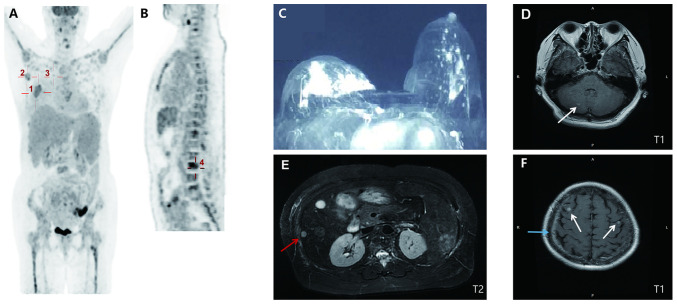
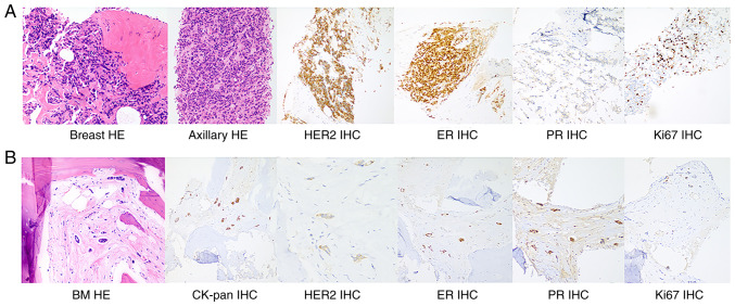
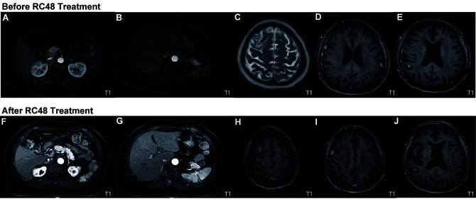
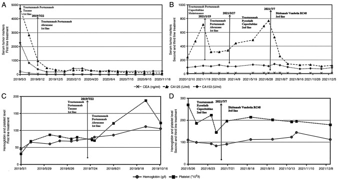
Breast cancer (BC) with overexpression of human epidermal growth factor receptor 2 (HER2) is closely associated with an elevated risk of multiple distant metastases and unfavorable prognosis. Disitamab Vedotin (RC48) is a newly developed antibody-drug conjugate targeting HER2, which is comprised of hertuzumab coupled to monomethyl auristatin E via a cleavable linker. Pre-clinical studies indicated its strong anti-tumor activity in HER2-positive and low HER2 expression models of BC. The present study reported on the case of a 60-year-old postmenopausal female who suffered from fatigue and was diagnosed with a right-sided BC tumor. The diagnosis was stage IV (cT4N3M1) hormone receptor (HR)-positive and HER2-positive invasive ductal carcinoma with systemic metastases (brain included). The patient initially responded well to 26 cycles of the first-line anti-HER2 targeted therapy plus chemotherapy (trastuzumab+pertuzumab+nab-paclitaxel) combined with whole-brain radiotherapy. However, both extracranial and intracranial lesions achieved progressive disease (PD), which eventually occurred during 5 sequential cycles of maintenance therapy. Subsequently, 4 cycles of second-line treatment (trastuzumab + pyrotinib + capecitabin) were continued until the levels of blood tumor markers CEA, CA15-3 and CA125 were elevated, and systemic PD was able to be attained (the brain metastases were rated as stable disease). Finally, the patient received RC48 as the third-line therapy and achieved a durable and effective clinical response. To date, the patient has benefited from 12 cycles of RC48 without any severe adverse effects. The overall survival was >3 years. The present study showcased that RC48 was effective and tolerable for a patient with HR- and HER2-positive BMBC.

Keywords: ADC; HER2-positive breast cancer; RC48; anti-HER2 target therapy; metastatic breast cancer.


Conflict of interest statement

The authors declare that they have no competing interests.

Figures

Figure 1.
Figure 1.
Baseline imaging presentation. (A) PET/CT (10 days after initial presentation; coronal section) indicated a malignant tumor in the right breast (30.1×25.3 mm2; SUVmax, 5.2; marked by cross 1) with right-sided axillary (20.7×11.2 mm2; SUVmax, 3.2; and 17.4×9.8 mm; SUVmax, 6.0; marked by cross 2) and internal mammary (19.1×11.5 mm2; SUVmax, 2.6; marked by cross 3) metastatic lymph nodes. (B) PET/CT (median sagittal section) indicated systemic bone metastases in spine (marked by cross 4), bilateral ribs and sternum. (C) Contrast-enhanced breast CT (at the initial presentation) indicated an irregular mass with an unclear margin in the areola area of the right breast (31×15×22 mm3; breast imaging-reporting and data system score of 5). (D) Cranial T1 weighted MRI (15 days after initial presentation) revealed nodular lesions in the right cerebellum (marked by the arrow). (E) Abdominal T2-weighted MRI (14 days after initial presentation) suggested metastases in the liver with multiple abnormal round signaling shadows (one is 8 mm in diameter, marked by the red arrow). (F) Cranial T1 weighted MRI revealed nodular lesions in bilateral frontal lobes (marked by white arrows) with leptomeningeal enhancement (marked by the blue arrow). SUVmax, maximum standardized uptake value; PET, positron emission tomography.
Figure 2.
Figure 2.
H&E staining and IHC staining of the breast mass, axillary lymph nodes and bone marrow metastases. (A) H&E staining of breast tissue and axillary lymph nodes suggested grade II invasive ductal carcinoma with the following IHC staining results: HER2 (+++), ER (++), PR (−) and Ki67 (20%). (B) Histology of bone marrow biopsy indicated atypical cells with the following IHC staining results: CK-pan (+), HER2 (+), ER (+), PR (+) and Ki67 (<5%), which are consistent with bone marrow metastasis (magnification, ×200). IHC, immunohistochemistry; HER2, human epidermal growth factor receptor 2; ER, estrogen receptor; PR, progesterone receptor; CK, cytokeratin; BM, bone marrow.
Figure 3.
Figure 3.
Comparison of MRI prior to (June, 2021) and after (December, 2021) third-line treatment based on the newest evidence. On contrast-enhanced abdominal T1-weighted MRI, abnormal small nodular signals were observed in (A and F) segment II of the left lobe and in segment VI of the right lobe of the liver, and (B and G) a wedge-shaped abnormal signal area was observed in the subcapsular of the spleen (10×13 mm2). Contrast-enhanced brain MRI indicated (Cand H) multiple brain metastases in the bilateral frontal lobes, (D and I) leptomeninges and (E and J) right parietal lobe. The results indicated that there was no obvious progression after administration of RC48 and the imaging findings were rated as stable disease.
Figure 4.
Figure 4.
Levels of three serum tumor markers measured (A) after the first-line therapy and (B) after the other treatment strategies had been applied. (A) The patient responded well to the initial 3 and 14 cycles of the first-line therapy, as the levels of all three tumor markers significantly decreased. (B) CA-125 levels continuously increased with the maintenance and second-line therapies. Single-dose RC48 significantly reduced the levels of the three tumor markers. Measurement of the hemoglobin level and platelet count (C) after the first-line treatment and (D) during the second- and third-line therapy. (C) After transfusion of red blood cells and injection of RHTI, first-line chemotherapy was initiated. Both hemoglobin levels and platelet count increased after 3 cycles of the PPH regimen and 5 cycles of the PHA regimen. (D) The hemoglobin level and platelet count of the patient of the present study remained stable during administration of RC48, which indicated that RC48 hardly caused damage to the hematopoietic function. Regimens: RHTI, recombinant human thrombopoietin injection; PPH, trastuzumab (6 mg/kg) on day 0 (the first dose, 8 mg/kg) + pertuzumab (420 mg) on day 0 (the first dose, 840 mg) + paclitaxel 120 mg on day 1, 150 mg on day 8, q3w; PHA, trastuzumab + pertuzumab + nab-paclitaxel 200 mg on days 1 and 8, q3w; RC48, Disitamab Vedotin.

Similar articles

Cited by

References

    1. Slamon DJ, Clark GM, Wong SG, Levin WJ, Ullrich A, McGuire WL. Human breast cancer: Correlation of relapse and survival with amplification of the HER-2/neu oncogene. Science. 1987;235:177–182. doi: 10.1126/science.3798106. - DOI - PubMed
    1. Moasser MM. The oncogene HER2: Its signaling and transforming functions and its role in human cancer pathogenesis. Oncogene. 2007;26:6469–6487. doi: 10.1038/sj.onc.1210477. - DOI - PMC - PubMed
    1. Harbeck N, Penault-Llorca F, Cortes J, Gnant M, Houssami N, Poortmans P, Ruddy K, Tsang J, Cardoso F. Breast cancer. Nat Rev Dis Primers. 2019;5:66. doi: 10.1038/s41572-019-0111-2. - DOI - PubMed
    1. Klos KJ, O'Neill BP. Brain metastases. Neurologist. 2004;10:31–46. doi: 10.1097/01.nrl.0000106922.83090.71. - DOI - PubMed
    1. Kennecke H, Yerushalmi R, Woods R, Cheang MC, Voduc D, Speers CH, Nielsen TO, Gelmon K. Metastatic behavior of breast cancer subtypes. J Clin Oncol. 2010;28:3271–3277. doi: 10.1200/JCO.2009.25.9820. - DOI - PubMed