. 2022 Sep 1;26(5):389-396.
doi: 10.4196/kjpp.2022.26.5.389.

HMGB1 increases RAGE expression in vascular smooth muscle cells via ERK and p-38 MAPK-dependent pathways

Affiliations

HMGB1 increases RAGE expression in vascular smooth muscle cells via ERK and p-38 MAPK-dependent pathways

Eun Jeong Jang et al. Korean J Physiol Pharmacol. .

Abstract

The increased expression of receptors for advanced glycation end-product (RAGE) is known as a key player in the progression of vascular remodeling. However, the precise signal pathways regulating RAGE expression in vascular smooth muscle cells (VSMCs) in the injured vasculatures are unclear. Given the importance of mitogen-activated protein kinase (MAPK) signaling in cell proliferation, we investigated the importance of MAPK signaling in high-mobility group box 1 (HMGB1)-induced RAGE expression in VSMCs. In HMGB1 (100 ng/ml)-stimulated human VSMCs, the expression of RAGE mRNA and protein was increased in association with an increase in AGE-induced VSMC proliferation. The HMGB1-induced RAGE expression was attenuated in cells pretreated with inhibitors for ERK (PD98059, 10 μM) and p38 MAPK (SB203580, 10 μM) as well as in cells deficient in ERK and p38 MAPK using siRNAs, but not in cells deficient of JNK signaling. In cells stimulated with HMGB1, the phosphorylation of ERK, JNK, and p38 MAPK was increased. This increase in ERK and p38 MAPK phosphorylation was inhibited by p38 MAPK and ERK inhibitors, respectively, but not by JNK inhibitor. Moreover, AGE-induced VSMC proliferation in HMGB1-stimulated cells was attenuated in cells treated with ERK and p38 MAPK inhibitors. Taken together, our results indicate that ERK and p38 MAPK signaling are involved in RAGE expression in HMGB1-stimulated VSMCs. Thus, the ERK/p38 MAPK-RAGE signaling axis in VSMCs was suggested as a potential therapeutic target for vascular remodeling in the injured vasculatures.

Keywords: HMGB1; MAPK; RAGE; Vascular remodeling; Vascular smooth muscle cells.


Conflict of interest statement

CONFLICTS OF INTEREST

The authors declare no conflicts of interest.

Figures

Fig. 1
Fig. 1. Effects of HMGB1 on RAGE expression in hVSMCs.
The cultured hVSMCs were stimulated with HMGB1 (100 ng/ml) for 0 to 48 h, and then the expression of RAGE mRNA and protein was determined by using RT-PCR and Western blot, respectively. GAPDH and β-Actin were used as internal controls. Blots are representative of 5 independent experiments. Bottom: Relative intensities were quantified and expressed as the means ± SEMs of 5 independent experiments. HMGB1, high-mobility group box 1; RAGE, receptors for advanced glycation end-product; hVSMCs, human vascular smooth muscle cells; MW, molecular weight. *p < 0.05, **p < 0.01 vs. corresponding value at 0 h.
Fig. 2
Fig. 2. Effects of HMGB1 on AGE-induced proliferation of hVSMCs.
The cultured hVSMCs were stimulated with HMGB1 (100 ng/ml) for 24 h, and then cell proliferation was induced by AGE (30 μg/ml) for 24 h in the presence or absence of anti-RAGE (10 μg/ml). Cell proliferation determined by an MTT assay was quantified and expressed as the means ± SEMs of 3–4 independent experiments. HMGB1, high-mobility group box 1; hVSMCs, human vascular smooth muscle cells. **p < 0.01 vs. corresponding control in non-AGE, ##p < 0.01 vs. corresponding control in vehicle, and ††p < 0.01 vs. corresponding anti-RAGE effects in vehicle.
Fig. 3
Fig. 3. Characteristics of ERK, JNK, and p38 MAPK phosphorylation in HMGB1-stimulated hVSMCs.
hVSMCs were stimulated with HMGB1 (100 ng/ml) for 0 to 24 h, and then the levels of phosphorylated and total ERK (A), JNK (B), and p38 MAPK (C) were determined by Western blotting. Blots are representative of 3–4 independent experiments. Bottom: Relative intensities of phosphorylated to total ERK, JNK, and p38 MAPK were quantified and expressed as the means ± SEMs of 3–4 independent experiments. MAPK, mitogen-activated protein kinase; HMGB1, high-mobility group box 1; hVSMCs, human vascular smooth muscle cells. *p < 0.05, **p < 0.01 vs. value in 0 h.
Fig. 4
Fig. 4. Involvement of MAPK signaling in HMGB1-induced RAGE expression in hVSMCs.
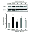
hVSMCs were treated with inhibitors of ERK (PD98059, 10 μM), JNK (SP600125, 10 μM), and p38 MAPK (SB203580, 10 μM) for 30 min, and then cells were stimulated with HMGB1 (100 ng/ml) for 24 h. The expression of RAGE in HMGB1-stimulated cells was determined by Western blot using β-Actin as an internal control. Blots are representative of 5 independent experiments. Bottom: Relative RAGE intensities to β-Actin were quantified, and data were expressed as the means ± SEMs of 5 independent experiments. MAPK, mitogen-activated protein kinase; HMGB1, high-mobility group box 1; RAGE, receptors for advanced glycation end-product; hVSMCs, human vascular smooth muscle cells. **p < 0.01 vs. corresponding value in control. #p < 0.05 vs. corresponding value in vehicle.
Fig. 5
Fig. 5. Individual role of MAPK signaling in HMGB1-induced RAGE expression in hVSMCs.
hVSMCs were transfected with siRNAs for (A) ERK (10 μM), (B) JNK (10 μM), and (C) p38 MAPK (10 μM) for 48 h, and then cells were stimulated with HMGB1 (100 ng/ml) for 24 h. RAGE expression in HMGB1-stimulated cells was determined by Western blot. β-Actin was used as internal controls. Blots are representative of 5 independent experiments. Bottom: Relative intensities of MAPK subfamilies and RAGE to β-Actin were quantified and expressed as the means ± SEMs of 5 independent experiments. MAPK, mitogen-activated protein kinase; HMGB1, high-mobility group box 1; RAGE, receptors for advanced glycation end-product; hVSMCs, human vascular smooth muscle cells. **p < 0.01 vs. corresponding value in negative control.
Fig. 6
Fig. 6. Crosstalk among MAPK subfamilies in HMGB1-stimulated hVSMCs.
To determine crosstalk among ERK, JNK, and p38 MAPK signaling, the phosphorylation of ERK and p38 MAPK in cells stimulated with HMGB1 was determined in the presence of various inhibitors for MAPKs. (A) hVSMCs were treated with inhibitors for p38 MAPK (SB203580, 10 μM) and JNK (SP600125, 10 μM) for 30 min, and then stimulated with HMGB1 (100 ng/ml) for 24 h. The levels of phosphorylated and total ERK were determined by Western blotting. Blots are representative of 5 independent experiments. (B) hVSMCs were treated with inhibitors for ERK (PD98059, 10 μM) and JNK (SP600125, 10 μM) for 30 min, and then stimulated with HMGB1 (100 ng/ml) for 24 h. The levels of phosphorylated and total p38 MAPK were determined by Western blotting. Blots are representative of 5 independent experiments. Bottom: Relative intensities of phosphorylated to total ERK and p38 MAPK were quantified and expressed as the means ± SEMs of 5 independent experiments. MAPK, mitogen-activated protein kinase; HMGB1, high-mobility group box 1; hVSMCs, human vascular smooth muscle cells. *p < 0.05, **p < 0.01 vs. corresponding controls. #p < 0.05 vs. corresponding vehicle.
Fig. 7
Fig. 7. Role of ERK and p38 MAPK signaling on HMGB1-augmented proliferation in AGE-stimulated VSMCs.
hVSMCs were pre-treated with HMGB1 (100 ng/ml) for 24 h in the presence of various inhibitors for MAPKs including ERK (PD98059, 10 μM), JNK (SP600125, 10 μM), and p38 MAPK (SB203580, 10 μM), and then stimulated with AGE (30 μg/ml) for 24 h. Cell proliferation was determined by an MTT assay. Relative cell proliferation was quantified and expressed as the means ± SEMs of 5 independent experiments. MAPK, mitogen-activated protein kinase; HMGB1, high-mobility group box 1; VSMCs, vascular smooth muscle cells; hVSMCs, human VSMCs. **p < 0.01 vs. corresponding control, #p < 0.05 vs. corresponding value in non-HMGB1, ††p < 0.01 vs. corresponding value in vehicle.

Similar articles

Cited by

References

    1. Land WG. Chronic allograft dysfunction: a model disorder of innate immunity. Biomed J. 2013;36:209–228. doi: 10.4103/2319-4170.117622. - DOI - PubMed
    1. Hreggvidsdottir HS, Ostberg T, Wähämaa H, Schierbeck H, Aveberger AC, Klevenvall L, Palmblad K, Ottosson L, Andersson U, Harris HE. The alarmin HMGB1 acts in synergy with endogenous and exogenous danger signals to promote inflammation. J Leukoc Biol. 2009;86:655–662. doi: 10.1189/jlb.0908548. - DOI - PubMed
    1. Kalinina N, Agrotis A, Antropova Y, DiVitto G, Kanellakis P, Kostolias G, Ilyinskaya O, Tararak E, Bobik A. Increased expression of the DNA-binding cytokine HMGB1 in human atherosclerotic lesions: role of activated macrophages and cytokines. Arterioscler Thromb Vasc Biol. 2004;24:2320–2325. doi: 10.1161/01.ATV.0000145573.36113.8a. - DOI - PubMed
    1. Cai Y, Sukhova GK, Wong HK, Xu A, Tergaonkar V, Vanhoutte PM, Tang EH. Rap1 induces cytokine production in pro-inflammatory macrophages through NFκB signaling and is highly expressed in human atherosclerotic lesions. Cell Cycle. 2015;14:3580–3592. doi: 10.1080/15384101.2015.1100771. - DOI - PMC - PubMed
    1. Zou H, Yang Y, Gao M, Zhang B, Ming B, Sun Y, Chen H, Tang X, Chen Z, Xiong P, Xu Y, Fang M, Tan Z, Gong F, Zheng F. HMGB1 is involved in chronic rejection of cardiac allograft via promoting inflammatory-like mDCs. Am J Transplant. 2014;14:1765–1777. doi: 10.1111/ajt.12781. - DOI - PubMed