. 2022 Dec;11(1):2147-2159.
doi: 10.1080/22221751.2022.2114852.

Serodiagnosis and therapeutic monitoring of New-World tegumentary leishmaniasis using synthetic type-2 glycoinositolphospholipid-based neoglycoproteins

Affiliations

Serodiagnosis and therapeutic monitoring of New-World tegumentary leishmaniasis using synthetic type-2 glycoinositolphospholipid-based neoglycoproteins

Sayonara M Viana et al. Emerg Microbes Infect. 2022 Dec.

Abstract

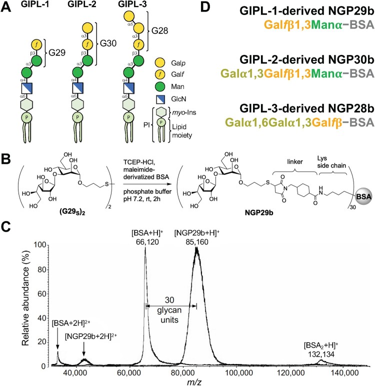
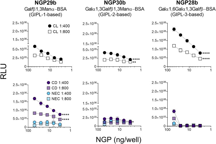
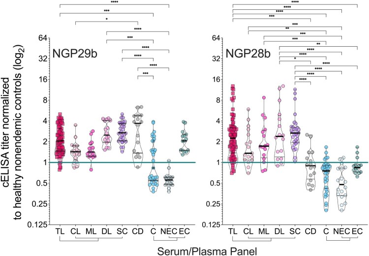
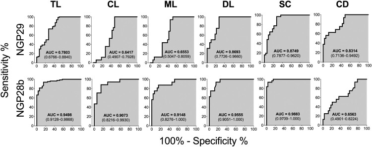
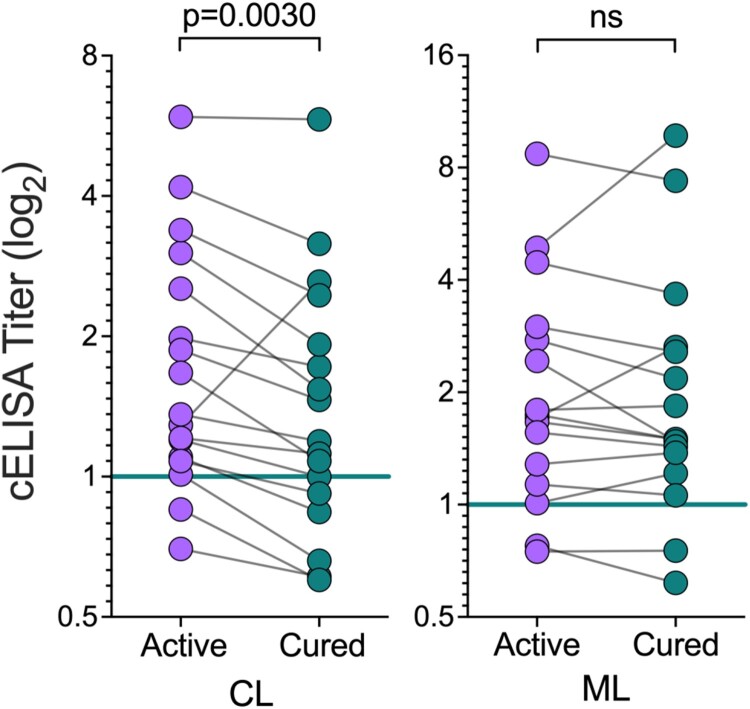
American tegumentary leishmaniasis (TL) caused by Leishmania braziliensis is characterized by a spectrum of clinical presentations, ranging from localized cutaneous ulcers (CL), mucosal (ML), or disseminated (DL) disease, to a subclinical (SC) asymptomatic form. Current diagnosis based on parasite culture and/or microscopy lacks sensitivity and specificity. Previous studies showed that patients with CL and ML have very high levels of Leishmania-specific anti-α-Gal antibodies. However, the native parasite α-Gal glycotope(s) is(are) still elusive, thus they have not yet been explored for a more accurate TL diagnosis. Using a chemiluminescent immunoassay, we evaluated the seroreactivity of TL patients across its clinical spectrum, and of endemic (EC) and nonendemic healthy controls (NEC) against three synthetic neoglycoproteins (NGP29b, NGP30b, and NGP28b), respectively comprising the L. major-derived type-2 glycoinositolphospholipid (GIPL)-1 (Galfβ1,3Manα), GIPL-2 (Galα1,3Galfβ1,3Manα), and GIPL-3 (Galα1,6Galα1,3Galfβ) glycotopes. Contrary to NGP29b and NGP30b, NGP28b exhibited high sensitivity and specificity to a CL serum pool. More importantly, NGP28b reacted strongly and specifically with individual sera from distinct clinical forms of TL, especially with SC sera, with 94% sensitivity and 97% specificity, by post-two-graph receiver-operating characteristic curve analysis. Contrary to NGP29b, NGP28b showed low cross-reactivity with Chagas disease and control (NEC/EC) sera. Additionally, seroreactivity of CL patients against NGP28b was significantly decreased after successful chemotherapy, indicating that L. braziliensis-specific anti-α-Gal antibodies may serve as an early biomarker of cure in CL. Our data also points towards the applicability of L. major type-2 GIPL-3-derived Galα1,6Galα1,3Galfβ glycotope for the serological diagnosis of American TL, particularly of the subclinical form.

Keywords: Leishmania braziliensis; anti-α-Gal antibodies; diagnostic and prognostic biomarkers; tegumentary leishmaniasis; α-Gal neoglycoproteins.


Figures

Figure 1
Figure 1
. Synthetic neoglycoproteins used in this study. (A) Schematic representation of type-2 GIPLs 1-3 of L. major. The terminal glycan moiety (G29, G30, or G28) targeted for chemical synthesis in each GIPL is indicated. Galp, galactopyranose; Galf, galactofuranose; Man, mannopyranose; GlcN, glucosamine; myo-Ins, myo-inositol; P, phosphate; PI, phosphatidylinositol. (B) Schematic representation of the synthesis of NGP29b containing the type-2 GIPL-1 terminal, nonreducing glycotope Galfβ1,3Manα. TCEP-HCl, Tris (2-carboxyethyl) phosphine hydrochloride; linker, 4-(succinimidomethyl)cyclohexane-1-carboxy group. The same conjugation was used for the synthesis of NGP30b and NGP28b [24]. (C) Representative MALDI-TOF-MS spectrum of NGP29b to confirm the covalent conjugation of the glycan units to the carrier protein, as recently described [32]. The same quality-control procedure was used for NGP30b and NGP28b, as previously described [24]. Doubly charged ([BSA+2H]+2 and [NGP29b+2H]+2) and singly charged ([BSA + H]+, [NGP29b + H]+, and [BSA2 + H]+) ions of BSA and NGP29b are indicated. The number of glycan units (n = 30) covalently attached to the BSA moiety is indicated. m/z, mass to charge ratio. (D) Composition of the synthetic NGP29b, NGP30b, and NGP28b. For simplicity, the glycan thiopropyl group (at the reducing end), the linker covalently attached to the lysine residue, and the number of glycan units shown in B, are not indicated.
Figure 2.
Figure 2.
Cross-titration of NGPs with serum pools from CL, Chagas disease, and nonendemic controls. cELISA tests were performed with NGP29b, NGP28b, or NGP30b at concentrations ranging from 50 to 3.13 ng/well, using a pool of sera (n = 15) from patients with active CL, caused by L. braziliensis. Pools of sera obtained from patients with chronic Chagas disease (CD) (n = 15), and from nonendemic healthy controls (NEC) (n = 15) were also evaluated. Each point represents the mean of duplicate values of the relative luminescence units (RLU) obtained for each sample and bars indicate SD. Statistical analysis: two-way Anova with main effects only and Dunnett’s multiple comparison test (with individual variances computed for each comparison). The CL and CD curve were compared with the NEC curve, at 1:400 and 1:800 serum pool dilution. *p < 0.05, **p < 0.01,****p < 0.0001; statistically non-significant differences are not shown.
Figure 3.
Figure 3.
Normalized IgG response of sera from patients with tegumentary leishmaniasis (TL) to L. major type-2 GIPL-1-derived NGP29b and GIPL-3-derived NGP28b. cELISA immunoassays were performed using NGPs at 5 ng/well and serum samples (1:800 dilution) from all TL samples (n = 80), with different clinical forms (CL; n = 17; ML n = 16; DL, n = 16; and SC, n = 31) plotted separately; Chagas disease (CD, n = 16); and all non-TL, seemingly healthy controls (NEC + EC; n = 33), also plotted separately (EC, n = 15; and NEC, n = 18). Each point represents the mean of triplicate relative luminescence units (RLU) values normalized to NEC serum pools. The cutoff value (cELISA titer = 1.000), calculated as described in Materials and Methods, is indicated by the continuous green line. Data are represented as violin plots (truncated) of individual points, with median (thick black line) and interquartile range (dotted black lines) values indicated. *p < 0.05, **p < 0.01, ****p < 0.0001, Kruskal Wallis followed by Dunn’s multiple comparison tests. Statistically non-significant differences between serum groups are not shown.
Figure 4.
Figure 4.
Receiver-operating characteristic (ROC) curves for NGP29b and NGP28b comparing the reactivity of sera from total TL patients or CL, ML, DL, SC, or CD patients versus control sera from endemic (EC) and nonendemic (NEC) individuals, using cELISA titers normalized to NECs. The area under the curve (AUC) is indicated in the gray area, and 95% confidence interval (CI) values are indicated in parentheses.
Figure 5.
Figure 5.
Seroreactivity to NGP28b of TL patients, before and after treatment. Sera from CL (n = 17) or ML (n = 16) patients, obtained before and 90 days after standard Sbv treatment, were probed by cELISA with NGP28b (5 ng/well). Each point represents the mean of triplicate RLU values normalized to a pool of sera from seemingly healthy NEC individuals. p Values were calculated using Wilcoxon matched-pairs test. Significance level: p < 0.05.

References

    1. Goto H, Lindoso JA.. Current diagnosis and treatment of cutaneous and mucocutaneous leishmaniasis. Expert Rev Anti Infect Ther. 2010;8(4):419–433. - PubMed
    1. Ramírez JL, Guevara P.. Persistent infections by Leishmania (Viannia) braziliensis. Mem Inst Oswaldo Cruz. 1997;92(3):333–338. - PubMed
    1. Amato VS, Tuon FF, Bacha HA, et al. . Mucosal leishmaniasis. Current scenario and prospects for treatment. Acta Trop. 2008;105:1–9. - PubMed
    1. Gaze ST, Dutra WO, Lessa M, et al. . Mucosal leishmaniasis patients display an activated inflammatory T-cell phenotype associated with a nonbalanced monocyte population. Scand J Immunol. 2006;63(1):70–78. - PubMed
    1. Scorza B, Carvalho E, Wilson M.. Cutaneous manifestations of human and murine leishmaniasis. Int J Mol Sci. 2017;18(6):1296–1296. - PMC - PubMed