Review
. 2023 Jan 1;96(1141):20220336.
doi: 10.1259/bjr.20220336. Epub 2022 Sep 12.

Sural nerve: imaging anatomy and pathology

Affiliations
Review

Sural nerve: imaging anatomy and pathology

Logan Joseph Jackson et al. Br J Radiol. .

Abstract

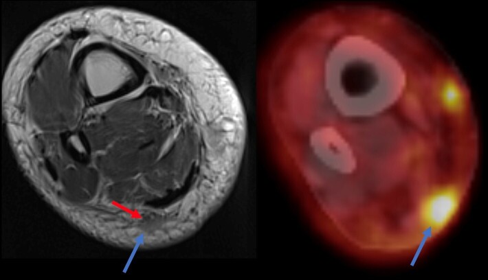
High resolution ultrasound (US) and magnetic resonance (MR) neurography are both imaging modalities that are commonly used for assessing peripheral nerves including the sural nerve (SN). The SN is a cutaneous sensory nerve which innervates the lateral ankle and foot to the base of the fifth metatarsal. It is formed by contributing nerves from the tibial and common peroneal nerves with six patterns and multiple subtypes described in literature. In addition to the SN being a cutaneous sensory nerve, the superficial location enables the nerve to be easily biopsied and harvested for a nerve graft, as well as increasing the susceptibility to traumatic injury. As with any peripheral nerves, pathologies such as peripheral nerve sheath tumors and neuropathies can also affect the SN. By utilizing a high frequency probe in US and high-resolution MR neurography, the SN can be easily identified even with the multiple variations given the standard distal course. US and MRI are also useful in determining pathology of the SN given the specific image findings that are seen with peripheral nerves. In this review, we evaluate the normal imaging anatomy of the SN and discuss common pathologies identified on imaging.


Figures

Figure 1.
Figure 1.
Illustrations showing the course and the origin of the sural nerve from branches of the tibial and common peroneal nerves and the innervated territory of the sural nerve and its components
Figure 2.
Figure 2.
Normal ultrasound image of the sural nerve (SN) in the leg. At the popliteal fossa, image (a) shows the origin of the medial sural cutaneous nerve (MSCN) from the tibial nerve (TN) with probe positioning. At the popliteal fossa, image (b) shows the origin of the lateral sural cutaneous nerve (LSCN) from the common peroneal nerve (CPN) with probe positioning. Image (c) shows the medial and lateral sural cutaneous nerves (MSCN, LSCN) just prior to their fusion in the mid to distal calf and image (d) shows the origin of the SN after fusion of the MSCN and LSCN. Image (e) is a longitudinal image of the SN in the calf. Image (f) shows a transverse gray scale and corresponding color Doppler images at the distal leg level showing the sural nerve (SN) satellite to the small saphenous vein (SSV). Transverse gray scale image (g) at the ankle level showing the sural nerve with probe positioning. The SN is posterior to the peroneal tendons and satellite to the SSV
Figure 3.
Figure 3.
Normal MRI anatomy of the sural nerve. The sural is formed by the fusion of the medial and lateral sural cutaneous branches (arrows in a, (b, c) at the mid to distal leg level then courses in the superficial posterolateral soft tissue adjacent the small saphenous vein to the ankle
Figure 4.
Figure 4.
60-year-old female with peroneal brevis tendon rupture and sural neuropathy. Transverse (a) and longitudinal (b) ultrasound cuts at the level of the ankle shows multifocal nodular thickening of the sural nerve (arrows) without nerve transection consistent with sural neuropathy
Figure 5.
Figure 5.
51-year-old male with symptoms of sural neuropathy and prior history of ankle ORIF for ankle fracture. Longitudinal (a) and axial (b, c) ultrasound cuts at the lateral distal leg level demonstrates focal nodular thickening of the sural nerve at the level of the ankle (blue arrows) adjacent to the soft tissue scar (red arrows) consistent with sural neuropathy without nerve transaction
Figure 6.
Figure 6.
60-year-old female with a biopsy confirmed Merkel cell carcinoma. There is a mass in the posterior subcutaneous fat in the distal calf (blue arrow) that is FDG avid consistent with a tumor deposit. The mass encases the sural nerve (red arrow). The patient had symptoms of sural neuropathy
Figure 7.
Figure 7.
50-year-old female with a palpable mass along the posterolateral mid calf. Axial T1 (a) and axial T2 (b) weighted images demonstrates a low signal intensity mass (arrows) showing diffuse enhancement (c). Coronal T1 -weighted image (d) shows the mass (asterix) along the course of the sural nerve (arrow). Pathology demonstrates desmoid fibromatosis
Figure 8.
Figure 8.
47-year-old female with a palpable mass along the lateral ankle. Axial T2 FS (a) shows a cystic high signal lesion abutting the sural nerve (arrow). Axial T1 (b), the lesion is low signal abutting and causing mass effect on the sural nerve (arrow). There is no enhancement of the lesion post contrast (c). More inferiorly (d), the lesion tracks to the anterior ankle joint and is consistent with a ganglion cyst. Coronal STIR (e) image shows the lesion and the mass effect on the sural nerve that is slightly hyperintense (arrows). Ultrasound images (f, g, h) show the cystic lesion superficial to the sural nerve (arrows). There is thickening and hypo echogenicity of a segment of the sural nerve suggestive of neuropathy
Figure 9.
Figure 9.
71-year-old male patient with a palpable abnormality in the popliteal fossa. Ultrasound (a, b) demonstrates a vascularized ovoid hypoechoic mass that is in communication with a superficial nerve branch (arrows). MRI demonstrates a T2 hyperintense mass lesion splaying the distal sciatic nerve at its bifurcation with mass effect on the common peroneal and tibial nerves origin (c). Sagittal T1 and coronal PD images demonstrate the origin of the mass from the lateral sural cutaneous branch (arrows in d and e), a branch from the CPN. Pathology revealed a peripheral nerve sheath tumor
Figure 10.
Figure 10.
36-year-old female referred for palpable painful lesion in the lateral ankle with a clinical suspicion for a foreign body. Ultrasound demonstrates an oval hypoechoic lesion in continuity with the sural nerve (a) and is superficial to the peroneal tendons (b). Doppler ultrasound demonstrates mild vascularity of the lesion (c). Pathology demonstrates a schwannoma of the sural nerve

References

    1. Riedl O, Frey M. Anatomy of the sural nerve: cadaver study and literature review. Plast Reconstr Surg 2013; 131: 802–10. doi: 10.1097/PRS.0b013e3182818cd4 - DOI - PubMed
    1. Wang F, Zhou D, Li W, Ge M, Zhu D. A new pattern of the sural nerve added to “anatomy of the sural nerve: cadaver study and literature review.” Plast Reconstr Surg Glob Open 2017; 5. doi: 10.1097/GOX.0000000000001628 - DOI - PMC - PubMed
    1. Steele R, Coker C, Freed B, Wright B, Brauer P. Anatomy of the sural nerve complex: unaccounted anatomic variations and morphometric data. Ann Anat 2021; 238: 151742. doi: 10.1016/j.aanat.2021.151742 - DOI - PubMed
    1. Puma A, Grecu N, Villa L, Butori C, Besson T, Cambieri C, et al. . Ultra-high-frequency ultrasound imaging of sural nerve: a comparative study with nerve biopsy in progressive neuropathies. Muscle Nerve 2021; 63: 46–51. doi: 10.1002/mus.27073 - DOI - PubMed
    1. Watabe LT, Santos P, Mendes F de A, Lima C de, Acioly MA. Fibular nerve repair by lateral sural cutaneous nerve graft: anatomic feasibility study and clinical results. World Neurosurg 2021; 146: e537–43. doi: 10.1016/j.wneu.2020.10.127 - DOI - PubMed