. 2022 Sep 28;96(18):e0092122.
doi: 10.1128/jvi.00921-22. Epub 2022 Aug 30.

Discovery and Genomic Characterization of a Novel Henipavirus, Angavokely Virus, from Fruit Bats in Madagascar

Affiliations

Discovery and Genomic Characterization of a Novel Henipavirus, Angavokely Virus, from Fruit Bats in Madagascar

Sharline Madera et al. J Virol. .

Abstract

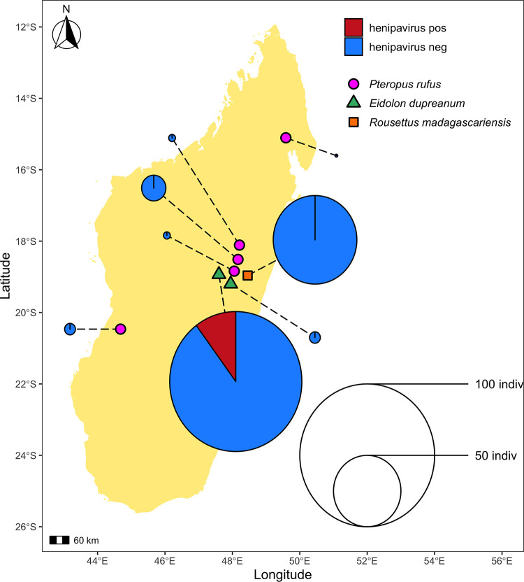
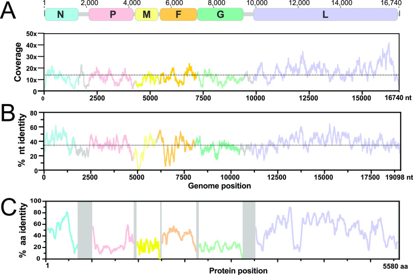
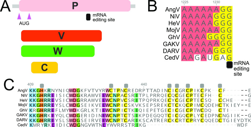
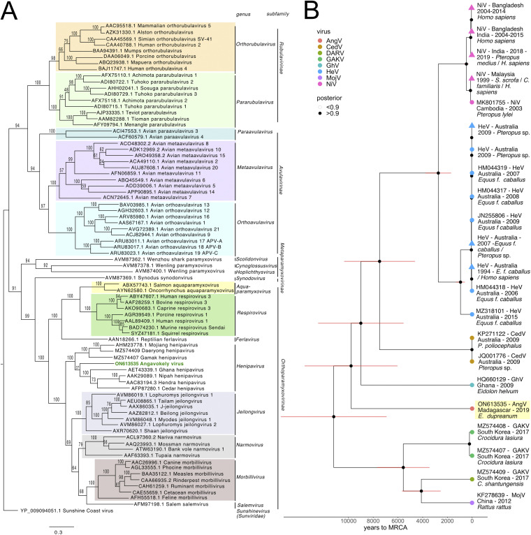
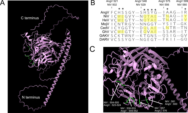
The genus Henipavirus (family Paramyxoviridae) currently comprises seven viruses, four of which have demonstrated prior evidence of zoonotic capacity. These include the biosafety level 4 agents Hendra (HeV) and Nipah (NiV) viruses, which circulate naturally in pteropodid fruit bats. Here, we describe and characterize Angavokely virus (AngV), a divergent henipavirus identified in urine samples from wild, Madagascar fruit bats. We report the nearly complete 16,740-nucleotide genome of AngV, which encodes the six major henipavirus structural proteins (nucleocapsid, phosphoprotein, matrix, fusion, glycoprotein, and L polymerase). Within the phosphoprotein (P) gene, we identify an alternative start codon encoding the AngV C protein and a putative mRNA editing site where the insertion of one or two guanine residues encodes, respectively, additional V and W proteins. In other paramyxovirus systems, C, V, and W are accessory proteins involved in antagonism of host immune responses during infection. Phylogenetic analysis suggests that AngV is ancestral to all four previously described bat henipaviruses-HeV, NiV, Cedar virus (CedV), and Ghanaian bat virus (GhV)-but evolved more recently than rodent- and shrew-derived henipaviruses, Mojiang (MojV), Gamak (GAKV), and Daeryong (DARV) viruses. Predictive structure-based alignments suggest that AngV is unlikely to bind ephrin receptors, which mediate cell entry for all other known bat henipaviruses. Identification of the AngV receptor is needed to clarify the virus's potential host range. The presence of V and W proteins in the AngV genome suggest that the virus could be pathogenic following zoonotic spillover. IMPORTANCE Henipaviruses include highly pathogenic emerging zoonotic viruses, derived from bat, rodent, and shrew reservoirs. Bat-borne Hendra (HeV) and Nipah (NiV) are the most well-known henipaviruses, for which no effective antivirals or vaccines for humans have been described. Here, we report the discovery and characterization of a novel henipavirus, Angavokely virus (AngV), isolated from wild fruit bats in Madagascar. Genomic characterization of AngV reveals all major features associated with pathogenicity in other henipaviruses, suggesting that AngV could be pathogenic following spillover to human hosts. Our work suggests that AngV is an ancestral bat henipavirus that likely uses viral entry pathways distinct from those previously described for HeV and NiV. In Madagascar, bats are consumed as a source of human food, presenting opportunities for cross-species transmission. Characterization of novel henipaviruses and documentation of their pathogenic and zoonotic potential are essential to predicting and preventing the emergence of future zoonoses that cause pandemics.

Keywords: Eidolon dupreanum; Madagascar; bat-borne virus; emerging zoonosis; henipavirus; novel virus.


Conflict of interest statement

The authors declare no conflict of interest.

Figures

FIG 1
FIG 1
Geographic location of sampling sites used in this study. Sampling sites grouped by bat species found depicted as follows: P. rufus (pink circles) Ambakoana (−18.51 S, 48.17 E)/Mahabo (−20.46 S, 44.68 E)/Mahialambo (−18.11 S, 48.21 E)/Makira (−15.11 S, 49.59 E)/Marovitsika (−18.84 S, 48.06 E) roosts; E. dupreanum (green triangles) Angavobe (−18.94 S, 47.95 E)/Angavokely (−18.93 S, 47.76 E) caves; R. madagascariensis (orange squares) Maromizaha cave (−18.96 S, 48.45). Dashed lines link each sampling site to corresponding pie charts, which show the percentage of HNV-positive specimens for all sampled species and sites. Dashed lines are included for visualization purposes only, and variation in length is not significant. Pies are size-weighted by the total bat population sampled at each site, corresponding to the legend. HNV-positive samples were only recovered from the E. dupreanum Angavokely site.
FIG 2
FIG 2
AngV genome organization. (A) Coding regions for each gene are shown and depicted in color; noncoding intergenic and terminal regions are highlighted in gray. Depicted genes represented as follows: nucleocapsid (N), phosphoprotein (P), matrix (M), fusion (F), glycoprotein (G), and RNA polymerase (L). Sequencing read depth supporting each position of the recovered genome sequence is plotted below the genomic schematic. Scanning nucleotide (B) and amino acid (C) pairwise identity to Nipah virus (GenBank accession number: AF212302). Dotted horizontal lines represent average read depth (14.29) or average nucleotide pairwise identity (36%).
FIG 3
FIG 3
Organization of the P gene of AngV. (A) Alternative transcriptional start sites (pink triangle) generate the P and C protein. Pseudotemplated addition of one or two guanine nucleotides at the putative mRNA editing site generates a V and W protein, respectively. (B) Sequence alignment of the putative mRNA editing site across members of the Henipavirus genus (cRNA depicted). (C) Amino acid alignment of the unique C-terminal region of the V protein following the addition of one guanine nucleotide to the putative mRNA editing site. Gray boxes denote conserved cysteine and histidine residues suggested to directly coordinate bound zinc ions (32). Individual nucleotides or amino acids are color coordinated if at least 75% conserved at the alignment position. Nucleotide or amino acid position numbers displayed represent the position within the AngV gene or protein. Virus name (abbreviation), followed by GenBank accession number: Angavokely virus (AngV) ON613535; Nipah virus (NiV) AF212302; Hendra virus (HeV) AF017149; Mojiang virus (MojV) KF278639; Ghanaian bat Henipavirus (GhV) HQ660129; Daeryong virus (DARV) MZ574409; Gamak virus (GAKV) MZ574407; Cedar virus (CedV) JQ001776. CedV is shown here only for comparison, as the CedV P protein is not believed to undergo RNA editing or to generate a functional V protein (8, 32).
FIG 4
FIG 4
(A) Phylogenetic analysis of the complete L protein sequences of members of the family Paramyxoviridae. We note that the sequence for the newly described Langya henipavirus (LayV) (12) was not yet available at the time of this writing and is, therefore, not included in the phylogenies. Tree is rooted with Sunshine Coast Virus (GenBank accession number: YP_009094051.1) as an outgroup, with outgroup branch length shrunk for ease of viewing. Novel HNV, AngV, is depicted in green. Subfamilies and genera are demarcated, excluding those unassigned to subfamily (genera Scolidonvirus, Cynoglossusvirus, Hoplichthysvirus). Bootstrap support is depicted and GenBank accession numbers displayed next to virus names. Scale bar represents substitutions per site. (B) Time-resolved Bayesian phylogeny computed in BEAST 2 incorporating all available Henipavirus whole-genome nucleotide sequences, with the addition of newly discovered GAKV, DARV, and AngV. Closely related sequences are collapsed at triangle nodes for NiV and HeV (phylogeny with uncollapsed branches available in Fig. S2 in the supplemental material). HPD intervals of 95% around the timing of each branching node are visualized as red horizontal bars. Posterior support of >0.9 is indicated by black coloring of the corresponding node, and distinct Henipavirus species are indicated by colored tip points, with AngV highlighted in yellow for further emphasis. The estimated time to MRCA for Angavokely virus and the previously described bat-borne HNVs is 9,794 (95% HPD 6,519 to 14,025) years ago.
FIG 5
FIG 5
AlphaFold-predicted AngV glycoprotein 3D structure and ephrin binding residue sequence alignment. (A) AlphaFold-predicted 3D structure of AngV glycoprotein. N and C termini are indicated in white text, and residues corresponding to ephrin binding sites in other HNVs that are not conserved in AngV are colored green. (B) Alignment of HNV ephrin binding residues. The position of previously described HNV ephrin binding residues are noted by a star, and residues conserved across most HNVs are highlighted in yellow. Amino acid position numbers displayed represent the position within the AngV or NiV glycoproteins. Virus name (abbreviations) followed by GenBank accession number: Angavokely virus (AngV) ON613535; Nipah virus (NiV) AF212302; Hendra virus (HeV) AF017149; Mojiang virus (MojV) KF278639; Cedar virus (CedV) JQ001776; Ghanaian bat Henipavirus (GhV) HQ660129; Gamak virus (GAKV) MZ574407; Daeryong virus (DARV) MZ574409. (C) AlphaFold detail from full structure prediction for AngV. Localization of ephrin binding sites conserved across most HNVs is colored green and labeled corresponding to position in the NiV and AngV genomes.

References

    1. Eaton BT, Broder CC, Middleton D, Wang L-F. 2006. Hendra and Nipah viruses: different and dangerous. Nat Rev Microbiol 4:23–35. 10.1038/nrmicro1323. - DOI - PMC - PubMed
    1. Sharma V, Kaushik S, Kumar R, Yadav JP, Kaushik S. 2019. Emerging trends of Nipah virus: a review. Rev Med Virol 29:e2010. 10.1002/rmv.2010. - DOI - PMC - PubMed
    1. Arunkumar G, Chandni R, Mourya DT, Singh SK, Sadanandan R, Sudan P, Bhargava B, Nipah Investigators People and Health Study Group . 2019. Outbreak investigation of Nipah virus disease in Kerala, India, 2018. J Infect Dis 219:1867–1878. 10.1093/infdis/jiy612. - DOI - PubMed
    1. Murray K, Selleck P, Hooper P, Hyatt a, Gould a, Gleeson L, Westbury H, Hiley L, Selvey L, Rodwell B. 1995. A morbillivirus that caused fatal disease in horses and humans. Science 268:94–97. 10.1126/science.7701348. - DOI - PubMed
    1. Hsu VP, Hossain MJ, Parashar UD, Ali MM, Ksiazek TG, Kuzmin I, Niezgoda M, Rupprecht C, Bresee J, Breiman RF. 2004. Nipah virus encephalitis reemergence, Bangladesh. Emerg Infect Dis 10:2082–2087. 10.3201/eid1012.040701. - DOI - PMC - PubMed

Publication types