. 2022 Sep;31(9):e4389.
doi: 10.1002/pro.4389.

Depicting the proton relay network in human aromatase: New insights into the role of the alcohol-acid pair

Affiliations

Depicting the proton relay network in human aromatase: New insights into the role of the alcohol-acid pair

Chao Zhang et al. Protein Sci. 2022 Sep.

Abstract

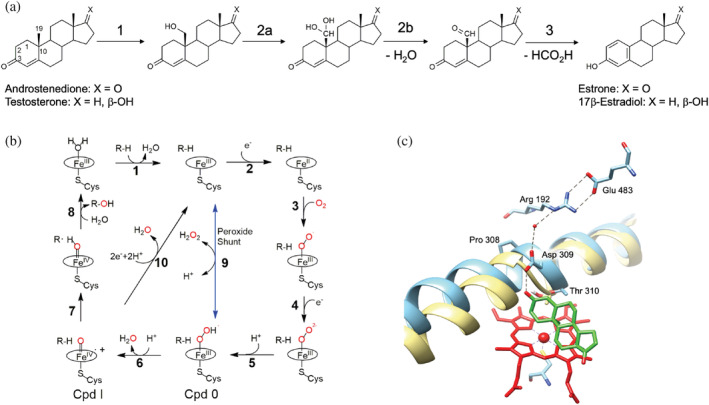
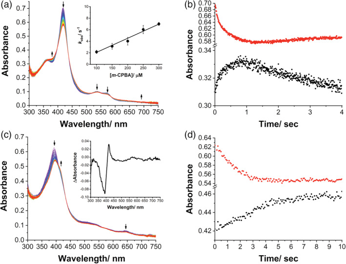
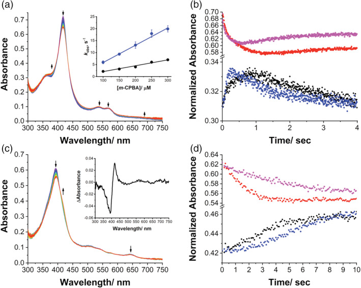
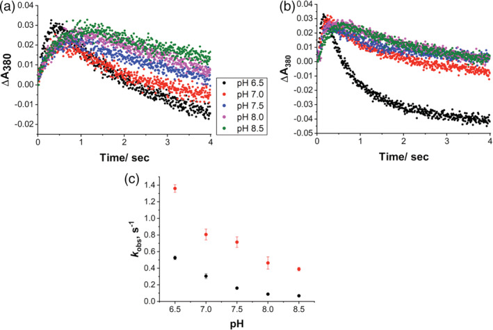
Human aromatase is the cytochrome P450 catalyzing the conversion of androgens into estrogens in a three steps reaction essential to maintain steroid hormones balance. Here we report the capture and spectroscopic characterization of its compound I (Cpd I), the main reactive species in cytochromes P450. The typical spectroscopic transitions indicating the formation of Cpd I are detected within 0.8 s when mixing aromatase with meta-chloroperoxybenzoic acid. The estrogen product is obtained from the same reaction mixture, demonstrating the involvement of Cpd I in aromatization reaction. Site-directed mutagenesis is applied to the acid-alcohol pair D309 and T310 and to R192, predicted to be part of the proton relay network. Mutants D309N and R192Q do not lead to Cpd I with an associated loss of activity, confirming that these residues are involved in proton delivery for Cpd I generation. Cpd I is captured for T310A mutant and shows 2.9- and 4.4-fold faster rates of formation and decay, respectively, compared to wild-type (WT). However, its activity is lower than the WT and a larger amount of H2 O2 is produced during catalysis, indicating that T310 has an essential role in proton gating for generation of Cpd 0 and Cpd I and for their stabilization. The data provide new evidences on the role of threonine belonging to the conserved "acid-alcohol" pair and known to be crucial for oxygen activation in cytochromes P450.

Keywords: alcohol-acid pair; aromatase; compound I; cytochromes P450; proton delivery.


Conflict of interest statement

The authors declare no conflicts of interest.

Figures

SCHEME 1
SCHEME 1
Reaction and catalytic mechanism of human aromatase. (a) The three‐step reaction catalyzed by aromatase allowing the conversion of androgens into estrogens. (b) Catalytic cycle of cytochromes P450 and possible uncoupling pathways. The peroxide shunt pathway can generate directly Cpd 0 (9) and Cpd I (6). (c) Active site of human aromatase (cyan, PDB ID 3S79) with the predicted proton relay network. The helix I is superimposed to the corresponding one in cytochrome P450cam (yellow, PDB ID 5CP4) to show the distortion in human aromatase due to the presence of Pro308. The heme is shown in red and the substrate androstenedione in green. Dashed black lines represent the predicted proton relay network.
FIGURE 1
FIGURE 1
Compound I formation in aromatase WT. (a) UV/Visible spectral changes in 0.8 s after mixing of 7 μM aromatase WT (final concentration) with 200 μM m‐CPBA (final concentration) in 100 mM potassium phosphate buffer, 10% glycerol, pH 7.5, at 4°C. Inset: plot of observed rate constants (k obs) for Cpd I formation against various concentrations of m‐CPBA at pH 7.5 and 4°C. (b) Kinetic traces of reaction between aromatase WT and m‐CPBA at 380 nm (black circles) and 418 nm (red circles) in 4.0 s. (c) Spectral changes observed upon mixing 7 μM aromatase (final concentration) in complex with the 19‐oxoandrostenedione with 200 μM m‐CPBA (final concentration) in 100 mM potassium phosphate buffer, 10% glycerol, pH 7.5, at 4°C. Inset: Difference spectrum obtained by subtracting the spectrum recorded after 4 s and the initial one. (d) Kinetic traces of reaction between aromatase WT in complex with the 19‐oxoandrostenedione and m‐CPBA at 380 nm (black circles) and 418 nm (red circles) in 4.0 s. WT, wild‐type.
FIGURE 2
FIGURE 2
Compound I formation in aromatase mutant T310A. (a) UV/Visible spectral changes in 0.8 s after mixing of 7 μM T310A (final concentration) with 200 μM m‐CPBA (final concentration) in 100 mM potassium phosphate buffer, 10% glycerol, pH 7.5, at 4°C. Inset: plot of observed rate constants (k obs) for Cpd I formation against various concentrations of m‐CPBA at pH 7.5 and 4°C for T310A (blue circles) and WT (black circles). (b) Kinetic traces of reaction between T310A and m‐CPBA at 380 nm (blue circles) and 418 nm (magenta circles) in 4.0 s. For comparison, the traces at 380 nm (black circles) and 418 nm (red circles) for WT are also shown. (c) Spectral changes observed upon mixing 7 μM T310A (final concentration) in complex with the 19‐oxoandrostenedione with 200 μM m‐CPBA (final concentration) in 100 mM potassium phosphate buffer, 10% glycerol, pH 7.5, at 4°C. Inset: Difference spectrum obtained by subtracting the spectrum recorded after 4 s and the initial one. (d) Kinetic traces of reaction between T310A in complex with 19‐oxoandrostenedione and m‐CPBA at 418 nm (black circles) and 394 nm (red circles) in 10.0 s. For comparison, the traces at 394 nm (black circles) and 418 nm (red circles) for WT are also shown. WT, wild‐type.
FIGURE 3
FIGURE 3
Effect of pH on Compound I formation and decay. (a) Kinetic traces at 380 nm of aromatase WT and (b) T310A at different pH. (c) Plot of the Cpd I decay constant (k 2) obtained for WT (black circles) and T310A (red circles) as a function of pH. WT, wild‐type.
FIGURE 4
FIGURE 4
Effect of pH on catalytic activity of WT and its mutants. The activity as a function of time was measured for (a) WT, (b) D309N, (c) R192Q, and (d) T310A. Reaction conditions: 0.5 μM enzyme, 0.5 μM CPR, 20 μM 19‐oxo ASD, 0.5 mM NADPH in 100 mM potassium phosphate buffer, and 10% glycerol, at 30°C. WT, wild‐type.

Similar articles

Cited by

References

    1. Guengerich FP. Common and uncommon cytochrome P450 reactions related to metabolism and chemical toxicity. Chem Res Toxicol. 2001;14:611–650. - PubMed
    1. de Montellano PRO. Cytochrome P450: Structure, mechanism, and biochemistry. 3rd ed. New York: Springer Science & Business Media, 2005.
    1. Denisov IG, Makris TM, Sligar SG, Schlichting I. Structure and chemistry of cytochrome P450. Chem Rev. 2005;105:2253–2277. - PubMed
    1. Guengerich FP. Human cytochrome P450 enzymes. In: Ortiz de Montellano PR, editor. Cytochrome P450: Structure, mechanism, and biochemistry. Cham: Springer International Publishing, 2015; p. 523–785.
    1. Di Nardo G, Gilardi G. Natural compounds as pharmaceuticals: The key role of cytochromes P450 reactivity. Trends Biochem Sci. 2020;45:511–525. - PubMed

Publication types

LinkOut - more resources