Review
. 2022 Sep;31(9):e4399.
doi: 10.1002/pro.4399.

The gateway to guanine nucleotides: Allosteric regulation of IMP dehydrogenases

Affiliations
Review

The gateway to guanine nucleotides: Allosteric regulation of IMP dehydrogenases

Rubén M Buey et al. Protein Sci. 2022 Sep.

Abstract

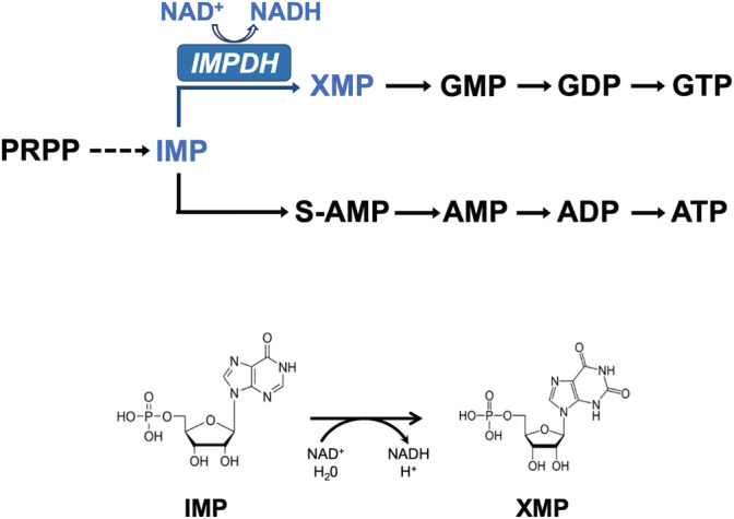
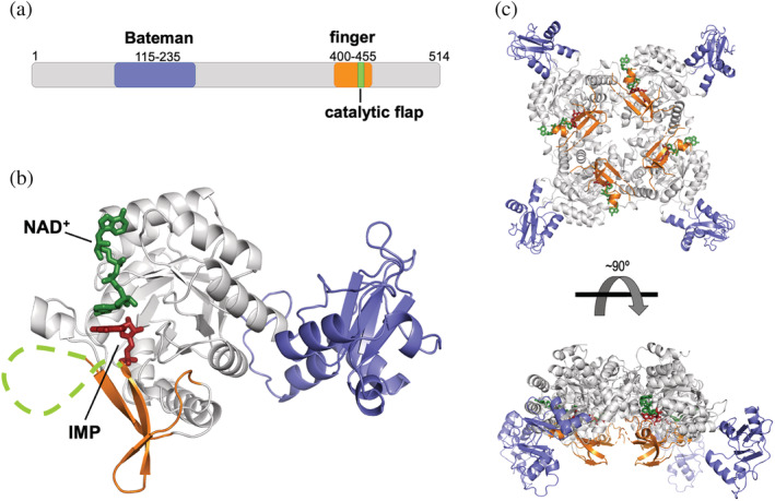
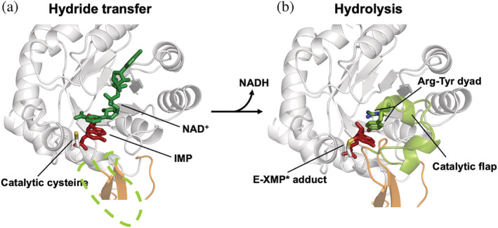
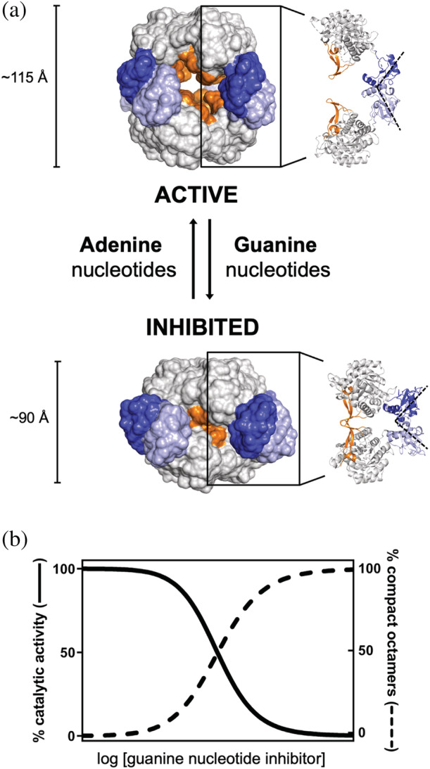
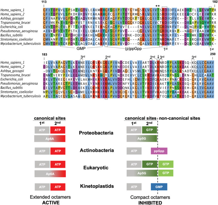
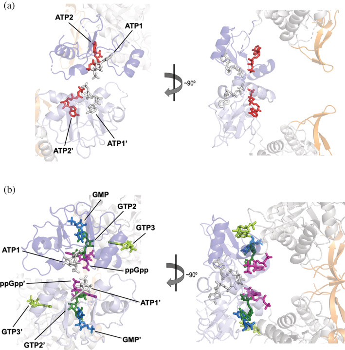
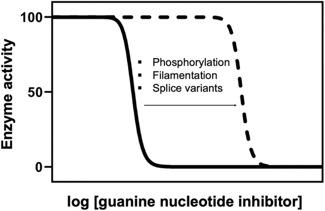
Inosine 5'-monophosphate dehydrogenase (IMPDH) is an evolutionarily conserved enzyme that mediates the first committed step in de novo guanine nucleotide biosynthetic pathway. It is an essential enzyme in purine nucleotide biosynthesis that modulates the metabolic flux at the branch point between adenine and guanine nucleotides. IMPDH plays key roles in cell homeostasis, proliferation, and the immune response, and is the cellular target of several drugs that are widely used for antiviral and immunosuppressive chemotherapy. IMPDH enzyme is tightly regulated at multiple levels, from transcriptional control to allosteric modulation, enzyme filamentation, and posttranslational modifications. Herein, we review recent developments in our understanding of the mechanisms of IMPDH regulation, including all layers of allosteric control that fine-tune the enzyme activity.

Keywords: IMP dehydrogenase; allosteric regulation; enzyme filamentation; protein structure and function; purine nucleotide biosynthesis.


Figures

FIGURE 1
FIGURE 1
Purine nucleotide biosynthesis simplified pathway. The reaction catalyzed by IMPDH is indicated in blue color. The formulas of the substrate IMP and product XMP involved in the chemical reaction are shown below
FIGURE 2
FIGURE 2
Structure of IMPDH. (a) Schematic representation of the structural and functional domains of IMPDH. Numbers correspond to the canonical variant of the human IMPDH1 enzyme. (b) Ribbon representation of a monomer of IMPDH with the two substrates (IMP in red sticks and NAD+ in green sticks) bound. The catalytic flap is shown with a discontinuous green line to indicate that it is unstructured. The structural model was generated by homology modeling, using PDBID 1MEW as template. (c) Ribbon representation of a tetramer of IMPDH with the two substrates bound. Color codes are the same for all three panels and they will be maintained throughout the rest of the figures in the manuscript (except Figure 10)
FIGURE 3
FIGURE 3
Conformations of the catalytic site of IMPDH. (a) Ribbon representation of the catalytic site of a monomer of IMPDH with the substrates bound (represented in green and red sticks) and the catalytic flap disordered (discontinuous green line). The side chain of the catalytic cysteine, shown in sticks, is positioned for the hydride transfer reaction. (b) A different conformation of the catalytic site, where the E‐XMP* covalent adduct is formed and the catalytic flap is positioned for the hydrolysis reaction. Structural models were generated by homology modeling using PDBID 1MEW and 3TSB/4XTD as templates, for (a) and (b) panels, respectively
FIGURE 4
FIGURE 4
Nucleotide‐controlled conformational switch of IMPDH. (a) Surface representation of the active and inhibited octameric conformations of IMPDH. Approximate dimensions along the quaternary symmetry axis are indicated. A ribbon representation of two opposite monomers within the octamer is shown on the right to indicate the relative positions of the functional domains with these octameric conformations. The Bateman domains of the monomers in the upper and lower tetramers are shown in dark and light blue, respectively. (b) Simulated plot representing the correlation between octamer compaction and catalytic inhibition with increasing concentrations of guanine nucleotides inhibitors. This plot represents a general trend observed for the IMPDH of different organisms
FIGURE 5
FIGURE 5
Diversity of nucleotide‐binding allosteric sites in the Bateman domain. Multiple protein sequence alignment of the Bateman domain of selected organisms. Boxes indicate key residues in the different nucleotide‐binding allosteric sites: canonical sites (first and second), third non‐canonical GTP eukaryotic site (third), (p)ppGpp bacterial site, and the GMP site in kinetoplastids. Selected residues are also marked with asterisks (phosphorylation sites) and an arrow (potential acetylation site). Numbers correspond to the canonical variant of the human IMPDH1 enzyme. The schemes below indicate which molecules bind to each of the allosteric sites in the different taxa
FIGURE 6
FIGURE 6
Diversity of allosteric modulators binding to the Bateman domain. (a) Conserved canonical adenine nucleotide‐binding sites in the active octameric extended conformation (PDBID 5MCP): two ATP molecules are shown in sticks, bound to the first (ATP1, light gray) and the second (ATP2, red) canonical sites. (b) Guanine nucleotide‐binding sites in the inhibited octameric compact conformation: ATP is shown in light gray sticks bound to the first canonical site (ATP1), GTP in dark green sticks bound to the second canonical site (GTP2), GTP in light green sticks bound to the third non‐canonical eukaryotic site (GTP3), ppGpp in pink sticks, and GMP in blue sticks bound to the kinetoplastidial‐specific site. PDBIDs used for creating this figure are 5TC3 (ATP1, GTP2, and GTP3), 7PMZ (ppGpp), and 6RFU (GMP). The Bateman domains of the monomers in the upper and lower tetramers are shown in dark and light blue, respectively
FIGURE 7
FIGURE 7
Dinucleoside polyphosphates bind to the Bateman domain of IMPDH. Structural superimposition of the mononucleotides ATP (light gray sticks) and GDP (dark green sticks) bound to the first and second canonical sites (extracted from PDBID 5TC3), respectively, and the dinucleoside polyphosphate Ap5G (orange sticks) simultaneously bound to both sites (extracted from PDBID 6RPU)
FIGURE 8
FIGURE 8
Fine‐tune of the allosteric regulation of IMPDH. Plot representing the reported effect of posttranslational modifications, enzyme filamentation, and splice variants on the allosteric inhibition of IMPDH by guanine‐nucleotides. These three mechanisms do not significantly affect enzyme activity but, instead, shift the sensitivity to the allosteric inhibition by guanine nucleotides
FIGURE 9
FIGURE 9
IMPDH filamentation in vertebrate cells. (a) Immunofluorescence micrograph showing human IMPDH1 filaments (green) upon overexpression in HeLa cells. Nuclei (blue) are stained with DAPI. (b, c) Negative‐stained electron microscopy micrographs showing spontaneous filamentation of human IMPDH1 in vitro in the presence of ATP (b) and filament bundling in macromolecular crowding conditions (150 mg/ml Ficoll‐70, (c)). Scale bars correspond to 5 μm (panel (a)) and 50 nm (panels (b) and (c))
FIGURE 10
FIGURE 10
Flat‐bowed conformational change. Structural comparison of tetramers with flat (green ribbons) and bowed (red ribbons) conformations. PDBID 1WXU (flat conformation; Ashbya gossypii IMPDH with no substrate in the catalytic site) versus PDBID 1XTI (bowed conformation; A. gossypii IMPDH bound to IMP, not shown in the figure). NAD is shown in blue spheres bound at the interface between two monomers (note that red and green color tones vary between the monomers)
FIGURE 11
FIGURE 11
Filamentation of human IMPDH2 tunes allosteric inhibition. (a) Schematic representation of the human IMPDH conformational changes in non‐polymerized IMPDH octamers upon allosteric inhibition induced by moderate GTP concentrations (orange‐colored GTP range): octamer compaction and tetramer bowing. (b) The polymer lattice forces human IMPDH2 tetramers to remain flat within compressed octamers (orange rectangles), remaining partially active at moderate concentrations of GTP (orange‐colored GTP range). Thus, polymerization shifts the sensitivity of human IMPDH2 to GTP. At very high GTP concentrations (dark red‐colored GTP range), tetramers acquire the bowed conformation (red ellipsoids) and disassemble. The substrate IMP, on the other hand, increases resistance to GTP‐induced compaction and depolymerization
FIGURE 12
FIGURE 12
Retinal variants are more resistant to GTP allosteric inhibition than the canonical variant of human IMPDH1. (a) Polymers of the canonical variant β of human IMPDH1 enzyme can accommodate extended active octamers with flat tetramers (green squares), as well as compact inhibited octamers with bowed tetramers (red ellipsoids), which are induced at moderate concentrations of GTP (orange‐colored GTP range). (b) The N‐terminal α‐helical extension (black rectangles) of the retinal variant β of human IMPDH1 forces the tetramers in the compressed octamers to remain flat, partially retaining catalytic activity, even at very high concentrations of GTP (dark red‐colored GTP range)
FIGURE 13
FIGURE 13
Pathogenic mutations in human IMPDH genes. Ribbon representation of a monomer of human IMPDH with mutations associated to either retinopathies (IMPDH1; indicated with (1) and side‐chain sticks colored in light pink) or neuropathies (IMPDH2; indicated with (2) and side‐chain stick colored in light green). Substrates IMP and NAD+ are shown as red and green sticks, respectively. The K+ ion necessary for catalysis is represented as a pink sphere. ATP and GTP molecules bound to the allosteric sites are also shown. It can be clearly seen that most of these mutations cluster around the allosteric sites, altering nucleotide binding and inhibition
FIGURE 14
FIGURE 14
Allosteric inhibitors of IMPDH. Structures of the reported allosteric inhibitors of Pseudomonas aeruginosa IMPDH (upper row) and human IMPDH2 enzyme (sanglifehrin A and sapannone A). The pendant side chain of sanglifehrin A, which binds specifically to a pocket in the Bateman domain of IMPDH2, is shown inside a rectangle. It should be noted the diversity of chemical scaffolds of the reported allosteric inhibitors that bind to the Bateman domain of IMPDH

Similar articles

Cited by

References

    1. Allison AC, Eugui EM. Mycophenolate mofetil and its mechanisms of action. Immunopharmacology. 2000;47:85–118. - PubMed
    1. Jayaram H, Cooney DA, Grusch M, Krupitza G. Consequences of IMP dehydrogenase inhibition, and its relationship to cancer and apoptosis. Curr Med Chem. 1999;6:561–574. - PubMed
    1. Braun‐Sand SB, Peetz M. Inosine monophosphate dehydrogenase as a target for antiviral, anticancer, antimicrobial and immunosuppressive therapeutics. Future Med Chem. 2009;2:81–92. - PubMed
    1. Cuny GD, Suebsuwong C, Ray SS. Inosine‐5′‐monophosphate dehydrogenase (IMPDH) inhibitors: A patent and scientific literature review (2002‐2016). Expert Opin Ther Pat. 2017;27:677–690. - PubMed
    1. Nagano N, Orengo CA, Thornton JM. One fold with many functions: The evolutionary relationships between TIM barrel families based on their sequences, structures and functions. J Mol Biol. 2002;321:741–765. - PubMed

Publication types

LinkOut - more resources