

An assessment of plantar fascia with ultrasound findings in patients with plantar fasciitis: a systematic review
- PMID: 36040577
- PMCID: PMC10063743
- DOI: 10.1007/s40477-022-00712-0
An assessment of plantar fascia with ultrasound findings in patients with plantar fasciitis: a systematic review
Abstract
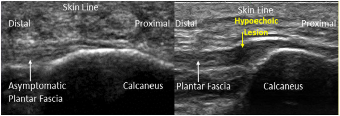
Introduction: Plantar fasciitis (PFS) is described by an intense pain over medial tubercle of calcaneus, increased with the first step after waking up, after rest and during weightbearing activity. It is the most common cause of plantar heel pain in adults with the prevalence estimated 10% of the general population. Ultrasound imaging is commonly being used to measure the PF thickness, evaluate the efficacy of different treatments and a guide therapeutic technique in patients with PFS. The objective of this study was to systematically review the studies that were previously published to evaluate the role of ultrasound in the assessment of PF in patients with PFS.
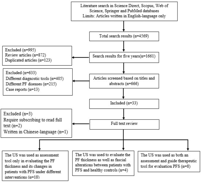
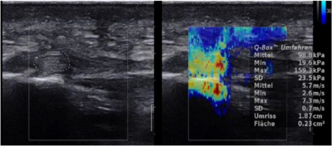
Methods: A systematic search was carried out over the last 5 years from 2017 to 2022 on basis the following electronic databases: Science Direct, Scopus, Web of Science, Springer and PubMed. The keywords that used in the searching were: ultrasound, sonography, ultrasonography, plantar fasciitis, imaging of plantar fascia, physiotherapy of plantar fasciitis, interventional treatment of plantar fasciitis, randomized controlled trial of plantar fasciitis and interventional ultrasound. The review focused on the assessment of PF in patients with PFS underwent different interventions using B-mode, shear wave elastography (SWE) and color Doppler ultrasound.
Results: During the search process, 1661 were recorded using the proper keywords from 2017 to 2022 in which 666 original articles were found after removing the review and duplicated articles. Of these, thirty articles met the inclusion criteria and included in this review. The articles have assessed the PF in patients with PFS under different conditions using different ultrasound modes. Twenty-six articles evaluated the effectiveness of different treatment on PF in patients with PFS using different ultrasound modes. In 8 of 26 articles, the ultrasound was used as both an assessment tool of PF and guide therapeutic technique in patients with PFS. In 18 articles, the ultrasound was used as only assessment tool to identify the PF thickness and its observation changes in patients with PFS. Four articles compared the PF thickness and its intrafascial changes between patients with PFS and healthy subjects.
Conclusion: The ultrasound can be a reliable tool in assessment the effect of different interventions on PF by evaluating its thickness, echogenicity and stiffness changes in patients with PFS. There were different methods and treatments were used among the studies.
Keywords: Imaging of plantar fascia; Interventional treatment of plantar fasciitis; Physiotherapy of plantar fasciitis; Plantar fasciitis; Sonography; Ultrasound.
© 2022. Società Italiana di Ultrasonologia in Medicina e Biologia (SIUMB).
Conflict of interest statement
The authors declare that they have no conflict of interest.
Figures
References
Publication types
MeSH terms
LinkOut - more resources
Full Text Sources
Miscellaneous
