. 2022 Aug 30;17(8):e0273663.
doi: 10.1371/journal.pone.0273663. eCollection 2022.

Solubility affects IL-1β-producing activity of the synthetic candidalysin peptide

Affiliations

Solubility affects IL-1β-producing activity of the synthetic candidalysin peptide

Taiki Mori et al. PLoS One. .

Abstract

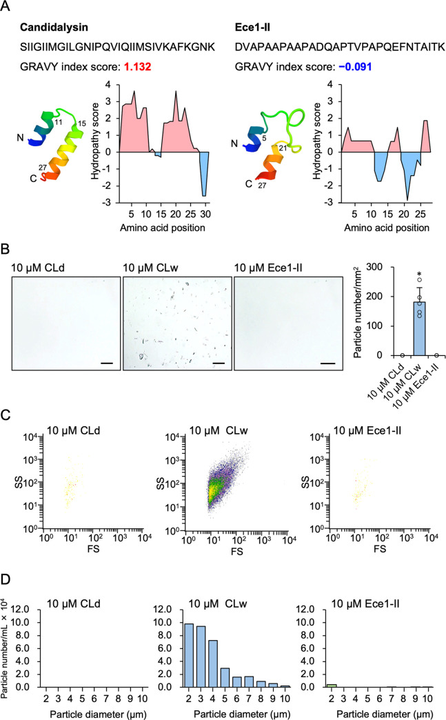
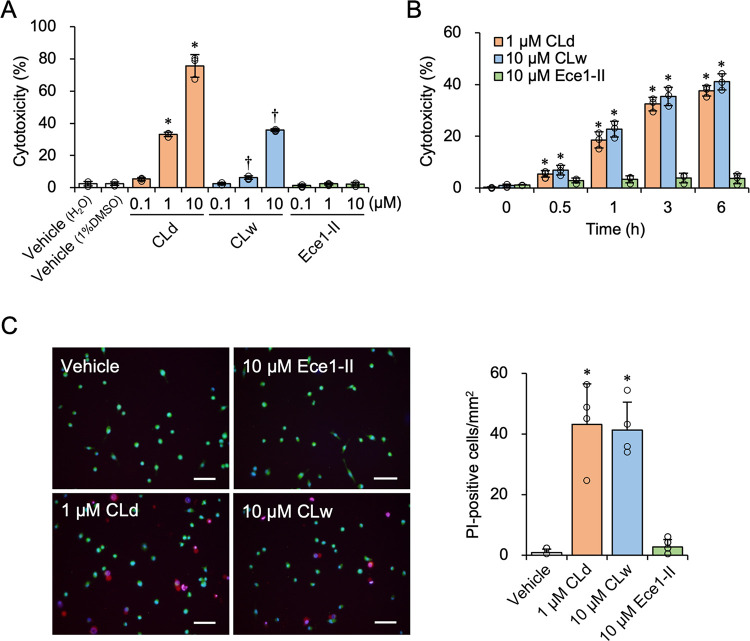
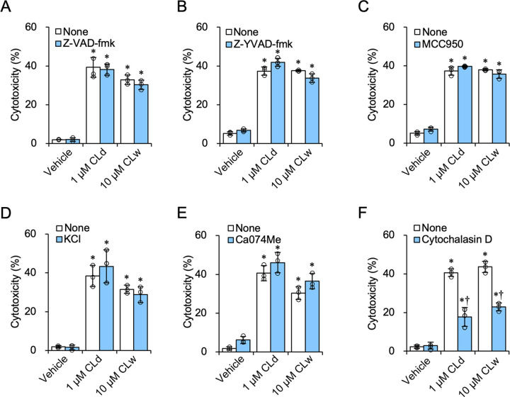
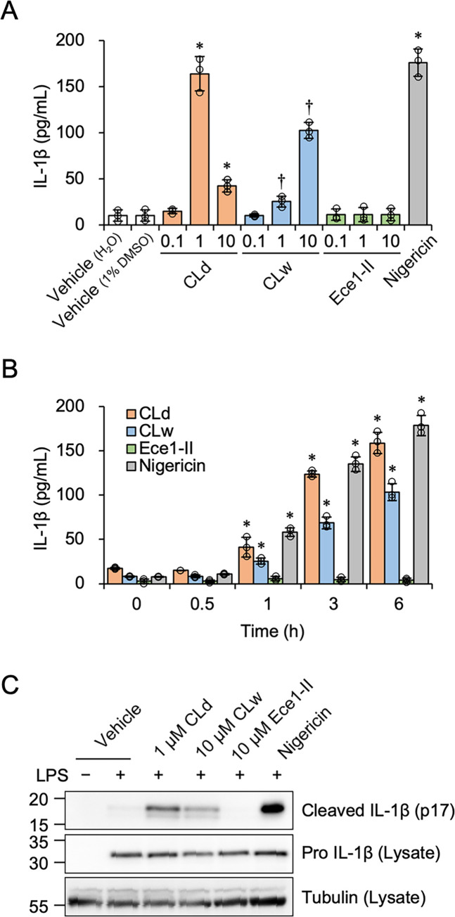
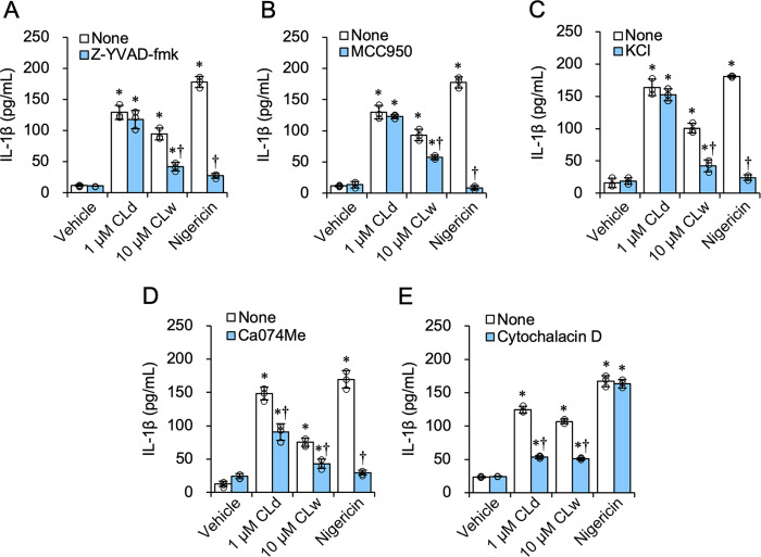
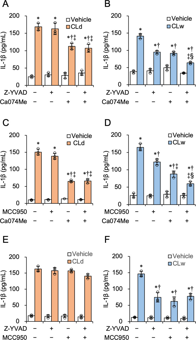
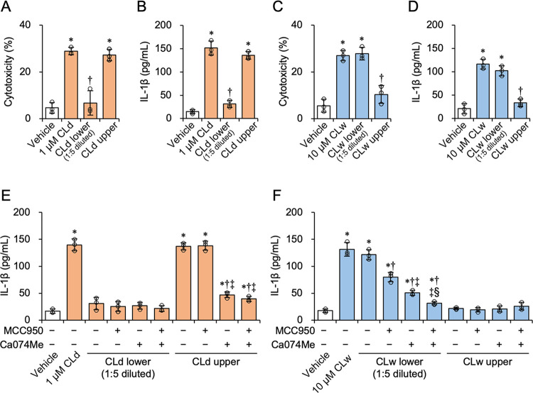
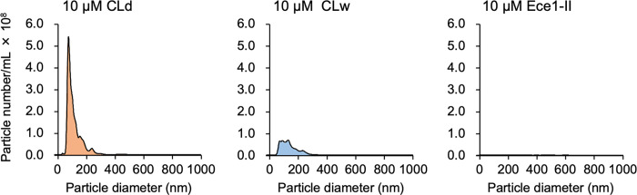
Candidalysin, a peptide toxin produced specifically from hyphae of Candida albicans, plays a crucial role in C. albicans pathogenesis in the oral cavity and vagina. Synthetic peptides have been widely used in previous studies to investigate the bioactivity of candidalysin. Although the solubility of the peptide, which is expected to have a hydrophobic property, has not been well characterized, candidalysin solutions are usually prepared in water. In this study, we prepared the synthetic peptide candidalysin in water (CLw) or in dimethyl sulfoxide (CLd) and compared their cytotoxicity and interleukin (IL)-1β-producing activity to determine whether the activity of the peptide would be affected. In addition, we evaluated whether the NOD-like receptor family pyrin domain-containing 3 (NLRP3) inflammasome pathway or other pathways were involved in their activities. Unexpectedly, we found that CLw was not completely solubilized and contained abundant insoluble microparticles. CLw was active at comparably high concentrations (≥ 10 μM). In contrast, CLd is completely solubilized and sufficiently active at low concentrations, that is, 1 μM or less. CLw showed weak cytotoxicity and NLRP3-dependent and cathepsin B-dependent IL-1β-producing activity, whereas CLd showed strong cytotoxicity and cathepsin B-dependent IL-1β-producing activity. Fractionation of CLw revealed that NLRP3-dependent activity was caused by insoluble microparticles. Furthermore, nanoparticle tracking of CLd revealed that the peptide was present as nanoparticles with a size of 96 nm. CLw contained a small amount of such nanoparticles. Thus, the bioactivities of the synthetic peptide candidalysin, especially the IL-1β-producing activity, are affected by the solubility of the peptide depending on the solvent employed. The NLRP3-dependent activity of the synthetic peptide is caused by insoluble microparticles and may not be the intrinsic activity of candidalysin.


Conflict of interest statement

The authors have declared that no competing interests exist.

Figures

Fig 1
Fig 1. Solubility of the synthetic candidalysin peptide.
A: Amino acid sequence, putative tertiary structure, GRAVY index score, and hydropathy plot with Kyte-Doolittle scale of candidalysin (left) and Ece1-II (right). A positive hydropathy score reflects an enrichment of hydrophobic amino acids. B: Microscopic images of 10 μM CLd, CLw, and the Ece1-II control peptide dissolved in water. Images were taken at 200× magnification. Scale bar, 100 μm. The quantified data of particle number/mm2 in the peptide solutions is shown in the chart. Data are presented as the mean ± SD (n = 5) and are representative of three independent experiments. *P < 0.05 compared with 10 μM Ece1-II by one-way ANOVA followed by Dunnett’s test (μc < μi). C: Plots of the particles detected in 10 μM CLd, CLw, and Ece1-II by the flowcytometric forward scatter (FS) and side scatter (SS) analyses. D: The number and size of microparticles in 10 μM CLd, CLw, and Ece1-II quantified using a microparticle counter. Data are representative of three independent experiments.
Fig 2
Fig 2. Cytotoxicity of CLd and CLw in differentiated THP-1 cells.
A: Cells were treated with varied concentrations of CLd, CLw, or Ece1-II for 3 h. The vehicle controls for CLd and CLw are a medium containing 1% DMSO and a water-added medium (H2O), respectively. Cytotoxicity was quantified by a LDH release assay. Data are presented as the mean ± SD (n = 3) and are representative of three independent experiments. *P < 0.05 compared with the vehicle (1% DMSO; μc < μi); P < 0.05 compared with the vehicle (H2O; μc < μi) by one-way ANOVA followed by Dunnett’s test. B: Cells were treated with 1 μM CLd, 10 μM CLw, or 10 μM Ece1-II for the indicated period (0−6 h). Cytotoxicity was quantified by an LDH release assay. Data are presented as the mean ± SD (n = 3) and are representative of three independent experiments. *P < 0.05 compared with 0 h by one-way ANOVA followed by Dunnett’s test (μc < μi). C: Cells were treated with 1 μM CLd, 10 μM CLw, or 10 μM Ece1-II for 3 h, followed by staining with PI (red, indicative of damaged cells), Calcein-AM (green, indicative of live cells), and Hoechst 33342 (blue, all cells). The vehicle control is a medium containing 1% DMSO. Fluorescence images were taken with a fluorescence microscope at 100× magnification. Scale bars, 100 μm. Quantified data (the number/mm2 of PI-positive cells) are presented as the mean ± SD (n = 4) and are representative of three independent experiments. *P < 0.05 compared with the vehicle by one-way ANOVA followed by Dunnett’s test (μc < μi).
Fig 3
Fig 3. Influence of the inhibitors on the cytotoxicity of CLd and CLw.
The differentiated THP-1 cells were pretreated with the following inhibitors: 20 μM of the pan-caspase inhibitor Z-VAD-fmk (A); 20 μM of the caspase-1 inhibitor Z-YVAD-fmk (B); 2 μM of the NLRP3 inhibitor MCC950 (C); 25 mM KCl for inhibition of K+ efflux (D); 20 μM of the cathepsin B inhibitor Ca074Me (E); and 20 μM of the phagocytosis inhibitor cytochalasin D (F) or with vehicle for 1 h. The vehicle control is a medium containing 2% DMSO. Cells were then treated with 1 μM CLd or 10 μM CLw for 3 h. Cytotoxicity was quantified by LDH release assay. Data are presented as the mean ± SD (n = 3) and are representative of three independent experiments. *P < 0.05 compared with the vehicle (μc < μi); P < 0.05 compared with the none (μc > μi) by one-way ANOVA followed by Dunnett’s test.
Fig 4
Fig 4. The IL-1β-producing activity of CLd and CLw in LPS-primed THP-1 macrophage-like cells.
A: Cells were treated with varied concentrations of CLd, CLw, Ece1-II, or 1 μM nigericin for 3 h. The vehicle controls for CLd and CLw are a medium containing 1% DMSO and a water-added medium (H2O), respectively. The IL-1β levels in the culture supernatants were assessed by ELISA. Data are presented as the mean ± SD (n = 3) and are representative of three independent experiments. *P < 0.05 compared with the vehicle (1% DMSO; μc < μi); P < 0.05 compared with the vehicle (H2O; μc < μi) by one-way ANOVA followed by Dunnett’s test. B: Cells were treated with 1 μM CLd, 10 μM CLw, 10 μM Ece1-II, or 1 μM nigericin for the indicated period (0−6 h). The IL-1β levels in the culture supernatants were assessed by ELISA. Data are presented as the mean ± SD (n = 3) and are representative of three independent experiments. *P < 0.05 compared with 0 h by one-way ANOVA followed by Dunnett’s test (μc < μi). C: Cells were treated with 1 μM CLd, 10 μM CLw, 10 μM Ece1-II, or 1 μM nigericin for 3 h. The vehicle control is a medium containing 1% DMSO. Cells that were not primed with LPS were also prepared. Culture supernatants were collected for the detection of the mature form of 17-kDa IL-1β (cleaved IL-1β; p17), and cell lysates were used for the detection of pro-IL-1β and tubulin by immunoblot analyses. Representative results of at least three independent experiments are shown. The uncropped images are shown in S1 Raw images.
Fig 5
Fig 5. Influence of the inhibitors on the IL-1β-producing activity of CLd and CLw.
LPS-primed THP-1 cells were pretreated with following inhibitors: 20 μM of the caspase-1 inhibitor Z-YVAD-fmk (A); 2 μM of the NLRP3 inhibitor MCC950 (B); 25 mM KCl for inhibition of K+ efflux (C); 20 μM of the cathepsin B inhibitor Ca074Me (D); and 20 μM of the phagocytosis inhibitor cytochalasin D (E) or with vehicle for 1 h. The vehicle control is a medium containing 2% DMSO. Cells were then treated with 1 μM CLd or 10 μM CLw for 3 h. The IL-1β levels in the culture supernatants were assessed by ELISA. Data are presented as the mean ± SD (n = 3) and are representative of three independent experiments. *P < 0.05 compared with the vehicle (μc < μi); P < 0.05 compared with the none (μc > μi) by one-way ANOVA followed by Dunnett’s test.
Fig 6
Fig 6. Influence of the simultaneous inhibition of the NLPR3 pathway and cathepsin B on the IL-1β-producing activity of CLd and CLw.
LPS-primed THP-1 cells were pretreated with following inhibitors: 20 μM of the caspase-1 inhibitor Z-YVAD-fmk and/or 20 μM of the cathepsin B inhibitor Ca074Me (A and B); 2 μM of the NLRP3 inhibitor MCC950 and/or 20 μM Ca074Me (C and D); and 20 μM Z-YVAD-fmk and/or 2 μM MCC950 (E and F) or with vehicle for 1 h. Cells were then treated with 1 μM CLd (A, C, and E) or 10 μM CLw (B, D, and F) for 3 h. The vehicle controls for CLd and CLw are a medium containing 1% DMSO (A, C, and E) and a water-added medium (B, D, and F), respectively. TheIL-1β levels in the culture supernatants were assessed by ELISA. Data are presented as the mean ± SD (n = 3) and are representative of three independent experiments. *P < 0.05 compared with the vehicle (μc < μi); P < 0.05 compared with the no inhibitor control (μc > μi); P < 0.05 compared with the single use of the first inhibitor (μc > μi); §P < 0.05 compared with the single use of the second inhibitor (μc < μi) by one-way ANOVA followed by Dunnett’s test.
Fig 7
Fig 7. Identification of bioactive fractions of CLd and CLw.
A-D: Fractionation of CLd and CLw was performed by centrifugation at 100 × g for 5 min at room temperature. The upper fractions were undiluted, and the lower fractions were diluted five times to restore to their original volume, being used equally with the uncentrifuged peptide solutions. The LPS-primed THP-1 macrophage-like cells were treated with the upper and lower fractions of CLd and CLw for 3 h. The vehicle controls for CLd and CLw are a medium containing 1% DMSO (A and B) and a water-added medium (C and D), respectively. Cytotoxicity was quantified by LDH release assay (A and C), and IL-1β levels in the culture supernatants were assessed by ELISA (B and D). Data are presented as the mean ± SD (n = 3) and are representative of three independent experiments. *P < 0.05 compared with the vehicle by one-way ANOVA followed by Dunnett’s test (μc < μi). E and F: For simultaneous inhibition assay, the LPS-primed THP-1 macrophage-like cells were pretreated with 2 μM of the NLRP3 inhibitor MCC950 and/or 20 μM Ca074Me or with vehicle for 1 h. Cells were then treated with the upper and lower fractions of CLd (E) and CLw (F) for 3 h. The vehicle controls for CLd and CLw are a medium containing 2% DMSO (E) and a medium containing 1% DMSO (F), respectively. The IL-1β levels in the culture supernatants were assessed by ELISA. Data are presented as the mean ± SD (n = 3) and are representative of three independent experiments. *P < 0.05 compared with the vehicle (μc < μi); P < 0.05 compared with the no inhibitor control (μc > μi); P < 0.05 compared with the single use of the first inhibitor (μc > μi); §P < 0.05 compared with the single use of the second inhibitor (μc < μi) by one-way ANOVA followed by Dunnett’s test.
Fig 8
Fig 8. Detection of nanoparticles in CLd and CLw.
Analyses of the number and size of nanoparticles in 10 μM CLd, CLw, and Ece1-II control peptide were performed using a NanoSight NS300 nanoparticle characterizer. Data are presented as the mean of five independent experiments.

Similar articles

Cited by

References

    1. König A, Hube B, Kasper L. The Dual Function of the Fungal Toxin Candidalysin during Candida albicans—Macrophage Interaction and Virulence. Toxins (Basel). 2020;12: 469. doi: 10.3390/toxins12080469 - DOI - PMC - PubMed
    1. Moyes DL, Wilson D, Richardson JP, Mogavero S, Tang SX, Wernecke J, et al.. Candidalysin is a fungal peptide toxin critical for mucosal infection. Nature. 2016;532: 64–68. doi: 10.1038/nature17625 - DOI - PMC - PubMed
    1. Richardson JP, Brown R, Kichik N, Lee S, Priest E, Mogavero S, et al.. Candidalysins Are a New Family of Cytolytic Fungal Peptide Toxins. Hogan DA, editor. MBio. 2022. doi: 10.1128/mbio.03510-21 - DOI - PMC - PubMed
    1. Brown R, Priest E, Naglik JR, Richardson JP. Fungal Toxins and Host Immune Responses. Front Microbiol. 2021;12: 1–19. doi: 10.3389/fmicb.2021.643639 - DOI - PMC - PubMed
    1. Westman J, Plumb J, Licht A, Yang M, Allert S, Naglik JR, et al.. Calcium-dependent ESCRT recruitment and lysosome exocytosis maintain epithelial integrity during Candida albicans invasion. Cell Rep. 2022;38: 110187. doi: 10.1016/j.celrep.2021.110187 - DOI - PMC - PubMed

Publication types

MeSH terms