. 2022 Oct 19;110(20):3374-3388.e8.
doi: 10.1016/j.neuron.2022.08.001. Epub 2022 Aug 29.

A locus coeruleus-dorsal CA1 dopaminergic circuit modulates memory linking

Affiliations

A locus coeruleus-dorsal CA1 dopaminergic circuit modulates memory linking

Ananya Chowdhury et al. Neuron. .

Abstract

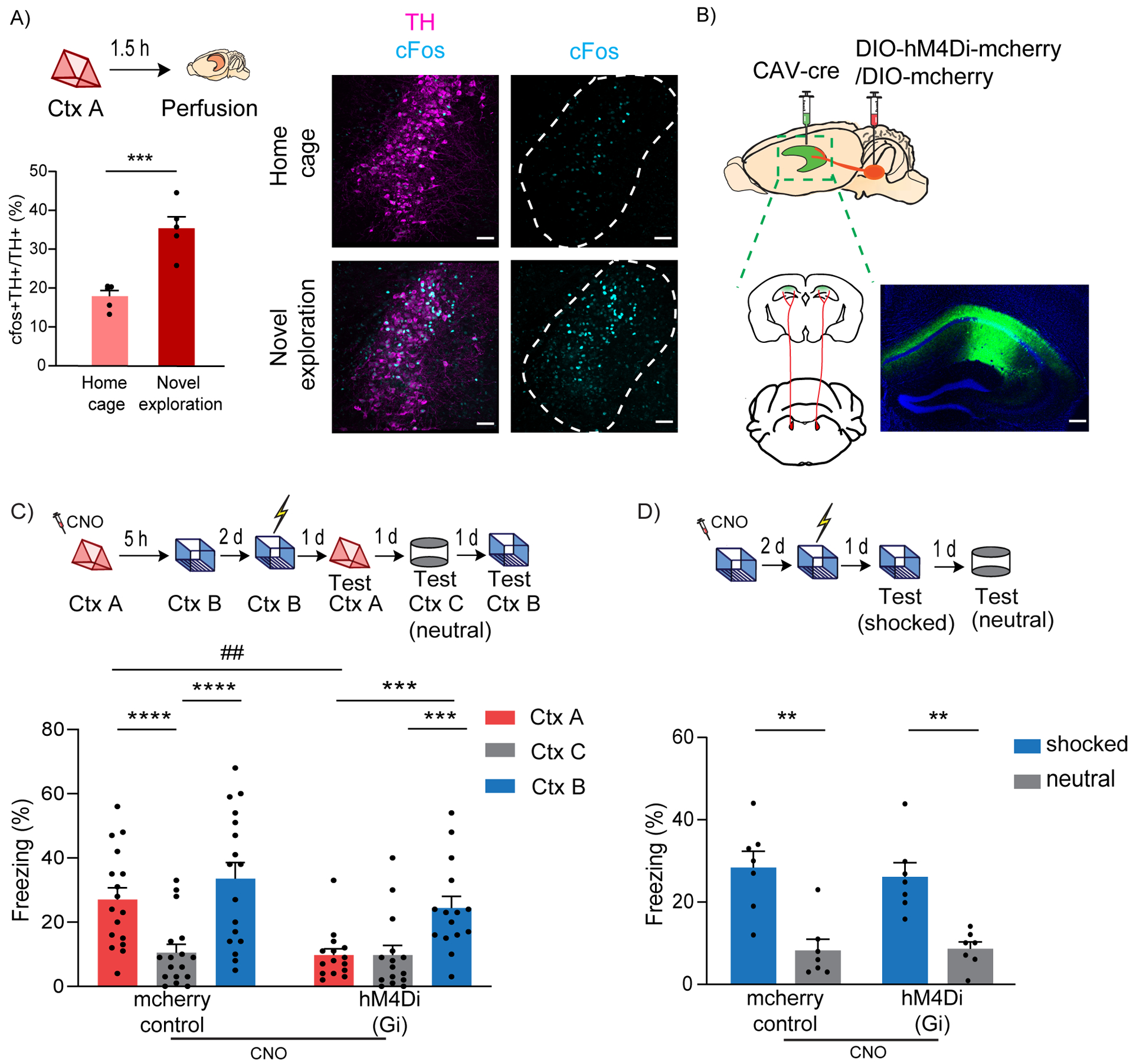
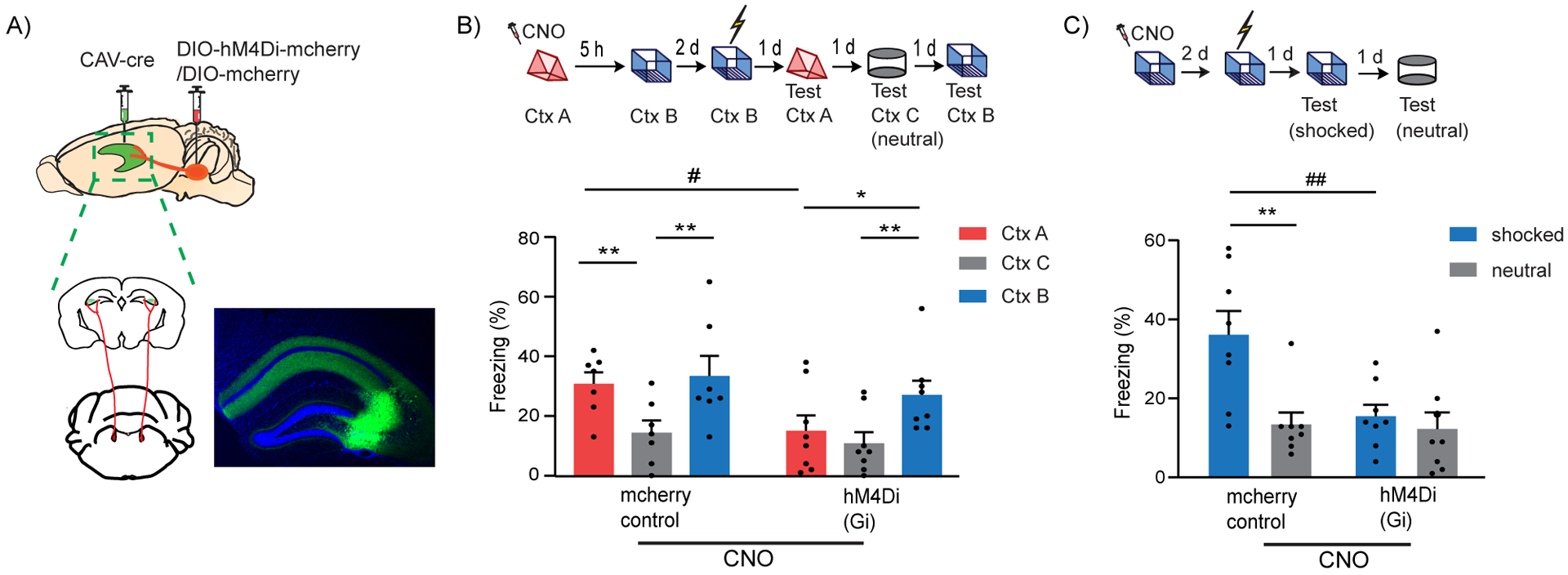
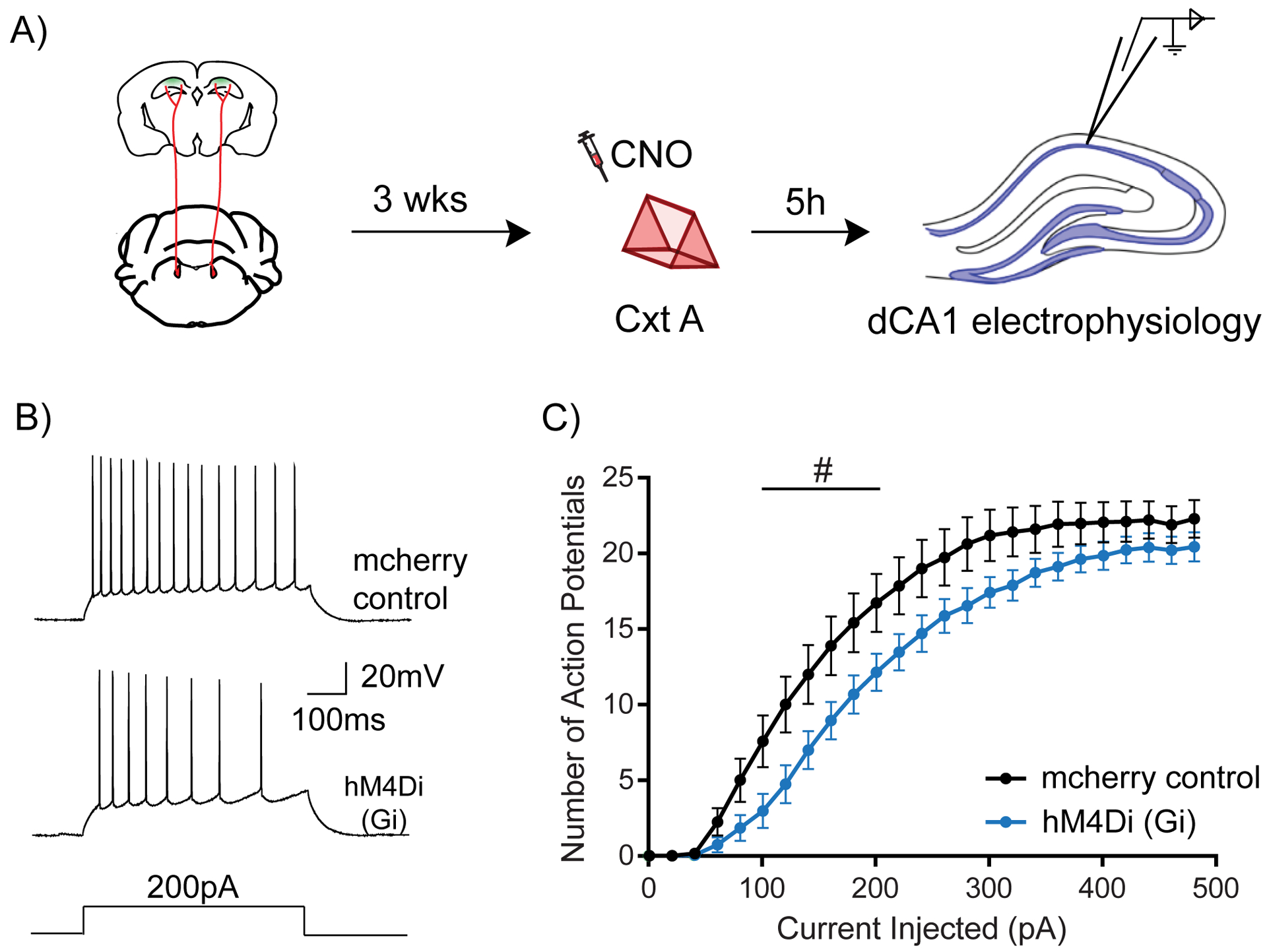
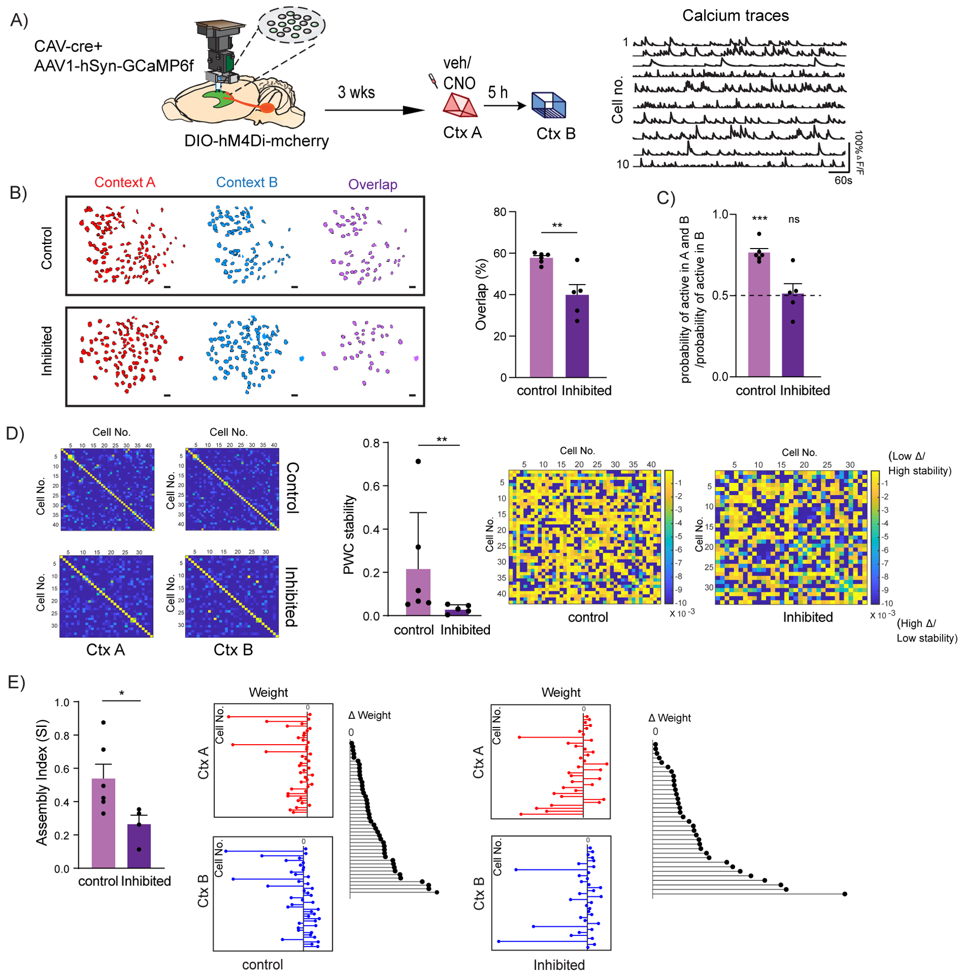
Individual memories are often linked so that the recall of one triggers the recall of another. For example, contextual memories acquired close in time can be linked, and this is known to depend on a temporary increase in excitability that drives the overlap between dorsal CA1 (dCA1) hippocampal ensembles that encode the linked memories. Here, we show that locus coeruleus (LC) cells projecting to dCA1 have a key permissive role in contextual memory linking, without affecting contextual memory formation, and that this effect is mediated by dopamine. Additionally, we found that LC-to-dCA1-projecting neurons modulate the excitability of dCA1 neurons and the extent of overlap between dCA1 memory ensembles as well as the stability of coactivity patterns within these ensembles. This discovery of a neuromodulatory system that specifically affects memory linking without affecting memory formation reveals a fundamental separation between the brain mechanisms modulating these two distinct processes.

Keywords: contextual memory; dopamine; dorsal hippocampus; ensembles; locus coeruleus; median raphe; memory linking; neuromodulation; neuronal excitability.


Conflict of interest statement

Declaration of interests The authors declare no competing interests.

Figures

Figure 1:
Figure 1:. LC to dCA1 projecting cells are required for contextual memory linking, but not for contextual conditioning
(A) Exploration of a novel context increased cFos expression in the TH+ cells of LC (unpaired t-test, n=5, ***p<0.001). Example images of TH and cFos staining in LC. Scale bar, 50μm. TH- magenta, cFos- cyan. The LC is outlined. (B) Schematics of experimental design for surgery. Example image for virus spread in HP estimated with AAV-DIO-GFP injected together with CAV-cre. Scale bar, 300μm. (C) Inhibition of LC cells projecting to dCA1 during exploration of context A impaired contextual memory linking tested with a 5 hours interval. (Control, n=17; LC inhibited, n=15. Two-way repeated measures ANOVA, Sidak post hoc. ##p <0.01, ***p<0.001, ****p<0.0001). Context A- Ctx A, Context B- Ctx B, Context C- Ctx C (neutral). CNO-Clozapine-N-oxide was given to all mice. * is used to depict significance within groups and # is used to show significance between groups for two-way RM ANOVA. (D) Inhibition of LC cells projecting to dCA1 did not affect contextual conditioning (Two-way RM ANOVA, Sidak posthoc test; control, n=7; LC inhibited, n=7, **p<0.001). CNO-Clozapine-N-oxide was given to all mice. All results are mean ± s.e.m.
Figure 2:
Figure 2:. LC to dCA3 projecting cells are required for contextual memory formation
(A) Schematics of experimental design for surgery. Example image for virus spread in HP estimated with AAV-DIO-GFP injected together with CAV-cre. Scale bar, 300μm. (B) Inhibition of LC cells projecting to dCA3 during exploration of context A impaired performance in contextual memory linking tested with a 5 hours interval. (Control, n=7; LC inhibited, n=8. Two-way repeated measures ANOVA, Sidak post hoc. #p <0.05, *p < 0.05, **p<0.01). Clozapine-N-oxide was given to all mice. * is used to depict significance within groups and # is used to show significance between groups for two-way RM ANOVA. (C) Inhibition of LC cells projecting to dCA3 impaired contextual conditioning (control, n=8; LC inhibited, n=8; Two-way RM ANOVA, Sidak posthoc test; **p<0.001, ##p<0.01). Clozapine-N-oxide was given to all mice. * is used to depict significance within groups and # is used to show significance between groups for two-way RM ANOVA. All results are mean ± s.e.m.
Figure 3:
Figure 3:. LC to dCA1 inhibition decreases novelty induced increases in neuronal excitability
(A) Experimental design for measuring dCA1 neuronal excitability. (B) Representative traces showing adaptive firing responses to a 200pA current injection in dCA1 pyramidal neurons. (C) Inhibition of LC cells projecting to dCA1 during context exploration reduced the firing rate of dCA1 neurons 5 hours later (Two-way repeated measures ANOVA; control n=15, LC inhibited n=15, #p<0.05). Clozapine-N-oxide was given to all mice. # is used to show significance between groups for two-way RM ANOVA. All results are mean + s.e.m.
Figure 4:
Figure 4:. LC to dCA1 inhibition decreases the overlap between dCA1 memory ensembles and affects their firing properties
(A) Schematics for miniscope setup and calcium signal imaging in dCA1. Example calcium traces. (B) Inhibition of LC neurons projecting to dCA1 reduced the percentage overlap between memory ensembles encoding contexts explored 5 hours apart (Control n=6, and LC inhibited n=5; unpaired t-test, **p<0.01). Percentage overlap was calculated as neurons active in both A and B over total cells active in A and B. Example plots of active neurons in contexts A and B and neuronal overlap between different conditions. Scale bars, 50μm. (C) Inhibition of LC neurons projecting to dCA1 reduced the likelihood to chance levels (dashed line) that a cell active in context B had previously been active in context A (Control n=6, and LC inhibited n=5; One-sample t-test over 0.5 as chance level, ***p< 0.001). (D) Within the overlapping neuronal population, inhibition of the LC neurons projecting to dCA1 reduced the stability of the coactivity maps between the two contexts visited (Control n=6, and LC inhibited n=5; Mann Whitney test, **p<0.01). Example PWC stability maps. (E) Inhibition of the LC neurons projecting to dCA1 reduced the stability of the dCA1 assemblies detected within the overlapping neuronal population (Control n=6, and LC inhibited n=4; unpaired t-test, *p<0.05). Representative images for weight distribution of the assemblies detected for context A (red), context B (blue), or delta between the two weights (black) with all neurons sorted in the same order in all 3 graphs. All results are mean + s.e.m.
Figure 5:
Figure 5:. Simulation of LC to dCA1 inhibition using a spiking network model
(A) Conceptual diagram of the spiking network model. The model includes excitatory neurons (gray) and subpopulations of interneurons (red). Synaptic inputs representing different memories terminate in overlapping dendrites. Two novel contexts are simulated as memories A and B, separated by 5h. (B) Firing rate of neurons when current input is applied directly to the somatic compartment of the 2-stage neurons under control (blue) and LC inhibition (orange) condition. Under conditions of LC blocking, the excitability of the neurons does not increase, n=50. (C) Simulation of LC to dCA1 inhibition resulted in a reduction of the overlapping neuronal population. Percentage overlap was calculated as neurons activated during both context A and B over the total active neurons in A and B. n=10 simulation trials, unpaired t-test, ****p<0.0001. The contour plots show population activation during encoding of memories in context A and B. The third column indicates the neurons which were active (ff > 10Hz) during both memories. (D) The sizes of activated populations (number of neurons with ff>10Hz) during the encoding of Ctx A and Ctx B, under different conditions. All results are mean + s.e.m.
Figure 6:
Figure 6:. Dopamine D1/D5 receptors in dCA1 modulate contextual memory linking
(A) Inhibition of Dopamine D1/D5 receptors in dCA1 during context A disrupted contextual memory linking. (Saline, n=12; SCH 0.5 mM, n=12; SCH 1.0 mM, n=7; two-way repeated measures ANOVA, Sidak post hoc, *p<0.05, **p<0.01, ***p<0.001, ****p<0.0001) (B) Inhibition of Dopamine D1/D5 receptors in dCA1 did not affect contextual memory formation at the doses used. (Saline, n=10; SCH 0.5 mM, n=10; SCH 1.0 mM, n=7; two-way repeated measures ANOVA, Sidak post hoc, ***p<0.001, ****p<0.0001). (C) Inhibition of β-adrenergic receptors in dCA1 did not affect contextual memory linking at the doses used. (Saline, n=14; Prop 5 mM, n=14; Prop 20 mM, n=15; two-way repeated measures ANOVA, Sidak post hoc, **p<0.01, ****p<0.0001) All results are mean + s.e.m.
Figure 7:
Figure 7:. Optogenetic D1 receptor activation in dCA1 rescues linking deficits caused by LC to dCA1 inhibition
(A) Schematics of the Opto-D1 construct. (B) Schematics of experimental design. Scale bars, 300μm. (C) Optogenetic activation of D1 receptor signaling in a fraction of all cell types in dCA1 rescued the contextual memory linking deficit caused by chemogenetic inhibition of LC cells projecting to dCA1. (GFP, n=8; opto-D1, n=11; two-way repeated measures ANOVA, Sidak post hoc,*p<0.05, **p<0.01, ***p<0.001, ****p<0.0001, ##p<0.01). * is used to depict significance within groups and # is used to show significance between groups for two-way RM ANOVA. Clozapine-N-oxide and light were given to all mice. (D) Optogenetic activation of the D1 receptor during context exploration rescued the reduction in dCA1 firing rate caused by chemogenetic inhibition of LC cells projecting to the dCA1. (GFP, n=11; opto-D1, n=10; two-way repeated measures ANOVA, factor: D1 activation F(1,19) = 4.50, #p<0.05). Clozapine-N-oxide and light were given to all mice. All results are mean + s.e.m.

Comment in

References

    1. Achterberg EJ, van Kerkhof LW, Servadio M, van Swieten MM, Houwing DJ, Aalderink M, Driel NV, Trezza V, and Vanderschuren LJ (2016). Contrasting Roles of Dopamine and Noradrenaline in the Motivational Properties of Social Play Behavior in Rats. Neuropsychopharmacology 41, 858–868. 10.1038/npp.2015.212. - DOI - PMC - PubMed
    1. Aharoni D, and Hoogland TM (2019). Circuit Investigations With Open-Source Miniaturized Microscopes: Past, Present and Future. Frontiers in cellular neuroscience 13, 141. 10.3389/fncel.2019.00141. - DOI - PMC - PubMed
    1. Aharoni D, Khakh BS, Silva AJ, and Golshani P (2019). All the light that we can see: a new era in miniaturized microscopy. Nat Methods 16, 11–13. 10.1038/s41592-018-0266-x. - DOI - PMC - PubMed
    1. Airan RD, Thompson KR, Fenno LE, Bernstein H, and Deisseroth K (2009). Temporally precise in vivo control of intracellular signalling. Nature 458, 1025–1029. 10.1038/nature07926. - DOI - PubMed
    1. Alejandre-García T, Kim S, Pérez-Ortega J, and Yuste R (2020). Intrinsic excitability mechanisms of neuronal ensemble formation. bioRxiv, 2020.2007.2029.223966. 10.1101/2020.07.29.223966. - DOI - PMC - PubMed

Publication types

LinkOut - more resources