. 2022 Nov;42(11):1185-1206.
doi: 10.1002/cac2.12356. Epub 2022 Aug 30.

AIMP1 promotes multiple myeloma malignancy through interacting with ANP32A to mediate histone H3 acetylation

Affiliations

AIMP1 promotes multiple myeloma malignancy through interacting with ANP32A to mediate histone H3 acetylation

Rongfang Wei et al. Cancer Commun (Lond). 2022 Nov.

Abstract

Background: Multiple myeloma (MM) is the second most common hematological malignancy. An overwhelming majority of patients with MM progress to serious osteolytic bone disease. Aminoacyl-tRNA synthetase-interacting multifunctional protein 1 (AIMP1) participates in several steps during cancer development and osteoclast differentiation. This study aimed to explore its role in MM.

Methods: The gene expression profiling cohorts of MM were applied to determine the expression of AIMP1 and its association with MM patient prognosis. Enzyme-linked immunosorbent assay, immunohistochemistry, and Western blotting were used to detect AIMP1 expression. Protein chip analysis, RNA-sequencing, and chromatin immunoprecipitation and next-generation sequencing were employed to screen the interacting proteins and key downstream targets of AIMP1. The impact of AIMP1 on cellular proliferation was determined using 3-(4,5-dimethylthiazol-2-yl)-2,5-diphenyltetrazolium bromide (MTT) assay in vitro and a xenograft model in vivo. Bone lesions were evaluated using tartrate-resistant acid phosphatase staining in vitro. A NOD/SCID-TIBIA mouse model was used to evaluate the effect of siAIMP1-loaded exosomes on bone lesion formation in vivo.

Results: AIMP1 expression was increased in MM patients and strongly associated with unfavorable outcomes. Increased AIMP1 expression promoted MM cell proliferation in vitro and in vivo via activation of the mitogen-activated protein kinase (MAPK) signaling pathway. Protein chip assays and subsequent experiments revealed that AIMP1 interacted with acidic leucine-rich nuclear phosphoprotein 32 family member A (ANP32A) to regulate histone H3 acetylation. In addition, AIMP1 increased histone H3 acetylation enrichment function of GRB2-associated and regulator of MAPK protein 2 (GAREM2) to increase the phosphorylation of extracellular-regulated kinase 1/2 (p-ERK1/2). Furthermore, AIMP1 promoted osteoclast differentiation by activating nuclear factor of activated T cells c1 (NFATc1) in vitro. In contrast, exosome-coated small interfering RNA of AIMP1 effectively suppressed MM progression and osteoclast differentiation in vitro and in vivo.

Conclusions: Our data demonstrate that AIMP1 is a novel regulator of histone H3 acetylation interacting with ANP32A in MM, which accelerates MM malignancy via activation of the MAPK signaling pathway.

Keywords: AIMP1; ANP32A; MAPK signaling; histone H3 acetylation; multiple myeloma; osteoclast differentiation; osteolytic lesions.


Conflict of interest statement

No potential conflicts of interest were disclosed.

Figures
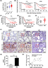
FIGURE 1
FIGURE 1
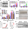
AIMP1 expression is higher in MM patients and negatively associated with poor survival. (A) AIMP1 mRNA levels were significantly higher in MM samples (GSE2658, n = 351) compared with the healthy individuals (Control, GSE5900, n = 22) and MGUS (GSE5900, n = 44) groups. (B) Higher AIMP1 expression was associated with shorter OS in the TT2 cohort (n = 351) and APEX cohort (n = 264). (C) Higher AIMP1 was associated with shorter EFS in the TT2 cohort (n = 351). (D) Higher AIMP1 expression was associated with shorter PFS in the APEX cohort (n = 264). (E) ELISA showed that serum AIMP1 levels were higher in MM patients than in healthy subjects (Control). (F) Representative images of IHC staining for AIMP1 and Ki67 antibodies in tissues samples from MM patients and healthy subjects (Control). (G) Quantitative analysis of IHC indicated that AIMP1 expression was higher in MM patients. (H) AIMP1 expression was positively associated with Ki67 expression. MM patient survival data was plotted using Kaplan‐Meier curve, and survival between patients with low and high expression was compared using a log‐rank test. Data are presented as the mean ± SD. *P < 0.05; **P < 0.01; ***P < 0.001. Abbreviations: AIMP1: Aminoacyl‐tRNA synthetase‐interacting multifunctional protein 1; MM: Multiple myeloma; MGUS: Monoclonal gammopathy of undetermined significance; OS: Overall survival; TT2: Total therapy 2; APEX: The assessment of proteasome inhibition for extending remission; EFS: Event‐free survival; PFS: Progression‐free survival; ELISA: Enzyme linked immunosorbent assay; IHC: Immunohistochemistry
FIGURE 2
FIGURE 2
Increased AIMP1 expression promotes MM cell proliferation in vitro and in vivo. (A) WB analysis of AIMP1 expression in AIMP1‐OE and AIMP1‐KD MM cells. (B) MTT assays were used to assess proliferation of AIMP1‐OE and AIMP1‐WT cells (n = 6). (C) MTT assays were used to assess proliferation of AIMP1‐KD and AIMP1‐WT cells (n = 6). (D) Exogenous AIMP1 protein (100 nmol/L) promoted proliferation of four MM cell lines. (n = 6). (E) Images of the harvested xenograft tumors established using AIMP1‐OE or AIMP1‐WT cells (n = 6). (F) Time course of tumor growth after implantation of AIMP1‐OE and AIMP1‐WT cells. (G) Mean tumor weight of NOD/SCID mice in AIMP1‐OE or AIMP1‐WT groups. (H) WB tested the expression of AIMP1 and Flag in AIMP1‐OE and AIMP1‐WT tumors. Data are presented as the mean ± SD; *P < 0.05; **P < 0.01; ***P < 0.001. Abbreviations: AIMP1: Aminoacyl‐tRNA synthetase‐interacting multifunctional protein 1; MM: Multiple myeloma; WB: Western blotting; MTT: 3‐(4,5‐Dimethylthiazol‐2‐yl)‐2,5‐Diphenyltetrazolium Bromide; WT: Wild type; OE: Overexpression; KD: Knockdown
FIGURE 3
FIGURE 3
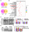
AIMP1 regulates MM cell proliferation via modulation of the MAPK signaling pathway. (A) Venn diagrams of co‐upregulated genes in two AIMP1‐OE MM cell lines and two AIMP1 protein‐treated MM cell lines compared with AIMP1‐WT cells. (B) KEGG pathway analysis of the RNA‐seq data indicated that AIMP1 was associated with the MAPK signaling pathway and osteoclast differentiation. (C) WB analysis of p‐ERK1/2 and ERK1/2 expressions in AIMP1‐OE and AIMP1‐KD cells. (D) Quantitative analysis of p‐ERK1/2 and ERK1/2 protein expressions in AIMP1‐OE and AIMP1‐KD cells compared with the AIMP1‐WT cells (n = 3). (E) AIMP1 protein activated p‐ERK1/2 expression in MM cells after treated for 2 h. (F) Quantitative analysis of p‐ERK1/2 and ERK1/2 protein expressions in AIMP1 protein‐treated cells compared with AIMP1‐WT cells (n = 3). Data are presented as the mean ± SD; *P < 0.05; P < 0.001. Abbreviations: AIMP1: Aminoacyl‐tRNA synthetase‐interacting multifunctional protein 1; MM: Multiple myeloma; OE: Overexpression; WT: Wild type; KD: Knockdown; KEGG: Kyoto Encyclopedia of genes and genes; RNA‐seq: RNA sequencing; MAPK: mitogen‐activated protein kinase; WB: Western blotting; p‐ERK1/2: Phosphorylated extracellular‐regulated kinase 1/2; ERK1/2: Extracellular‐regulated kinase 1/2
FIGURE 4
FIGURE 4
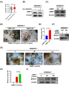
AIMP1 induces osteoclast differentiation via activation of NFATc1. (A) AIMP1 signaling in patients with serious bone lesions (n = 54) compared with patients with mild bone lesions (n = 42). (B) Co‐culture and WB assay indicated that AIMP1 was secreted from MM cells and impacted AIMP1 expression in RAW264.7 cells. (C) WB assay confirmed overexpression of AIMP1 in RAW264.7 cells. (D) TRAP staining revealed that increased AIMP1 expression promoted osteoclast differentiation (pointed with red arrows) in RAW 264.7 macrophages treated with RANKL and M‐CSF (n = 3). (E) Quantitative analysis of multinucleated osteoclasts. (F) WB assay confirmed that increased AIMP1 expression activated NFATc1 expression in RAW264.7 cells. (G) TRAP staining revealed that AIMP1 protein promoted osteoclast differentiation (pointed with red arrows) in RAW264.7 macrophages (n = 3). (H) Quantitative analysis of multinucleated osteoclasts. (I) NFATc1 expression was stimulated by AIMP1 protein. Data are presented as the mean ± SD; *P < 0.05; ***P < 0.001. Abbreviations: AIMP1: Aminoacyl‐tRNA synthetase‐interacting multifunctional protein 1; NFATc1: Nuclear factor of activated T cells c1; TRAP: Tartrate‐Resistant Acid Phosphatase; RANKL: Receptor activator of nuclear factor‐κB ligand; M‐CSF: Macrophage Colony stimulating Factor
FIGURE 5
FIGURE 5
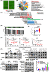
AIMP1 interacts with ANP32A to regulate histone H3 acetylation. (A) The Chip scan map of AIMP1 protein. The red arrows point to the positive control (GST, the concentrations: 10, 50, 100, 200 ng/μL), and the blue arrow indicates the negative control (BSA protein). (B) Pathway analysis of potential proteins in the KEGG database. (C) GO enrichment analysis of the proteins interacting with AIMP1 based on the GO database. (D) Top 15 potential AIMP1‐associated proteins, including ANP32A. (E) ANP32A mRNA levels were significantly elevated in the plasma cells from MM patients (n = 351) relative to those from MGUS patients (n = 44) and healthy individuals (Control, n = 22). (F) Increased ANP32A expression was associated with poor OS in the TT2 (n = 351), HOVON65 (n = 288) and APEX cohorts (n = 264). MM patient survival data were plotted using a Kaplan‐Meier curve, and the difference in survival between high and low expression groups were compared using a log‐rank test. (G) Co‐IP assays confirmed the physical interaction between AIMP1 and ANP32A. (H) WB analysis of acetyl‐H3 and histone‐H3 in AIMP1‐OE and AIMP1‐KD cells. (I) AIMP1 protein stimulated the expression of acetyl‐H3. (J) Interfering ANP32A by siRNA in AIMP1‐OE cells decreased acetyl‐H3 expression. (K) Reduced ANP32A repressed MM cell proliferation. Data are presented as the mean ± SD; *P < 0.05; ***P < 0.001. Abbreviations: AIMP1: Aminoacyl‐tRNA synthetase‐interacting multifunctional protein 1; ANP32A: Acidic leucine‐rich nuclear phosphoprotein 32 family member A; KEGG: Kyoto Encyclopedia of genes and genes; GO: Gene Ontology; MM: Multiple myeloma; MGUS: Monoclonal gammopathy of undetermined significance; HOVON65: The Dutch‐Belgian Cooperative Trial Group for Hematology Oncology Group‐65; APEX: The assessment of proteasome inhibition for extending remission; Co‐IP: Co‐immunoprecipitation; WB: Western blotting; OE: Overexpression; KD: Knockdown
FIGURE 6
FIGURE 6
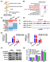
Elevated AIMP1 increases acetyl‐H3 enrichment function of GAREM2. (A) Schematic of the CHIP‐seq procedure. (B) KEGG pathway analysis of the data obtained from CHIP‐seq. (C) Heatmap of the acetyl‐H3‐mediated enrichment function of 10 genes, including GAREM2, in AIMP1‐OE and AIMP1‐WT cells. (D) Genome browser view of acetyl‐H3 signaling on GAREM2 in AIMP1‐OE and AIMP1‐WT cells. (E) ChIP‐qPCR showed increased acetyl‐H3 enrichment function of GAREM2 in AIMP1‐OE cells compared with AIMP1‐WT cells. (F) siGAREM2 downregulated p‐ERK1/2 expression. (G) MTT assay tested the effects of siGAREM2 on cell proliferation. Data are presented as the mean ± SD; *P < 0.05; **P < 0.01; ***P < 0.001. Abbreviations: AIMP1: Aminoacyl‐tRNA synthetase‐interacting multifunctional protein 1; GAREM2: GRB2‐associated and regulator of MAPK protein 2; CHIP‐seq: Chromatin Immunoprecipitation and next‐generation sequencing; OE: Overexpression; WT: Wild type; KD: Knockdown; p‐ERK1/2: Phosphorylated extracellular‐regulated kinase 1/2; ERK1/2: Extracellular‐regulated kinase 1/2
FIGURE 7
FIGURE 7
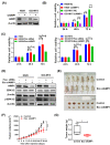
siAIMP1‐loaded exosomes inhibit MM cell proliferation in vitro and in vivo. (A) WB confirmation of AIMP1 expression in siAIMP1‐transfected cells. (B) MTT assay examined the effects of siAIMP1 on MM cell proliferation. (C) MTT assay indicated that siAIMP1‐loaded exosomes inhibited the proliferation of AIMP1‐WT H929 and OCI‐MY5 cells (siAIMP1‐loaded exosomes: 0.1 μg/200 μL/well). (D) WB analysis showed that siAIMP1‐loaded exosomes inhibited p‐ERK1/2 expression in MM cells (1 × 10⁶ MM cells treated with 20 μg siAIMP1‐load exosomes, 0.2 nmol/L free siRNA, or no treatment, all after 48 h). (E) Images of tumors harvested from the PDX mouse models in Control and Exo siAIMP1 groups. (F) Time course of tumor growth in Control and Exo siAIMP1 groups. (G) Mean tumor weight of the mice in Control and Exo siAIMP1 groups. Data are presented as the mean ± SD; *P < 0.05; **P < 0.01; ***P < 0.001. Abbreviations: AIMP1: Aminoacyl‐tRNA synthetase‐interacting multifunctional protein 1; MM: Multiple myeloma; WB: Western blotting; MTT: 3‐(4,5‐Dimethylthiazol‐2‐yl)‐2,5‐Diphenyltetrazolium Bromide; p‐ERK1/2: Phosphorylated extracellular‐regulated kinase 1/2; ERK1/2: Extracellular‐regulated kinase 1/2; PDX: Patient‐derived tumor xenograft
FIGURE 8
FIGURE 8
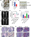
siAIMP1‐loaded exosomes inhibit osteoclast differentiation and reduce bone destruction. (A) TRAP staining revealed that siAIMP1‐loaded exosomes inhibited osteoclast differentiation (pointed with red arrows) in RAW264.7 macrophages treated with RANKL and M‐CSF (siAIMP1‐load exosomes: 2 μg/1 mL/well). (B) Quantitative analysis of multinucleated osteoclasts. (C) Images of NOD/SCID‐TIBIA mice in Control and Exo siAIMP1 groups. (D) Representative micro‐CT images of the bones in Control and Exo siAIMP1 groups. (E) BMD of NOD/SCID‐TIBIA mice in Control and Exo siAIMP1 groups. (F) BV/TV of NOD/SCID‐TIBIA mice in Control and Exo siAIMP1 groups. (G) H&E staining of histological sections of the bones in Control and Exo siAIMP1 groups. (H) TRAP staining of histological sections of bones (pointed with red arrows) in Control and Exo siAIMP1 groups. Data are presented as the mean ± SD; *P < 0.05; **P < 0.01. Abbreviations: AIMP1: Aminoacyl‐tRNA synthetase‐interacting multifunctional protein 1; TRAP: Tartrate‐Resistant Acid Phosphatase; BMD: Bone mineral density; BV/TV: Bone volume / Total volume; H&E: Hematoxylin‐eosin
FIGURE 9
FIGURE 9
Schematic depiction of the potential role of AIMP1 in MM. Abbreviations: AIMP1: Aminoacyl‐tRNA synthetase‐interacting multifunctional protein 1; ANP32A: Acidic leucine‐rich nuclear phosphoprotein 32 family member A; GAREM2: GRB2‐associated and regulator of MAPK protein 2; ERK1/2: Extracellular‐regulated kinase 1/2

Similar articles

Cited by

References

    1. Bianchi G, Munshi NC. Pathogenesis beyond the cancer clone(s) in multiple myeloma. Blood. 2015;125(20):3049–58. - PMC - PubMed
    1. Rajkumar SV, Dimopoulos MA, Palumbo A, Blade J, Merlini G, Mateos MV, et al. International Myeloma Working Group updated criteria for the diagnosis of multiple myeloma. Lancet Oncol. 2014;15(12):e538–48. - PubMed
    1. Rome S, Noonan K, Bertolotti P, Tariman JD, Miceli T. Bone Health, Pain, and Mobility: Evidence‐Based Recommendations for Patients With Multiple Myeloma. Clin J Oncol Nurs. 2017;21(5 Suppl):47–59. - PubMed
    1. Yang Y, Li Y, Gu H, Dong M, Cai Z. Emerging agents and regimens for multiple myeloma. J Hematol Oncol. 2020;13(1):150. - PMC - PubMed
    1. Westhrin M, Kovcic V, Zhang Z, Moen SH, Nedal TMV, Bondt A, et al. Monoclonal immunoglobulins promote bone loss in multiple myeloma. Blood. 2020;136(23):2656–66. - PubMed

Publication types